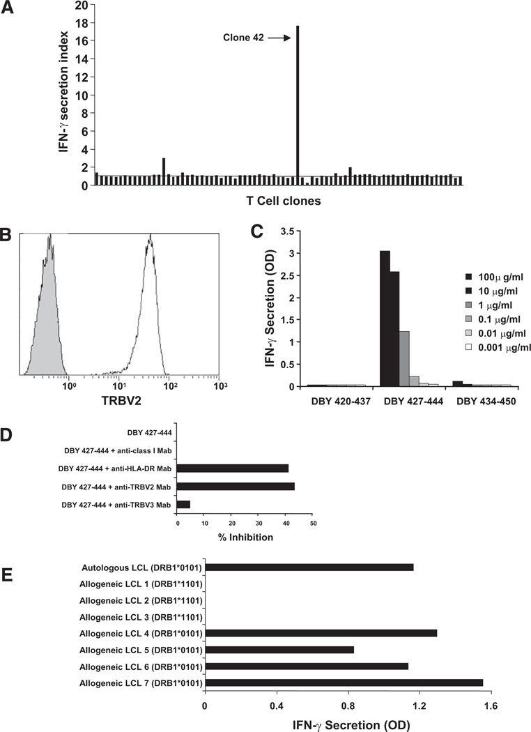
FIGURE 2.
The CD4+ T-cell response to DBY 427–444 is restricted by human leukocyte antigen (HLA)-DRB1*0101 molecules. (A) CD4+ T-cell clones established from the patient blood collected 31 months posttransplant were stimulated with donor Epstein-Barr virus (EBV) B-cells pulsed with peptide DBY 427–444. IFN-γ secretion was measured after overnight culture. Clone 42 displayed the high reactivity towards the antigenic peptide. (B) Flow cytometry analysis of clone 42 stained with an anti-TRBV2 monoclonal antibody (white histogram) or with an isotype control antibody (gray histogram). (C) The reactivity of clone 42 stimulated with increasing doses of peptide DBY 427–444 or 2 overlapping 18-mer DBY peptides was measured by IFN-γ secretion. (D) CD4+ T-cell clone 42 stimulation with autologous EBV-immortalized B cells (autologous cells were prepared from the patient’ blood when hematopoietic chimerism was 100%) pulsed with DBY peptide 427–444 or a control peptide (DBX 201–218) was assessed by measuring IFN-γ secretion in the culture supernatant. On the target side, antigen recognition was blocked with anti-HLA-DR mAb (G46.6, BD biosciences) but not anti-HLA class I mAb (W6/32, Sigma Aldrich). On the T-cell side, antigen recognition was blocked with anti-TCRB2 mAb but not with anti-TCRB3 mAb. (E) Clone 42 was stimulated with a series of third-party allogeneic Epstein-Barr virus (EBV)-B cells (LCL) sharing the expression of HLA-DRB1*0101 or HLA-DB1*1101 with the patient after pulsing with DBY 427–444. IFN-γ secretion was measured in the culture supernatant after overnight incubation.

