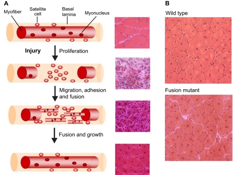
Fig. 2.
Muscle regeneration in adult mouse muscle. (A) Each myofiber in adult muscle is surrounded by a basal lamina sheath underneath which lie satellite cells in close apposition to the fiber. In response to injury, segmental necrosis of the myofiber occurs and satellite cells begin to proliferate and form myoblasts. These myoblasts differentiate and then migrate, adhere and fuse with one another to form multiple myotubes within the basal lamina sheath. Myoblasts/myotubes fuse with the stumps of the surviving myofiber and myotubes also fuse with each other to repair the injured myofiber. Regenerated myofibers are easily identified by the presence of centrally located nuclei. For each stage of regeneration in the schematic, a representative mouse muscle section is shown in cross-section and stained with Hematoxylin and Eosin to illustrate the morphological features of the tissue. (B) Cross-sections of regenerated myofibers 14 days after injury from wild-type and mutant mice stained with Hematoxylin and Eosin. Smaller myofibers are observed in the fusion mutant. Note the presence of centrally nucleated myofibers, a hallmark of muscle regeneration.

