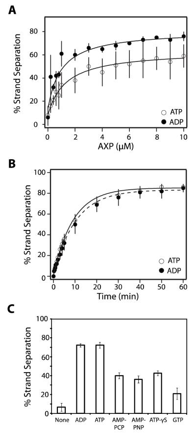
FIGURE 3. Nucleotides Stimulate Strand Separation Activity in the Absence of IHF.
Panel A. The strand separation assay was performed for 15 minutes as described except that IHF was omitted and the indicated concentration of nucleotide was included in the reaction mixture. Each data point represents the average of at least three separate experiments with standard deviation indicated with a bar. The solid lines represent the best fit of the data to equation 2. Panel B. The strand separation assay was performed in the absence of IHF and the presence of 10 μM nucleotide for the indicated time. Each data point represents the average of at least three separate experiments with standard deviation indicated. The solid lines represent the best fit of the data to equation 1. Panel C. The strand separation assay was performed as described except that IHF was omitted and ATP was replaced with the indicated nucleotide at a concentration of 10 μM AMP-PNP, β,γ-imidoadenosine 5′-triphosphate, AMP-PCP, β,γ-methylene-adenosine 5′-triphosphate. Each bar represents the average of at least three separate experiments with standard deviation indicated. We note that there is some day-to-day variability in the observed extent to which the strand separation reaction is stimulated by ATP (but not ADP); this is most evident in the data presented in panel A. Notwithstanding, the ensemble of data indicate that there is little difference between the two nucleotides in their capacity to stimulate strand separation (compare Figures 2, 3, 4, 5, and 6).

