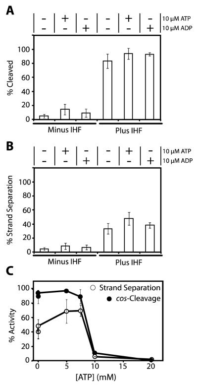
FIGURE 6. Coupled cos-Cleavage Endonuclease and Strand Separation Activities of λ Terminase.
Panel A. The cos-cleavage assay was performed as described in Experimental Procedures in the absence or presence of 50 nM IHF and nucleotide, as indicated. The samples were heated prior to loading onto the gel to ensure physical separation of the nicked strands. Each bar represents the average of at least three separate experiments with standard deviation indicated. Panel B. The in situ strand separation assay was performed as described in Experimental Procedures in the absence or presence of 50 nM IHF and nucleotide, as indicated. The samples were not heated prior to loading onto the gel and only those strands separated by terminase are observed. Each bar represents the average of at least three separate experiments with standard deviation indicated. Panel C. The cos-cleavage and strand separation assays were performed as described except that nucleotide was added to the reaction mixture as indicated. Each data point represents the average of at least three separate experiments with standard deviation indicated with a bar.

