Abstract
Recently, RccHanTM:WIST (Wistar Hannover) rats were introduced to toxicity studies in Japan. The present study was performed to obtain control data for general toxicological parameters as an aid for interpretation of results in toxicity studies using this strain of rats. Four test groups comprising of 25 male and 25 female RccHanTM:WIST rats were housed for 2, 4, 13 or 26 weeks from 6 weeks of age and observed and examined for clinical observation, body weight, food consumption, urinalysis, hematology, blood chemistry, organ weight, necropsy and/or histopathology. Ophthalmological examination was not conducted in this study, and the data in this report were obtained from an ongoing 104-week background study in RccHanTM:WIST rats. These data were compared with the historical control data of CD(SD) (Sprague-Dawley) and/or F344 (Fischer) rats. The body weights of RccHanTM:WIST rats were lower than those of CD(SD) rats and higher than those of F344 rats. The ophthalmological examination revealed a greater incidence of focal corneal opacity. Histopathology revealed focal mineralization of the cornea and Berlin blue-positive pigmentation in the epididymal interstitium as well as hepatocytes. Other than the above, some minor differences were found in urinalysis, hematology, blood chemistry and organ weights as compared with CD(SD) rats.
Keywords: RccHanTM:WIST rats, background data, general toxicity parameters
Introduction
A few years ago, RccHanTM:WIST (Wistar Hannover) rats were introduced to Japan as experimental animals for toxicity testing of pharmaceuticals etc., being ostensibly able to achieve a high survival rate in long-term experiments and easy to handle due to their relatively small body size and gentle nature. Wistar Hannover rats have been widely used in Europe as experimental animal models in toxicity studies in rodents. In many institutes including our laboratory in Japan, CD(SD) (Sprague-Dawley) rats are used predominantly as experimental models in toxicity studies, and F344 (Fischer) rats have been used as traditional animal models for assessing carcinogenic potential of various kinds of compounds. However, an epoch-making decision was made by NTP (National Toxicology Program), that is, the animal model in rat carcinogenicity studies was changed from F344 rats to Wistar Hannover rats1. This decision exerted an impact to a certain extent on researchers in the field of toxicology, and Wistar Hannover rats began to be used in toxicity studies in Japan, but relatively little data are available. In addition, the original Wistar Hannover rats were reported to have a serious defect of thyroid function and morphology2,3, and breeders made efforts to improve this defect via genetic cleaning. Under these circumstances, we face an urgent task to collect background data on Wistar Hannover rats. In this paper, we report historical control data of general toxicity parameters to clarify the biological nature of RccHanTM:WIST rats for up to week 26 of experiments (32 weeks of age), and the data were also compared with data for CD(SD) and/or F344 rats, though comparison with F344 rats was limited to body weight and food consumption, since this strain of rats is used infrequently in general toxicity studies in our laboratory.
Materials and Methods
Animals, animal husbandry and group composition
One hundred and ten male and 110 female RccHanTM:WIST SPF rats were obtained from Japan Laboratory Animals, Inc. (Tokyo, Japan) at 4 weeks of age. The animals were quarantined and acclimatized for 2 weeks and used at 6 weeks of age. They were housed individually in stainless steel wire mesh cages in an animal room that was maintained at 23 ± 3 °C and 50 ± 20% relative humidity and with air ventilation 10 to 15 times per hour and a 12-hour light dark cycle. Pelleted diet (radiation sterilized CR-LPF, Oriental Yeast Co., Ltd., Tokyo, Japan) and drinking water (tap water, via automatic water supply system) were provided ad libitum. Four test groups were created, 2-, 4-, 13- and 26-week experimental groups, and each group consisted of 25 animals of each sex.
Clinical observations, body weight and food consumption
Clinical observation was performed at least once a day, and body weight was measured twice a week until week 13 and once a week thereafter. Food consumption was measured once a week.
Ophthalmology
Ophthalmological examination was not conducted in this study, and the data in this report were obtained from an ongoing 104-week background study in RccHanTM:WIST rats, which was started at the same time as the present study. In that study, examinations were performed on 100 males and 100 females in weeks 4, 13 and 26 of the experiment under the same conditions as the present study. A mydriatic agent (Mydrine® P, Santen Pharmaceutical Co., Ltd., Osaka, Japan) was applied to dilate the pupil, the anterior portion and optic media were then examined using a slit lamp (SL-15, Kowa Co., Ltd., Aichi, Japan) and the fundus was examined using an ophthalmoscope (Omega 200, Heine Optotechnik, Herrsching, Germany).
Urinalysis
In weeks 4, 13 and 26, all animals in the 26-week experimental group were examined. The following parameters were examined in 4-hour urine: pH, protein, ketone body, glucose, occult blood, bilirubin and urobilinogen (Multistix® test paper, Siemens Medical Solutions Diagnostics, Tarrytown, NY, USA), color and sediment. The following parameters were examined in 20-hour urine: osmotic pressure (AUTO & STAT OM-6030, Arkray, Inc., Kyoto, Japan), sodium, potassium and chloride (Automatic Electrolyte Analyzer PVA-α II, A&D Co., Ltd., Kanagawa, Japan). Water intake (24-hour) and urinary volume were also measured.
Hematology
At the time of necropsy at weeks 2, 4, 13 and 26, blood samples were collected from the abdominal aorta under diethyl ether anesthesia into blood collection tubes (SB-41, Sysmex Corporation, Hyogo, Japan) containing EDTA-2 K. Prior to blood sample collection, the animals were deprived of food overnight. The parameters determined were red blood cell count (RBC), hemoglobin (HGB), hematocrit (HCT), mean corpuscular volume (MCV), mean corpuscular hemoglobin (MCH), mean corpuscular hemoglobin concentration (MCHC), reticulocyte ratio, platelet count, white blood cell count (WBC) and leukocyte differentiation (ADVIA® 120 Hematology System, Siemens Healthcare Diagnostics Inc., Deerfield, IL, USA). In addition, prothrombin time (PT), activated thromboplastin time (APTT) and fibrinogen (ACL Elite Pro System, Instrumentation Laboratory, Lexington, MA, USA) were determined in the plasma obtained by centrifuging the blood samples treated with 3.8 w/v% sodium citrate.
Blood chemistry
At the same time as hematology, the following parameters were determined in the serum: ALP, total cholesterol (T-CHO), triglyceride (TG), phospholipid (PL), total bilirubin (T-BIL), glucose (GLU), blood urea nitrogen (BUN), creatinine (CRNN), sodium (Na), potassium (K), chloride (Cl), calcium (Ca), inorganic phosphorus (P), total protein (TP), albumin (ALB) (Clinical Laboratory System TBA-120FR, Toshiba Medical Systems Corporation, Tokyo, Japan), globulin, A/G ratio and protein fraction (QuickScan Densitometer, Helena Laboratories, Inc., Saitama, Japan). In addition, the following parameters were determined in the plasma obtained from blood treated with heparin: aspartate aminotransferase (AST), alanine aminotransferase (ALT), lactate dehydrogenase (LDH) and creatine phosphokinase (CPK) (Clinical Laboratory System TBA-120FR, Toshiba Medical Systems Corporation, Tokyo, Japan).
Pathology
After collection of blood samples, the animals were exsanguinated and subjected to complete necropsy. Then, weights of the following organs were measured: brain, pituitary, thyroids (including parathyroids), adrenals, thymus, spleen, heart, lung (including bronchus), salivary glands (submandibular plus sublingual glands), liver, kidneys, testes/ovaries, prostate/uterus and seminal vesicles. Based on the above wet weight (absolute weight) and body weight at necropsy, organ weight per 100 g body weight (relative weight) was calculated. In addition to the above organs, the following organs were fixed with neutral buffered 10% formalin (however, eyeballs and optic nerves were fixed with fixatives containing 3% glutaraldehyde and 2.5% formalin, and testes and epididymides were fixed with Bouin’s solution and preserved in neutral buffered 10% formalin): spinal cord, sciatic nerves, Harderian glands, submandibular lymph node, mesenteric lymph node, thoracic aorta, trachea, tongue, esophagus, stomach, duodenum, jejunum, ileum, cecum, colon, rectum, pancreas, urinary bladder, epididymides, vagina, mammary gland, sternum (including bone marrow), femur (including bone marrow), femoral skeletal muscle, skin and any gross lesions. All of these organs/tissues were embedded in paraffin, sectioned and stained with hematoxylin and eosin (HE) and examined histopathologically.
The historical control data of CD(SD) [Crl:CD(SD)] rats fed with CR-LPF and F344 [F344/DuCrlCrlj] rats fed with CRF-1 were gathered from the control groups in toxicity studies conducted in our laboratory during the last 5 years.
The experiment was conducted in compliance with the laws or guidelines relating to animal welfare including Standards Relating to the Care and Management, etc. of Experimental Animals (Notification No. 6 of the Prime Minister’s Office, Japan, March 27, 1980) and Guidelines for Animal Experimentation (the Japanese Association for Laboratory Animal Science, May 22, 1987).
Results
Mortality and clinical signs
During the 26-week experimental period, no deaths occurred in either sex, and no abnormalities of the clinical signs were observed. Additionally, RccHanTM:WIST rats were relatively restless and ropable upon handling in comparison with CD(SD) rats.
Body weights
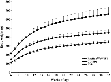
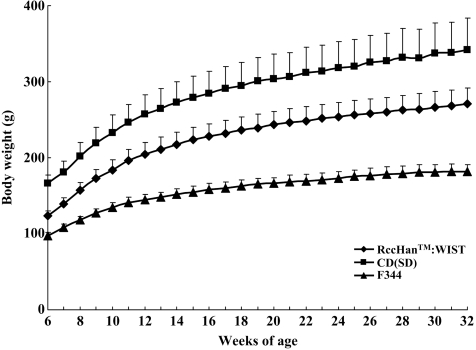
The growth curve of the RccHanTM:WIST rats was lower than that of the CD(SD) rats and higher than that of the F344 rats for both sexes during the experimental period (Figs. 1 and 2). The mean value for body weight of the RccHanTM:WIST rats at 32 weeks of age was 451 g in males and 271 g in females, and these values were approximately 70% to 80% of those of the CD(SD) rats (mean value: 660 g in males and 342 g in females) and approximately 130% to 150% of those of the F344 rats (mean value: 342 g in males and 182 g in females).
Fig. 1.
Body weight changes in male RccHanTM:WIST rats. For comparison, background data of body weight in our laboratory of CD(SD) rats and F344 rats are shown.
Fig. 2.
Body weight changes in female RccHanTM:WIST rats. For comparison, background data of body weight in our laboratory of CD(SD) rats and F344 rats are shown.
Food consumption
The food consumption (g/animal/day) of the male RccHanTM:WIST rats was lower than that of the CD(SD) rats and higher than that of the F344 rats during the experimental period (data not shown), while the food consumption of the female RccHanTM:WIST rats was comparable to or slightly lower than that of the CD(SD) rats and higher than that of the F344 rats. On the other hand, when food consumption was expressed as grams per 100 g body weight per day, the food consumption of the RccHanTM:WIST rats was comparable to that of the F344 rats and higher than that of the CD(SD) rats in both males and females.
Ophthalmology
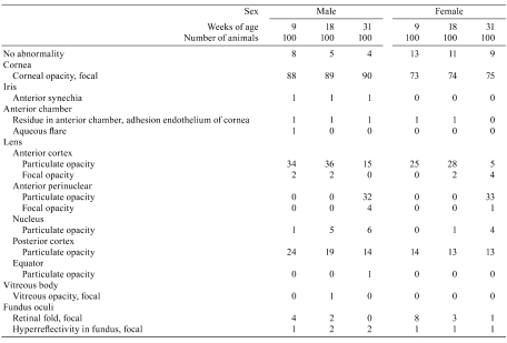
In the cornea of the RccHanTM:WIST rats, focal opacity was found by examination using a slit lamp in 88/100 males and 73/100 females at week 4 of the experiment (9 weeks of age), 89/100 males and 74/100 at week 13 of the experiment (18 weeks of age) and 90/100 males and 75/100 females at week 26 of the experiment (31 weeks of age; Table 1), and its incidence was much higher than in the CD(SD) rats, in which the incidence of this lesion is not more than about 32% (data not shown). As might be expected, when the examination was conducted using an ophthalmoscope instead of a slit lamp, the incidence of corneal opacity decreased to less than several percent in both sexes (data not shown).
Table 1. Incidences of Ophthalmological Data in Nontreated RccHanTM:WIST Rats.
As for the lens, focal opacity was observed in only a few females up to week 26 of the experiment by examination using a slit lamp, and this is similar to the observations in CD(SD) rats. In weeks 4 and 13 of the experiment (up to 18 weeks of age), particulate opacity in the lens was observed predominantly in the anterior cortex in both sexes; however, in week 26 of the experiment (31 weeks of age), opacity was observed predominantly in the anterior perinuclear region in both sexes, and this phenomenon with aging was similar to that of CD(SD) rats.
In the fundus oculi, focal retinal folds were observed in a small number of animals at each age, and the incidence tended to be reduced with age. Focal hyperreflectivity of the fundus oculi was observed in a small number of animals at each age. As for frequency of their occurrence, no difference was observed between the RccHanTM:WIST rats and CD(SD) rats.
Urinalysis
The results are shown in Tables 2-1 to 2-3.
Table 2-1. Urinalysis Data for Nontreated RccHanTM:WIST Rats.
Table 2-2. Urinalysis Data for Nontreated RccHanTM:WIST Rats.
Table 2-3. Urinalysis Data for Nontreated RccHanTM:WIST Rats.
Urine volume and one day’s output of electrolytes of the male RccHanTM:WIST rats were lower than in the CD(SD) rats (mean urine volume in CD(SD) male rats at week 26 of the experiment: 15.0 mL/24 h, n=168), but water intake was comparable. In the qualitative parameters, a tendency for lower pH was seen in RccHanTM:WIST rats compared with CD(SD) rats. The number of rats with positive protein increased with age in both male and female RccHanTM:WIST rats and was similar to that in CD(SD) rats. In the qualitative parameters, there were no findings specific to RccHanTM:WIST rats in comparison with CD(SD) rats.
Hematology
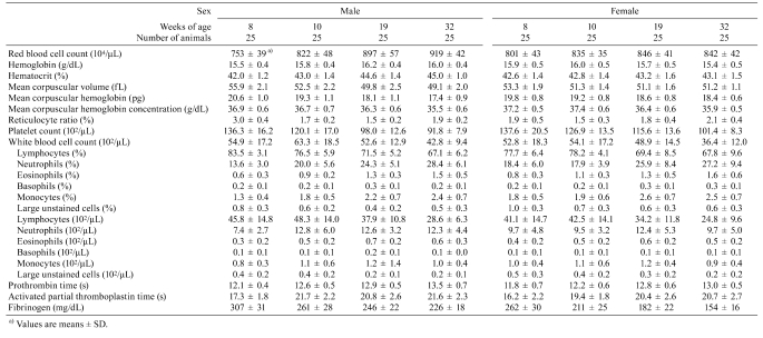
The results are shown in Table 3.
Table 3. Hematological Data for Nontreated RccHanTM:WIST Rats.
In both sexes of RccHanTM:WIST rats, tendencies for a decrease in white blood cell count, lymphocyte count, platelet count and fibrinogen were observed with age, and they were similar to those of the CD(SD) rats.
A majority of the parameters of the RccHanTM:WIST rats were comparable to those of the CD(SD) rats, but the white blood cell count was remarkably lower than in both sexes of CD(SD) rats (mean WBC counts in CD(SD) rats at 10, 19 and 32 weeks of age: 92.6, 102.1 and 92.7 × 10²/μL for males and 79.4, 65.4 and 54.4 × 10²/μL for females, respectively, n=130–160/age/sex); however, the count of each leukocyte fraction was comparable between the two strains.
Blood chemistry
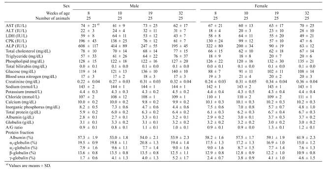
The results are shown in Table 4.
Table 4. Blood Chemical Data for Nontreated RccHanTM:WIST Rats.
In both sexes of RccHanTM:WIST rats, ALP activity and inorganic phosphorus decreased with age, and this phenomenon is well-known in rats. In addition, a tendency for a decrease in CPK activity and tendencies for an increase in glucose and gamma globulin were observed in both sexes with age.
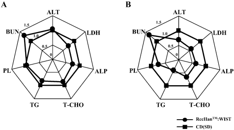
Plasma ALP activity at each age of RccHanTM:WIST rats was lower than in CD(SD) rats in both sexes (mean activity in CD(SD) rats at 10, 19 and 32 weeks of age: 715, 309 and 268 IU/L for males and 419, 151 and 101 IU/L for females, respectively, n=150–260/age/sex). In addition, ALT and LDH activities, total cholesterol, triglyceride and phospholipid levels in the female RccHanTM:WIST rats were lower than in the CD(SD) rats at 32 weeks of age, and the BUN levels in both sexes were higher than in the CD(SD) rats at each age (Fig. 3).
Fig. 3.
Comparison of blood chemical data of male (A) and female (B) RccHanTM:WIST rats and CD(SD) rats. Each parameter of RccHanTM:WIST rats is shown as the relative value over each value of CD(SD) rats at 32 weeks of age.
Organ weights
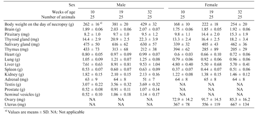
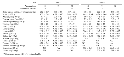
The results are shown in Tables 5 and 6.
Table 5. Absolute Organ Weight Data for Nontreated RccHanTM:WIST Rats.
Table 6. Relative Organ Weight Data for Nontreated RccHanTM:WIST Rats.
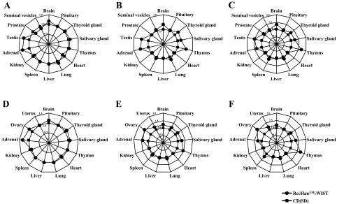
The relative weights of the adrenals (both sexes), testes and ovaries in the RccHanTM:WIST rats tended to be higher than in the CD(SD) rats at 10, 19 and 32 weeks of age, and the relative weight of the thymus also tended to be higher than in the CD(SD) rats in both sexes at 19 and 32 weeks of age (Fig. 4).
Fig. 4.
Comparison of relative organ weights of male (A, B and C) and female (D, E and F) RccHanTM:WIST rats and CD(SD) rats at 8 (A and D), 19 (B and E) and 32 (C and F) weeks of age. Each parameter of RccHanTM:WIST rats is shown as the relative value over each value of CD(SD) rats.
Necropsy
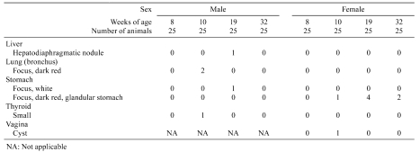
The results are shown in Table 7.
Table 7. Gross Pathological Findings in Nontreated RccHanTM:WIST Rats.
There were no gross findings specific to the RccHanTM:WIST rats.
Histopathology
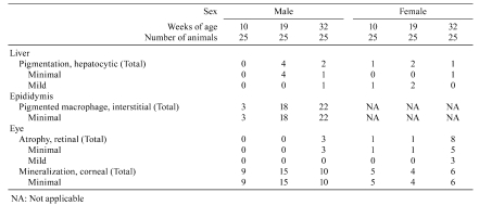
The characteristic findings in the RccHanTM:WIST rats compared with CD(SD) rats are shown in Table 8.
Table 8. Histopathological Characteristics of Nontreated RccHanTM:WIST Rats.
Histopathological findings characteristic to RccHanTM:WIST rats were observed in the eye, liver and epididymis. In the eye, a greater incidence of corneal mineralization was observed at each age (Fig. 5) and was more predominant in males than females; however, the incidence and severity were comparable among ages. In the liver, Berlin blue-positive pigmentation of periportal hepatocytes was observed in a few animals at each age. In most cases, Kupffer cells also contained the pigments. Pigment-laden macrophages were also noted in the epididymal interstitium, and the incidence was increased with age; almost all males were affected at week 26 of the experiment (22 out of 25 males). The pigments in the epididymis were also positive for Berlin blue and weakly positive for the Schmorl reaction (Fig. 6).
Fig. 5.
Corneal mineralization in the eyes of RccHanTM:WIST rats at 19 weeks of age (A to C, arrowheads). H.E. staining, ×400.
Fig. 6.
Histopathological findings in the epididymis of RccHanTM:WIST rats at 32 weeks of age. (A) Pigment-laden macrophages were observed in the epididymal interstitium. H.E. staining, ×400. (B and C) The pigments in the epididymis were also positive for Berlin blue (B) and weakly positive for the Schmorl reaction (C). ×400.
Although the original Wistar Hannover rats were reported to have serious defects of thyroid function and morphology2,3, there were no dysplastic lesions in the thyroid gland in any animal in the present study at least up to week 26 of the experiment.
Discussion
The body weights of the male and female RccHanTM:WIST rats were lower than those of the CD(SD) rats and higher than those of the F344 rats and fell between the two strains. The ophthalmological examination revealed a greater incidence of focal corneal opacity in RccHanTM:WIST rats when viewed with a slit lamp. Histopathology revealed focal mineralization of the cornea, and thus, it was considered that mineralization was involved in corneal opacity. Since the serum calcium levels were comparable among strains, it was not due to a high serum calcium level. In comparison with CD(SD) rats, clinical pathology in the RccHanTM:WIST rats revealed lower values for urine volume, one day’s output of electrolytes into the urine, peripheral white blood cell count without differential leukocyte ratio and plasma levels of ALP, ALT and LDH activities in males and/or females. The reasons for these differences were obscure, but they might be related to the small body size of the RccHanTM:WIST rats. BUN levels were higher in RccHanTM:WIST rats than in CD(SD) rats, but there was no evidence of renal tubular damage in histopathology of the kidney. Therefore, these were considered to be minor differences. In addition to the aforementioned corneal mineralization, histopathology revealed Berlin blue-positive pigments in the periportal hepatocytes, and this finding in the Wistar Hannover GALAS rat has also been described in other papers4. Pigment-laden macrophages were also found in the epididymal interstitium. The pigments were Berlin blue-positive and weakly positive for the Schmorl reaction, but identification could not be done. Measurement of organ weights revealed higher values for the relative weights of the adrenals, testes and ovaries in RccHanTM:WIST rats in comparison with CD(SD) rats, and again, this might be related to the small body size of the RccHanTM:WIST rats. It is likely that a certain large size is necessary to exert the function for the endocrine and reproductive organs.
We are now conducting a 104-week experiment in RccHanTM:WIST rats, and control data will be reported in the near future.
Acknowledgments
The authors would like to thank Japan Laboratory Animals, Inc. for providing the animals and Mr. Pete Aughton, D.A.B.T., ITR Laboratories Canada Inc., for language editing of this paper.
References
- 1.King-Herbert A, Thayer K. NTP workshop: Animal models for the NTP rodent cancer bioassay: Stocks and strains-Should we switch? Toxicol Pathol. 34: 802–805 2006. [DOI] [PMC free article] [PubMed] [Google Scholar]
- 2.Shimoi A, Kuwayama C, Miyauchi M, Kakinuma C, Kamiya M, Harada T, Ogihara T, Kurokawa M, Mizuguchi K. Vacuolar change in the thyroid follicular cells in BrlHan:WIST@Jcl(GALAS) rats. J Toxicol Pathol. 14: 253–257 2001. [Google Scholar]
- 3.Doi T, Namiki M, Ashina M, Toyota N, Kokoshima H, Kanno T, Wako Y, Tayama M, Nakashima Y, Nasu M, Tsuchitani M. Morphological and endocrinological characteristics of the endocrine systems in Wistar Hannover GALAS rats showing spontaneous dwarfs. J Toxicol Pathol. 17: 197–203 2004. [Google Scholar]
- 4.Miyata K, Sukata T, Kushida M, Ogata K, Suzuki M, Ozaki K, Uwagawa S. Spontaneous iron accumulation in hepatocytes of a 7-week-old female rat. J Toxicol Pathol. 22: 199–203 2009. [DOI] [PMC free article] [PubMed] [Google Scholar]


