SUMMARY
The replication of eukaryotic chromosomes is organized temporally and spatially within the nucleus through epigenetic regulation of replication origin function. The characteristic initiation timing of specific origins is thought to reflect their chromatin environment or sub-nuclear positioning, however the mechanism remains obscure. Here we show that the yeast Forkhead transcription factors, Fkh1 and Fkh2, are global determinants of replication origin timing. Forkhead regulation of origin timing is independent of local levels or changes of transcription. Instead, we show that Fkh1 and Fkh2 are required for the clustering of early origins and their association with the key initiation factor Cdc45 in G1-phase, suggesting that Fkh1 and Fkh2 selectively recruit origins to emergent replication factories. Fkh1 and Fkh2 bind Fkh-activated origins, and interact physically with ORC, providing a plausible mechanism to cluster origins. These findings add a new dimension to our understanding of the epigenetic basis for differential origin regulation and its connection to chromosomal domain organization.
Keywords: Replication origin timing, chromatin, Forkhead, Fox, centromere, telomere, chromosome-conformation, Cdc45, epigenetics, transcription, nuclear architecture
INTRODUCTION
Chromatin structure and organization influence most every genomic process (reviewed in (Jenuwein and Allis, 2001; Misteli, 2007)). Modification of chromatin structure to accommodate one genomic task inevitably alters the landscape for other processes. To function concurrently, fundamental processes such as DNA replication and transcription must be coordinated to preserve the accuracy and integrity of both, failure of which may lead to genome instability and developmental defects (reviewed in (Gondor and Ohlsson, 2009; Hiratani et al., 2009; Knott et al., 2009a; Mechali, 2010)). Epigenetic regulation of replication origin activation is thought to play a role in coordinating DNA replication with other genomic tasks, however our current understanding of how chromosomal replication is regulated by chromatin, let alone organized in three dimensions, is mostly correlative and sparse on mechanism.
Chromosomal DNA replication is governed primarily through regulation of replication initiation at origins. Origin DNA binds ORC and these are joined, in G1-phase, by inactive MCM helicase complexes resulting in assembly of pre-replicative complexes (pre-RCs), which are competent to initiate replication. Upon S-phase entry, Dbf4-dependent kinase (DDK) stimulates the loading of Cdc45 and Cyclin-dependent kinase stimulates the loading of additional factors to convert pre-RCs into active replisomes (reviewed in (Bell and Dutta, 2002)). However, not all pre-RCs initiate replication synchronously at the onset of S-phase, nor do all potential origins fire in every cell across a population. Instead, a subset of pre-RCs initiates replication early while clustering into foci, each containing multiple replisomes, that constitute replication factories (Kitamura et al., 2006; Meister et al., 2007). The dynamic nature of the replication foci suggests that as early replicons terminate, these factories are disassembled, allowing the next subset(s) of pre-RCs to initiate replication and establish new factories (Sporbert et al., 2002). The process is not purely stochastic. Whether in yeast or mammalian cells, certain origins reproducibly initiate more efficiently (ie, the frequency of initiation per cell cycle, ≤1) and/or earlier than others (across a population of cells), thereby giving rise to characteristic replication timing patterns of chromosomes (reviewed in (Diller and Raghuraman, 1994; Weinreich et al., 2004)).
Replication timing generally correlates with gene activity and chromatin structure, with earlier replicating regions being transcriptionally active and euchromatic, and later replicating regions being transcriptionally silent and heterochromatic (reviewed in (Gilbert, 2002; Gondor and Ohlsson, 2009; MacAlpine and Bell, 2005; Mechali, 2010; Weinreich et al., 2004)). These correlations suggest that origins may be subject to similar modes of regulation by local chromatin structure as promoters. Indeed, transcription factors can stimulate origin activity (Chang et al., 2004; Danis et al., 2004; Marahrens and Stillman, 1992), and active origins frequently co-localize with transcription start sites of active genes in Drosophila and mammalian cells (Cadoret et al., 2008; Karnani et al., 2010; MacAlpine et al., 2010; Sequeira-Mendes et al., 2009). The role of transcription factors here is thought to involve the recruitment of chromatin remodelers or modifiers that position nucleosomes or otherwise increase accessibility of origins to trans-acting factors (Flanagan and Peterson, 1999; Hu et al., 1999; Li et al., 1998; Lipford and Bell, 2001). Similar to their effects on transcription, local histone deacetylation typically delays or suppresses origin firing, whereas histone acetylation advances or stimulates origin activity (Aggarwal and Calvi, 2004; Aparicio et al., 2004; Goren et al., 2008; Knott et al., 2009b; Pappas et al., 2004; Stevenson and Gottschling, 1999; Vogelauer et al., 2002; Weber et al., 2008). However, distinct aspects of chromatin structure may affect origin timing versus efficiency. Recent studies indicate that histone acetylation is required for pre-RC assembly (Miotto and Struhl, 2007), and multiple, acetylated lysines in histone H3 and H4 N-termini are required for efficient origin activity (Eaton et al., 2011; Unnikrishnan et al., 2010). The mechanism of temporal control is less clear. Early firing is thought to represent a default state, with deacetylated chromatin imposing a delay.
Recently, we reported that the Rpd3L histone deacetylase delays the activation of ~100 origins throughout the yeast genome (~1/3 of the active origins) (Knott et al., 2009b). With this dataset we used classification-regression trees to identify annotated protein binding-sites (from (Harbison et al., 2004) whose presence or absence near origins was predictive of origin regulation by Rpd3L. This and further analysis identified binding sites of Forkhead transcription factors, Fkh1 and Fkh2, as being depleted near Rpd3L-regulated origins (data not shown). Fkh1 and Fkh2 have been well characterized for their role in regulating G2/M-phase specific transcription of a group of genes known as the CLB2 cluster (reviewed in (Murakami et al., 2010)), but have no known role in DNA replication. In this study, we show that Fkh1 and Fkh2 regulate the initiation timing of most of the earliest origins in the yeast genome through a novel mechanism involving origin clustering in G1-phase.
RESULTS
Fkh1 and Fkh2 control genome-wide initiation dynamics of replication origins
To test whether Fkh1 and Fkh2 influence replication origin function, we examined genome-wide origin-firing using BrdU immunoprecipitation analyzed by DNA sequencing (BrdU-IP-Seq), in cells arrested in early S-phase with hydroxyurea (HU). In this analysis, BrdU peak size is proportional to origin efficiency in HU: early-efficient origins produce large peaks while late and/or dormant origins yield smaller or no peaks (Knott et al., 2009b). Because Fkh1 and Fkh2 play partially complementary, yet opposing roles in regulation of G2/M-phase regulated genes (Murakami et al., 2010), we analyzed single as well as double deletion mutants of FKH1 and FKH2. Furthermore, because the double mutant cells exhibit slow, pseudohyphal growth, which complicates their analysis, we also examined these cells with over-expression of C-terminally truncated FKH2 (+pfkh2ΔC), which largely restores CLB2 cluster gene regulation (Reynolds et al., 2003). Consistent with this, we found that expression of Fkh2ΔC in fkh1Δ fkh2Δ cells suppressed their pseudohyphal growth and restored nearly normal growth rate (Fig. S1A and data not shown).
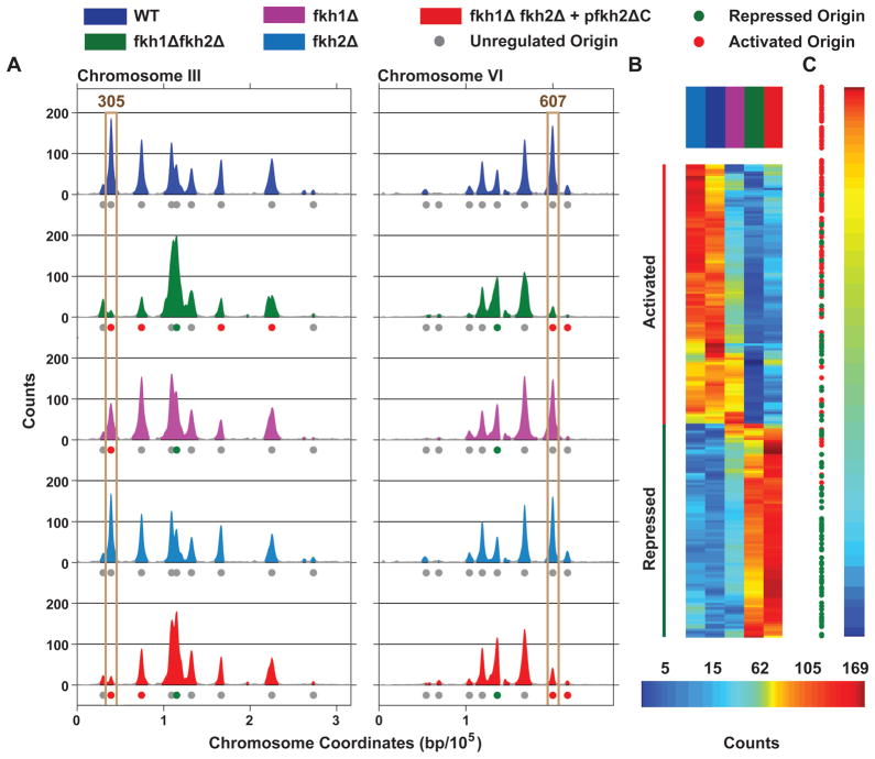
In wild-type (WT) cells, 295 peaks of BrdU incorporation were detected genome-wide (Fig. 1A and Data S1). Combined deletion of FKH1 and FKH2 had an unprecedented effect on origin activity throughout the genome, with the activities of the archetypal early origins ARS305 and ARS607 being strongly reduced (Fig. 1A). Genome-wide, of the 352 origins that were detected to fire in WT and/or fkh1Δ fkh2Δ cells, 106 (30%) origins were significantly decreased in activity (Fkh-activated) and 82 (23%) were significantly increased (Fkh-repressed) (Data S1). Deletion of FKH1 significantly (FDR<0.005) altered the activity of specific origins, with 35 being Fkh-activated and 16 Fkh-repressed, whereas deletion of FKH2 had no significant effect on the replication pattern (Fig. 1A, S1B, C, and Data S1). Fortuitously, expression of fkh2ΔC, while complementing the pseudohyphal growth defects due to transcriptional deregulation, did not complement the origin deregulation of fkh1Δ fkh2Δ cells, with virtually all of the same origins being identified as Fkh-activated (95) or Fkh-repressed (80) (Fig. 1A, S1B, C, and Data S1). This result demonstrates that the C-terminus of Fkh2 is required for origin regulation, and suggests that the effects on origins are independent of transcriptional regulation by Fkh1 and Fkh2. We took advantage of the ability of fkh2ΔC expression to complement the transcriptional defects, but not the replication defects, and to improve the growth of the double mutant cells to facilitate further analyses of fkh1Δ fkh2Δ cells.
Figure 1.
Analysis of early S-phase BrdU incorporation. A. BrdU incorporation plots of chromosomes III and VI are shown; plot colors and symbols correspond to the strain key above. Origins discussed in the text are boxed. B. Two-dimensional clustering of peak counts at Fkh-regulated origins is shown; columns (color-keyed above) correspond to strains and rows to origins. C. All detected origins (in rows) are arranged from maximum to minimum counts in WT, with the positions of Fkh-regulated origins indicated. See also Figure S1 and Data S1.
Two-dimensional clustering of the Fkh-regulated origins based on their peak sizes allows a global comparison of origin activities in the WT, single and double mutant strains. This analysis reveals the extensive deregulation of fkh1Δ fkh2Δ and fkh1Δ fkh2Δ +pfkh2ΔC cells, the strong similarity between replication patterns in the WT and fkh2Δ cells, and the intermediate phenotype of fkh1Δ cells (Fig. 1B). These data indicate that Fkh1 and Fkh2 play a major and complementary role in selecting certain origins for early activation, while repressing the activation of others. Fkh1 is sufficient to maintain normal (early) origin regulation in the absence of Fkh2, whereas Fkh2 only partially compensates for the absence of Fkh1.
To appraise the global relationship between origin activities and regulation by Fkh1 and/or Fkh2 (Fkh1/2), we arranged origins according to their WT activity levels (in HU) and plotted the positions of Fkh-activated and -repressed origins (Fig. 1C). Fkh-activated origins were strongly enriched among earlier-firing origins while Fkh-repressed origins were strongly enriched among later-firing (or inefficient) origins (p<0.001, hypergeometric test). These results show that Fkh1 and Fkh2 are largely responsible for differential origin firing dynamics throughout the genome.
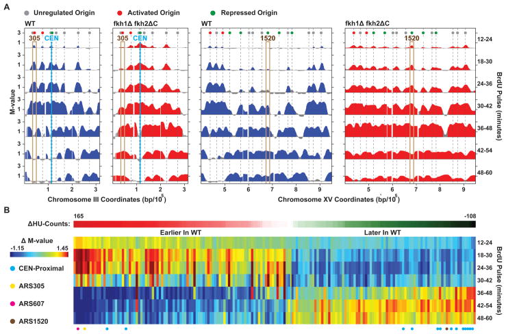
To examine in more detail the effect of Fkh1 and Fkh2 on temporal origin-firing dynamics, we analyzed replication throughout an unperturbed, synchronous S-phase. Total DNA content analysis showed similar overall replication kinetics in WT and fkh1Δ fkh2Δ +pfkh2ΔC cells (hereon fkh1Δ fkh2ΔC) (Fig. S2A). We next used BrdU pulse labeling combined with BrdU-IP analyzed by microarray (BrdU-IP-chip) to analyze origin-firing dynamics. At Fkh-activated ARS305 in WT cells, substantial BrdU incorporation occurred during the 12–24min through 30–42min pulses, and ceased by the 36–48min pulse, consistent with the early and synchronous replication of this origin (Fig. 2A). In fkh1Δ fkh2ΔC cells, however, BrdU incorporation at ARS305 was delayed and reduced in comparison, occurring mainly after replication had ceased in the WT (Fig. 2A). ARS607 and numerous other early origins showed similar delay of activity in fkh1Δ fkh2ΔC cells (Data S2). These data confirm the results of the analysis with HU and demonstrate that Fkh1/2 are required for the early activation of many origins throughout the yeast genome.
Figure 2.
Temporal analysis of DNA replication by BrdU pulse-labeling. A. BrdU incorporation plots of chromosome III and a region of XV are shown. Origins discussed in the text are boxed. B. The matrix shows differences (WT-fkh1Δ fkh2ΔC) in BrdU incorporation (Δ M-value) at all Fkh-regulated origins (columns) across time (rows); the origins are arranged from left to right by their differences (WT-fkh1Δ fkh2ΔC) in BrdU incorporation in HU (Δ HU Counts). Specific origins are indicated below. See also Figure S2 and Data S2.
The data also indicate that Fkh1/2 normally repress the earlier firing of many origins (Data S2). For example, examination of the late-replicating region of chromosome XV demonstrates that several later-firing origins, such as ARS1520, initiated replication earlier in the mutant cells (Fig. 2A). To address the formal possibility that the observed differences in origin activation timing derive from a change in origin activation efficiency, we performed two-dimensional gel electrophoresis analysis of replication initiation structures of Fkh-activated origin ARS305 and Fkh-repressed origin ARS1520. Both origins exhibit high efficiency in both WT and fkh1Δ fkh2ΔC cells (Fig. S2B). These data confirm that that Fkh1/2 establish the temporal program of origin activation.
For a global view of the impact of Fkh1/2 regulation on the temporal program, we clustered the Fkh-regulated origins according to their peak-count differences in the HU analysis, and plotted the differences in their levels of BrdU-incorporation between WT and mutant for each interval in the time-course (Fig. 2B). This analysis shows global correspondence between the change in origin activity in HU and the change in origin activity in the time course in the fkh1Δ fkh2ΔC cells, with Fkh-activated origins firing earlier and Fkh-repressed origins firing later in WT cells. Thus, Fkh1/2 play a major role in determining the characteristic firing times of replication origins throughout much of the yeast genome.
Fkh-regulation involves establishment of replication timing domains
Comparison of the WT and mutant chromosomal replication profiles reveals additional features of interest, including even earlier replication of centromere (CEN)-proximal sequences, such that these became the earliest replicating region of each chromosome (Fig. 2A and Data S2). Plotting CEN-proximal origins (ie, within 25kb) in the time-course clustergram shows that many of these origins initiated earlier in the mutant cells and were among the most strongly affected of the Fkh-repressed origins (Fig. 2B). Another striking feature of the mutant replication profiles is the delayed replication of most telomere (TEL)-proximal sequences (Data S2), particularly those with active origins, as evident on the right arm of chromosome III (Fig. 2A). These results further demonstrate the global role of Fkh1/2 in determining genome replication timing and suggest a function in chromosomal organization.
We wondered whether the distribution of Fkh-regulated origins along chromosomes might provide additional clues about their functional organization. Chromosomal plots of Fkh-regulated origins (ignoring non-regulated origins) show frequent, linearly contiguous groups of Fkh-activated and -repressed origins, suggesting a non-random distribution (Fig. S3A). To test this notion rigorously, we applied a permutation test that determines the likelihood that the contiguous groups are random. The result shows that the distribution of Fkh-activated and -repressed origins is non-random and that origins of each class frequently cluster linearly along the chromosome with other members of their class (p<0.01, Fig. S3B). Together with the CEN- and TEL-specific effects, these results are consistent with Fkh1/2 establishing domains of replication timing.
Fkh1/2 bind and function in cis to Fkh-activated origins
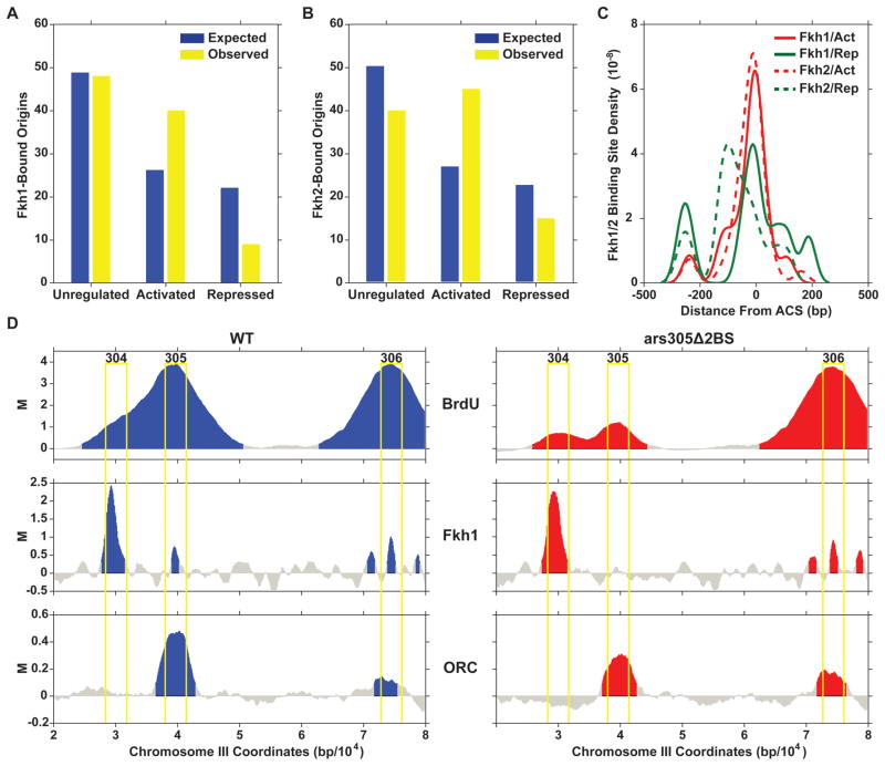
Fkh1 and Fkh2 exhibit similar DNA sequence binding specificities in vitro and bind extensively throughout the genome, with significant overlap of binding sites (data not shown and (Harbison et al., 2004; Hollenhorst et al., 2001; MacIsaac et al., 2006). To examine the relationship of Fkh1 and Fkh2 binding with origin regulation, we analyzed the distribution of putative Fkh1 and Fkh2 binding sites within 500bp of Fkh-activated, -repressed, and -unregulated origins (see Methods). This analysis shows that Fkh1 and Fkh2 binding sites are enriched near Fkh-activated origins and depleted near Fkh-repressed origins (Fig. 3A, B, hypergeometric test, p<0.01), as expected if Fkh1/2 act through direct binding near Fkh-activated origins. Fkh1 was most enriched, being ~four-fold enriched at Fkh-activated versus -repressed origins, consistent with a predominant role for Fkh1 rather than Fkh2 in origin regulation as indicated by the single mutant analysis above.
Figure 3.
Analysis of Fkh1 and Fkh2 binding sites near origins. A and B. Frequencies of expected and actual Fkh1 (A) and Fkh2 (B) consensus binding sites near Fkh-activated, Fkh-unregulated, and Fkh-repressed origins are shown. C. Frequency distribution plots of Fkh1 and Fkh2 consensus binding sites relative to ACS position are shown. D. M-values for BrdU-IP-chip and for ChIP-chip of Fkh1 and ORC binding along the ARS305 region in WT cells harboring ARS305 or ars305Δ2BS.
The enrichment of Fkh1/2 binding sites near origins may explain the selection of these origins for early activation, however, Fkh1/2 bind near some origins that are not Fkh-activated suggesting that Fkh1/2 binding in the vicinity is not sufficient for origin activation. To determine more precisely how Fkh1 and Fkh2 localize in relation to Fkh-regulated origins, we calculated the distance from each origin’s ARS-consensus sequence (ACS), which binds ORC, to the likeliest Fkh1 and Fkh2 binding site within 500bp and plotted the results as a frequency distribution (see Methods). The distribution reveals extraordinary proximity of Fkh1 and Fkh2 consensus sites to ACSs of Fkh-activated origins, with frequent overlap of the Fkh1/2 binding sites and ACSs (Fig. 3C). In contrast, Fkh1 and especially Fkh2 showed poorer alignment and binding density with those few Fkh-repressed origins proximal to Fkh1/2 binding sites. These results suggest that the positioning and/or number of these sites may be important for origin regulation
To test directly whether Fkh1/2 regulate origin function through binding in cis to the affected origin, we mutated two putative Fkh1/2 binding sites near ARS305 (ars305Δ2BS). Combined mutation of these sites significantly reduced BrdU incorporation at ARS305, but not at more distal origins, indicating that Fkh1/2 regulate ARS305 directly through binding in cis (Fig. 3D). Crucially, mutation of these binding sites eliminated Fkh1 binding to the ARS305 region without eliminating ORC binding (Fig. 3D). These results also eliminate concerns that origin deregulation results from mis-expression of a replication factor(s) in fkh1Δ fkh2ΔC cells. Overall, these results demonstrate that Fkh1/2 binding positively influences origin activity.
Fkh-dependent origin regulation is not correlated with transcription levels or changes
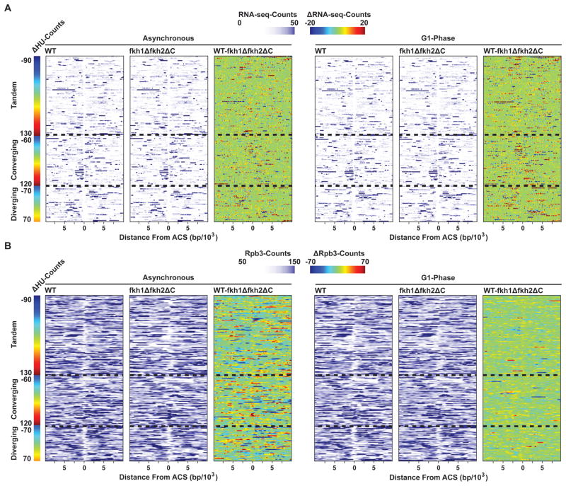
The notion of a mechanistic link between replication origin timing and transcriptional state, together with the well-characterized roles of Fkh1 and Fkh2 as transcriptional regulators, suggested that altered transcription, particularly of genes proximal to Fkh-regulated origins, might explain the altered origin firing. Although expression of Fkh2ΔC suppressed pseudohyphal growth, indicating that normal transcriptional regulation had been at least partially restored, we nonetheless wished to determine whether differences in transcription of genes proximal to the affected origins could account for the differences in origin activity. Accordingly, we analyzed global RNA transcript levels using strand-specific RNA quantification by sequencing (RNA-Seq) and RNA Polymerase II (Pol II) occupancy using chromatin immunoprecipitation analyzed by sequencing (ChIP-Seq) of the Pol II core subunit Rpb3 in WT and fkh1Δ fkh2ΔC cells, in unsynchronized cells and cells synchronized in G1-phase, when replication timing is established (Dimitrova and Gilbert, 1999; Raghuraman et al., 1997). Up-regulation of CLB2 in G1-phase fkh1Δ fkh2ΔC cells, which is consistent with the role of Fkh1 in CLB2 repression, and significant overlap between genes identified by the different methods validated both analyses (Table S1). A permutation test indicates that genes deregulated in fkh1Δ fkh2ΔC cells are not significantly co-localized with or proximal to Fkh-regulated origins (see Methods). We also plotted RNA transcript levels and Rpb3 occupancy, as well as their differences in fkh1Δ fkh2ΔC cells, within 10kb of Fkh-regulated origins (Fig. 4). Visual inspection of these plots show no obvious correlation with the effects on origin activities, regardless of the magnitude or directionality (positive or negative) of effect, the orientation of the immediately flanking genes, or the cell cycle stage. Linear regression analysis also shows no consistent correlation between the effects on origin activity and the expression levels of the immediately flanking genes (see Methods). These findings demonstrate that origin regulation by Fkh1/2 does not involve proximal changes in transcription.
Figure 4.
Transcription analysis surrounding Fkh-regulated origins in unsynchronized and G1-synchronized cells. RNA-Seq (A) and Rpb3 ChIP-Seq (B) read counts of WT, fkh1Δ fkh2ΔC, and WT-fkh1Δ fkh2ΔC differences (Δ), within 10kb of each Fkh-regulated origin, are aligned by each origin’s predicted or verified ACS. Origins are grouped according to the orientation of the flanking genes, and arranged by differences (WT-fkh1Δ fkh2ΔC) in BrdU incorporation in HU (Δ HU Counts).
Cdc45 preferentially associates with Fkh-activated origins in G1-phase
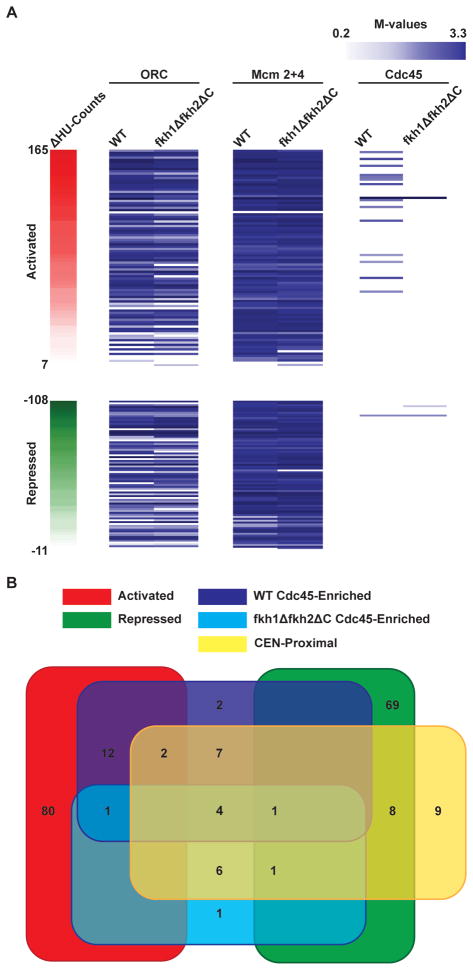
We wondered whether Fkh1/2 regulate replication timing by modulating the binding of replication factors to origins. To determine whether Fkh1/2 influence ORC binding or MCM loading, we used ChIP analyzed by microarray (ChIP-chip) to examine ORC binding in unsynchronized cells and Mcm2+4 binding in G1-synchronized cells. This results show no significant differences in ORC or Mcm2+4 binding between WT and fkh1Δ fkh2ΔC cells (Fig. 5A), contrary to the idea that Fkh1/2 affect origin-firing by modulating ORC binding or pre-RC assembly.
Figure 5.
Genome-wide binding of replication initiation factors to Fkh-regulated origins. A. M-values from ChIP-chip analysis of ORC, Mcm2+4, and Cdc45 at Fkh-regulated origins (in rows) are arranged by differences (WT-fkh1Δ fkh2ΔC) in BrdU incorporation in HU (Δ HU Counts). B. Venn diagram of Cdc45 binding within different origin classes is shown.
Origin initiation requires the DDK-dependent recruitment of Cdc45 to pre-RCs. However, Cdc45 associates specifically, albeit relatively weakly, with several early replication origins in G1-phase (prior to DDK activation), presaging their characteristic early S-phase activity (Aparicio et al., 1999). This suggests that these origins gain an early advantage (by G1-phase) in their ability to recruit Cdc45 to enable early initiation. Examination of Cdc45 binding by ChIP-chip shows Cdc45 association with many early origins, including Fkh-activated origins, such as ARS305 and ARS607, and a number of CEN-proximal origins (Fig. 5A, B). Of 29 origins that bind Cdc45 in WT G1-phase cells, 15 are Fkh-activated and 14 are CEN-proximal (on 11 CENs), while only one is Fkh-repressed. Strikingly, in the fkh1Δ fkh2ΔC cells, Cdc45 binding is lost from the Fkh-activated origins, which become significantly later firing, leaving only 14 origins binding Cdc45 (Fig. 5B). Of these 14, 12 are CEN-proximal, which as shown above, remain early firing. Thus, Cdc45 origin-binding in G1-phase is robustly associated with early initiation. These findings support the idea that Fkh1/2 influence origin function by regulating access to the pool of replication factors such as Cdc45, whereas CEN-proximal origins have access to Cdc45 independently of Fkh1/2.
Fkh1/2 are required for selective clustering of Fkh-activated origins in G1-phase
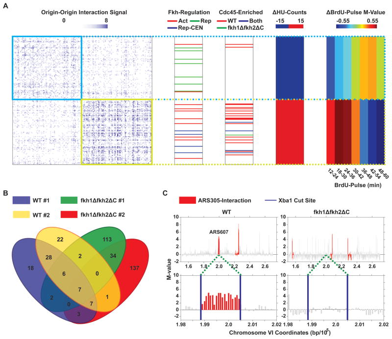
The organization of selected origins into subnuclear domains or replication foci by Fkh1/2 may explain their preferential access to limiting or sequestered initiation factors like Cdc45. In accord with this, a global analysis of intra- and inter-chromosomal interactions of the yeast genome using a variation of 4C (Chromosome Conformation Capture-on-Chip) suggests that early origins cluster in G1-phase (Duan et al., 2010). We analyzed this origin interaction data to determine whether origin clustering was associated with Fkh-regulation and/or Cdc45 binding in G1-phase. Two-dimensional clustering based on origin interaction frequencies resulted in two main clusters of interacting origins, with 89 and 92 origins, respectively (Fig. 6A). One cluster contains most of the Cdc45-bound origins, the most statistically significant Fkh-activated origins, and CEN-proximal origins. This cluster also contains earlier-firing origins on average than the other main cluster and is depleted of non-CEN proximal, Fkh-repressed origins (hypergeometric test, p<0.005). These findings suggest that Fkh-regulation involves selective origin clustering.
Figure 6.
Chromosome-conformation capture analyses of origin interactions. A. Two-dimensional clustering of origin-origin interaction frequencies is shown, with origins in columns and rows of the matrix. Columns to the right indicate Cdc45 ChIP-chip binding, average BrdU ΔHU-counts, and ΔBrdU-pulse M-values. The top 5% (based on p values) of Fkh-activated and Fkh-repressed origins are indicated. B. Venn diagram of overlap between experimental replicates is shown. C. Plots of the ARS607 region including relevant XbaI sites are shown. See also Figure S4.
To test whether Fkh1/2 have a role in origin clustering, we used 4C to analyze the trans associations of Fkh-activated origin ARS305 with other genomic sequences (for scheme, see Fig. S5A). We validated this analysis by comparing overlap between experimental replicates of WT and mutant cells, with and without crosslinking, and by analyzing the number of intra- versus inter-chromosomal interactions detected (Fig. S5B). As expected, and consistent with the results of (Duan et al., 2010), intrachromosomal interactions were enriched versus interchromosomal interactions (p<0.001). We detected 48 ARS305-interacting loci in both WT replicates (of 71 and 72 in the replicates), and 41 ARS305-interacting loci in both fkh1Δ fkh2ΔC replicates (of 164 and 189 in the replicates) (Fig. 6B). The larger number of detected interactions with lower overlap between them in the fkh1Δ fkh2ΔC replicates is consistent with a decrease in specificity of ARS305 interactions in the mutant cells. Most of the 48 sites in WT cells were not detected in the mutant cells, indicating that their interaction with ARS305 is Fkh1/2-dependent. For example, ARS305 interacted with ARS607 (as shown previously (Duan et al., 2010)) in both WT replicates and in neither fkh1Δ fkh2ΔC replicate (Fig. 6C), indicating that Fkh1/2 are required for interaction in G1-phase between these early-firing, Fkh-activated origins. These results indicate that Fkh1/2 play a role in determining the long-range chromatin contacts made by ARS305, and support the idea that Fkh1/2 function in origin regulation through origin clustering.
Fkh1 and Fkh2 interact with ORC
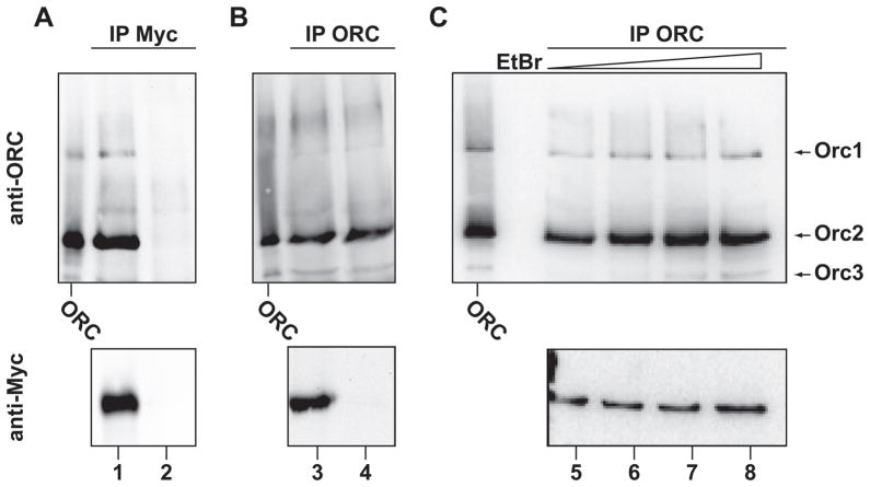
The binding of Fkh1/2 adjacent to many Fkh-activated origins, including ARS305 and ARS607 (data not shown and (Harbison et al., 2004; Keich et al., 2008)), led us to hypothesize that Fkh1/2 bound near origins might stabilize origin contacts in trans through interaction with ORC bound at other Fkh-activated origins. Immunoprecipitation (IP) of Myc-tagged Fkh1 or Fkh2 from soluble cell extracts resulted in co-precipitation of ORC (Fig. 7A, lanes 1 and 2, data not shown for Fkh2); Orc2 was robustly detected, Orc1 and Orc3 were weakly detected, and Orc4-Orc6 were obscured by co-migrating immunoglobulin heavy chain (data not shown). Reciprocal IP of ORC using a polyclonal antibody co-precipitated Fkh1 (Fig. 7B, lanes 3 and 4). Taken together, these results demonstrate a physical interaction (direct or indirect) between ORC and Fkh1. These interactions persisted in the presence of the DNA-intercalating agent, ethidium bromide, indicating that the interactions are likely not DNA-mediated (Fig. 7C, lanes 5–8). Together with the close proximity of Fkh1/2 binding sites with origin ACSs, these results support the idea that Fkh1/2 interact with ORC to bridge replication origins in trans.
Figure 7.
Co-IP of Fkh1 with ORC. Soluble extracts from FKH1-MYC (lanes 1, 3, 5–8) and untagged (lanes 2, 4) cells were subjected to IP with anti-Myc antibody (A) and anti-ORC antibody (B and C). IPs were analyzed by immunoblotting with anti-Myc and anti-ORC antibodies. C. Ethidium bromide (EtBr) was included in the IPs at 10, 40, and 100 μg/mL in lanes 6, 7, and 8, respectively. ORC protein was included as standard.
DISCUSSION
Fkh1/2 establish replication-timing domains through origin clustering
Our findings reveal a novel, global mechanism for the regulation of origin initiation timing, involving the spatial organization of replication origins by Fkh1/2. Previous studies have concluded that yeast origins are early by default, and that late timing is imposed by flanking sequences of a repressive nature (Ferguson and Fangman, 1992; Friedman et al., 1996). However, our findings show that Fkh1/2 actively program the timing of most of the earliest origins throughout the genome. Thus, we propose that Fkh1/2 establish early replication timing at Forkhead-activated origins by recruiting these origins into clusters where Cdc45 is (and likely other replication factors are) concentrated. The enrichment and distinct positioning relative to the ACS of Fkh1/2 binding sites likely explains the selective preference for Fkh-activated origins. Clustering may involve interaction of Fkh1/2 bound adjacent to an origin with ORC bound to a distal, second origin. Likewise, Fkh1/2 bound near the second origin might interact with a third origin, and so forth, providing a mechanism to cluster several origins together. This congregation of origins and initiation factors provides a kinetic advantage in assembling the factors needed for replication initiation upon S-phase entry, which transforms these origin clusters into early replication factories. The ensuing dynamics of the replication process, involving spooling of DNA through the replication factories (Kitamura et al., 2006), eventually repositions more distal, unfired origins, bringing them in proximity of the concentration of the replication factor(ie)s and thereby allowing them to gain access as early replicons terminate and are released. This is expected to result in an increasingly stochastic pattern of replication initiation as S-phase proceeds and many unfired origins compete for limited access. However, later-replicating regions also exhibit well-defined replication patterns indicating preferred origin timing and usage. Indeed, chromosomes IV, XII, XIV, and XV each have distinctly late-replicating regions >200kb in length, encompassing groups of contiguous Fkh-repressed origins, which lose this unique character in the absence of Fkh1/2 (Fig. 2A and Data S2).
Origin clusters may define replication-timing domains. The organization of mammalian chromosomes into spatial domains correlates strongly with replication timing (Ryba et al., 2010). Analysis of global 4C in yeast shows clustering of early origins (in G1-phase), and we have now shown that the early origin cluster contains Fkh-activated and Cdc45-bound origins (in G1-phase). We have confirmed that Fkh-activated origins ARS305 and ARS607 interact in trans, and critically, show that this interaction depends on Fkh1/2. In addition, Fkh-activated and Fkh-repressed origins often occur in separate, linearly contiguous groups along chromosomes, suggesting the formation of distinct domains. This may involve the anchoring of intrachromosomal chromatin loops by Fkh1/2 bound near origins, perhaps through interaction with ORC, particularly in the case of Fkh-activated origins, which are enriched for Fkh1/2 binding. In the case of Fkh-repressed origins, a dearth of Fkh1/2 binding sites presumably reduces the likelihood that these origins join the Fkh-activated clusters, which may permit other mechanisms, such as deacetylation or localization to the nuclear periphery, to define replication timing of these regions. Alternatively, the later timing may be a consequence of conformational or spatial constraints imposed by the chromosomal architecture established by Fkh1/2 clustering of Fkh-activated origins.
In the absence of Fkh1 and Fkh2, CEN-proximal origins dominate the early replication landscape, suggesting that CENs confer early replication intrinsically. CENs normally cluster and occupy a characteristic interior position in the nucleus (Jin et al., 1998) that we suggest overlaps with the pool of replication factor(ie)s. Consequently, CEN-proximal origins have favorable access to this pool and initiate early, independently of Fkh1/2. Thus, CEN-proximal origins may act as organizing sites for early-replicating origin clusters that include non-CEN-proximal origins. More distal Fkh-activated origins may utilize Fkh1/2 to cluster with CEN-proximal origins, thereby drawing these more distal origins into the pool. This is consistent with the finding that CEN-proximal origins localize to the large, early-replicating cluster in the global 4C data together with the earliest Fkh-activated origins. Thus, the advanced replication timing of CEN-proximal origins (and perhaps other Fkh-repressed origins) in cells lacking Fkh1/2 may result from reduced competition from Fkh-activated origins for limiting replication factor(ie)s, rather than a direct repressive function of Fkh1/2. Incidentally, CEN-proximity may explain the finding in yeast that plasmid-borne origins typically replicate early, as these studies were performed with CEN-harboring plasmids (Ferguson and Fangman, 1992; Friedman et al., 1996).
In contrast to CENs, TELs form several clusters that occupy the nuclear periphery (Gotta et al., 1996; Heun et al., 2001). The normally late replication of TEL-proximal regions is consistent with the notion that the dynamic nature of the replication process eventually relocates these distal regions to the interior of the nucleus, which ultimately enables their access to replication factor(ie)s. In the absence of Fkh1 and Fkh2, most of the active telomeric origins are further delayed. We imagine that the delayed activation of Fkh-activated origins located along distal chromosomal arms results in a corresponding delay in the relocation to TEL-proximal origins to the vicinity of replication factor(ie)s. Alternatively, Fkh1/2 may act directly to regulate TEL-proximal origins. Further study will be required to understand the regulation of CEN- and TEL-proximal origin timing.
Multiple, separable roles for Fkh1 and Fkh2 in regulation of the genome
A clear finding of this study is the mechanistic independence of Fkh-origin regulation from transcription. There is no correlation between the observed changes in replication timing and transcriptional levels of proximal genes. Importantly, expression of Fkh2 lacking its C-terminus in fkh1Δ fkh2Δ cells significantly restores transcriptional regulation of CLB2 cluster genes (only CLB2 remained deregulated and only in G1-phase cells) without restoring origin regulation, directly demonstrating a separation of these Fkh1/2 functions. Nevertheless, our results do not rule out the possibility that the function of Fkh1/2 in origin clustering may also underlie transcriptional control not elicited under our growth conditions.
As transcriptional regulators, Fkh1 and Fkh2 exhibit opposing, as well as partially complementary functions (Murakami et al., 2010). Fkh1 and Fkh2 also demonstrate distinct abilities to regulate origins, suggesting that the features that distinguish Fkh1 and Fkh2 functions in transcription also impinge on their functions as origin regulators. Whereas Fkh2 plays the lead role in transcriptional regulation, Fkh1 plays the lead role in origin regulation. Fkh1 differs from Fkh2 most significantly in the presence of a C-terminal extension in Fkh2, which regulates its interaction(s) with transcriptional co-activator(s) (Darieva et al., 2010; Darieva et al., 2003; Koranda et al., 2000; Pic-Taylor et al., 2004; Reynolds et al., 2003). This domain is also required for Fkh2’s function in origin regulation, suggesting that proper regulation of co-activator interactions is critical, and that factors interacting with Fkh2 but not with Fkh1 may disrupt origin regulation. Mcm1, which binds cooperatively with Fkh2, but not Fkh1 (Boros et al., 2003; Koranda et al., 2000; Kumar et al., 2000; Pic et al., 2000), is an intriguing candidate, as it has been reported to modulate origin function (Chang et al., 2004). We note that Mcm1 binding sites are not enriched near Fkh-activated origins (data not shown). Thus, consistent with the lack of effect on origin firing of FKH2 deletion, it is possible that Fkh2 normally plays no role in origin regulation, and only substitutes (partially) in Fkh1’s absence.
Fkh1, but not Fkh2, also regulates donor preference in yeast mating-type switching (Sun et al., 2002). Mating-type switching involves homologous recombination between the MAT locus (recipient) and one of two silent mating-type loci (donor) distally located on opposite arms of the same chromosome, HMLα and HMRa. This mechanism presumably necessitates chromosomal looping of either arm to juxtapose the donor and recipient loci. Remarkably, in MATa cells, HMLα is preferentially selected as the donor in over 90% of cells, which ensures efficient mating-type switching. This preference depends on Fkh1 binding to the recombination enhancer (RE), which is proximal to HMLα. Our finding that Fkh1/2 mediate long-range origin interactions suggest that Fkh1 mediates a stable, long-range interaction between MATa and the RE to specify the recombination between MATa and HMLα, which conspicuously, like early origin clustering, occurs during G1-phase. The role of Fkh1 in regulating recombination over long distances together with Fkh1/2’s role in regulating replication initiation timing through long-range origin clustering suggests that establishing long-range chromatin contacts may be a common mechanism of Fkh1/2 function, likely extending to transcriptional control.
Our proposed mechanism of origin clustering may also explain how the long-range interaction necessary for recombinational donor preference is established. Dormant origins are closely associated with the RE (ARS304) and with MAT (ARS313 and ARS314). Thus, interactions between Fkh1 bound to the RE and ORC bound to the distal ARS313 or ARS314 may stabilize long-range contacts between these loci; similar interactions between ORC bound to ARS304 and Fkh1 bound near MAT may also participate (though an RE-like element has not been identified near MAT). The dormancy of these origins is consistent with the idea that these loci form a separate chromosomal domain dedicated for recombination, which delays replication (by inhibiting initiation and allowing passive replication from distal, flanking origins). Exactly how such domains are dedicated to one function over another will require more investigation, but may reflect combinatorial regulation by Fkh1/2 together with other factors, along with defined sub-nuclear localization of these activities.
The findings presented here provide a clearer understanding of the epigenetic basis for differential origin regulation and its connection to the spatial organization of chromosomes. Rather than a direct connection with transcription, the results indicate that the organization of origins into functional clusters determines their activation kinetics. Our study identifies Fkh1 and Fkh2 as factors that participate in the establishment of the three-dimensional structure of the yeast genome and the epigenetic regulation of genome replication. This regulation through structure may be analogous to epigenetic mechanisms of transcriptional memory wherein gene looping or sub-nuclear localization is correlated with the maintenance of a transcriptional state or a potentiated state primed for rapid response (Misteli, 2007). Furthermore, this organization may contribute to a coordination of replication and transcription, perhaps with consequence for genome stability (Knott et al., 2009a). Indeed, this study’s findings provide a new handle to investigate the consequences of deregulating replication timing on gene regulation or genome stability. The identification of yeast members of the conserved Fox transcription factor family as physical mediators of chromosomal architecture and epigenetic regulation suggest conservation of this function, which may link replication timing control and the role of Fox proteins in metazoan development.
EXPERIMENTAL PROCEDURES
Cultures were grown in YEPD at 23°C and harvested at O.D.~1; pfkh2ΔC-harboring cells were pre-grown in synthetic drop-out medium for plasmid maintenance and transferred to YEPD for a few generations prior to synchronization and analysis. For BrdU-IP-Seq analysis, 50mL G1-blocked cells were released into 200mM HU and 400 μg/mL BrdU for 1hr and harvested. For BrdU-IP-chip time-course, 500mL G1-blocked cells were released and 50mL aliquots were incubated for 12min with BrdU (800 μg/mL) and harvested. Additional details are given in the Extended Experimental Procedures.
Supplementary Material
Acknowledgments
J. Aparicio, M. Arbeitman, C. Fox, and M. Michael for critical evaluation of the manuscript; M. Arbeitman for sharing equipment; S.P. Bell for purified ORC and antibody; J. Dalton for help with microscopy; C. Fox for plasmids, strains, antibody, and helpful discussions; R. Kalhor for the 4C protocol and invaluable advice; U. Keich for helpful discussion; and D. Kowalski and R. Young for strains. This work was funded by a pilot grant from the USC Epigenome Center sponsored by the Whittier Foundation (to O.M.A), NIH grants 5R01-GM065494 and 3R01-GM065494-S1(to O.M.A.), and P50-HG02790 (to S.R.V.K. and S.T.).
Footnotes
All original and processed data files can be found at http://www.ncbi.nlm.nih.gov/geo/under accession number GSE33704.
Publisher's Disclaimer: This is a PDF file of an unedited manuscript that has been accepted for publication. As a service to our customers we are providing this early version of the manuscript. The manuscript will undergo copyediting, typesetting, and review of the resulting proof before it is published in its final citable form. Please note that during the production process errors may be discovered which could affect the content, and all legal disclaimers that apply to the journal pertain.
References
- Aggarwal BD, Calvi BR. Chromatin regulates origin activity in Drosophila follicle cells. Nature. 2004;430:372–376. doi: 10.1038/nature02694. [DOI] [PubMed] [Google Scholar]
- Aparicio JG, Viggiani CJ, Gibson DG, Aparicio OM. The Rpd3-Sin3 histone deacetylase regulates replication timing and enables intra-S origin control in Saccharomyces cerevisiae. Mol Cell Biol. 2004;24:4769–4780. doi: 10.1128/MCB.24.11.4769-4780.2004. [DOI] [PMC free article] [PubMed] [Google Scholar]
- Aparicio OM, Stout AM, Bell SP. Differential assembly of Cdc45p and DNA polymerases at early and late origins of DNA replication. Proc Natl Acad Sci U S A. 1999;96:9130–9135. doi: 10.1073/pnas.96.16.9130. [DOI] [PMC free article] [PubMed] [Google Scholar]
- Bell SP, Dutta A. DNA replication in eukaryotic cells. Annu Rev Biochem. 2002;71:333–374. doi: 10.1146/annurev.biochem.71.110601.135425. [DOI] [PubMed] [Google Scholar]
- Boros J, Lim FL, Darieva Z, Pic-Taylor A, Harman R, Morgan BA, Sharrocks AD. Molecular determinants of the cell-cycle regulated Mcm1p-Fkh2p transcription factor complex. Nucleic Acids Res. 2003;31:2279–2288. doi: 10.1093/nar/gkg347. [DOI] [PMC free article] [PubMed] [Google Scholar]
- Cadoret JC, Meisch F, Hassan-Zadeh V, Luyten I, Guillet C, Duret L, Quesneville H, Prioleau MN. Genome-wide studies highlight indirect links between human replication origins and gene regulation. Proc Natl Acad Sci U S A. 2008;105:15837–15842. doi: 10.1073/pnas.0805208105. [DOI] [PMC free article] [PubMed] [Google Scholar]
- Chang VK, Donato JJ, Chan CS, Tye BK. Mcm1 promotes replication initiation by binding specific elements at replication origins. Mol Cell Biol. 2004;24:6514–6524. doi: 10.1128/MCB.24.14.6514-6524.2004. [DOI] [PMC free article] [PubMed] [Google Scholar]
- Danis E, Brodolin K, Menut S, Maiorano D, Girard-Reydet C, Mechali M. Specification of a DNA replication origin by a transcription complex. Nat Cell Biol. 2004;6:721–730. doi: 10.1038/ncb1149. [DOI] [PubMed] [Google Scholar]
- Darieva Z, Clancy A, Bulmer R, Williams E, Pic-Taylor A, Morgan BA, Sharrocks AD. A competitive transcription factor binding mechanism determines the timing of late cell cycle-dependent gene expression. Mol Cell. 2010;38:29–40. doi: 10.1016/j.molcel.2010.02.030. [DOI] [PMC free article] [PubMed] [Google Scholar]
- Darieva Z, Pic-Taylor A, Boros J, Spanos A, Geymonat M, Reece RJ, Sedgwick SG, Sharrocks AD, Morgan BA. Cell cycle-regulated transcription through the FHA domain of Fkh2p and the coactivator Ndd1p. Curr Biol. 2003;13:1740–1745. doi: 10.1016/j.cub.2003.08.053. [DOI] [PubMed] [Google Scholar]
- Diller JD, Raghuraman MK. Eukaryotic replication origins: control in space and time. Trends Biochem Sci. 1994;19:320–325. doi: 10.1016/0968-0004(94)90070-1. [DOI] [PubMed] [Google Scholar]
- Dimitrova DS, Gilbert DM. The spatial position and replication timing of chromosomal domains are both established in early G1 phase. Mol Cell. 1999;4:983–993. doi: 10.1016/s1097-2765(00)80227-0. [DOI] [PubMed] [Google Scholar]
- Duan Z, Andronescu M, Schutz K, McIlwain S, Kim YJ, Lee C, Shendure J, Fields S, Blau CA, Noble WS. A three-dimensional model of the yeast genome. Nature. 2010;465:363–367. doi: 10.1038/nature08973. [DOI] [PMC free article] [PubMed] [Google Scholar]
- Eaton ML, Prinz JA, MacAlpine HK, Tretyakov G, Kharchenko PV, MacAlpine DM. Chromatin signatures of the Drosophila replication program. Genome Res. 2011;21:164–174. doi: 10.1101/gr.116038.110. [DOI] [PMC free article] [PubMed] [Google Scholar]
- Ferguson BM, Fangman WL. A position effect on the time of replication origin activation in yeast. Cell. 1992;68:333–339. doi: 10.1016/0092-8674(92)90474-q. [DOI] [PubMed] [Google Scholar]
- Flanagan JF, Peterson CL. A role for the yeast SWI/SNF complex in DNA replication. Nucleic Acids Res. 1999;27:2022–2028. doi: 10.1093/nar/27.9.2022. [DOI] [PMC free article] [PubMed] [Google Scholar]
- Friedman KL, Diller JD, Ferguson BM, Nyland SV, Brewer BJ, Fangman WL. Multiple determinants controlling activation of yeast replication origins late in S phase. Genes Dev. 1996;10:1595–1607. doi: 10.1101/gad.10.13.1595. [DOI] [PubMed] [Google Scholar]
- Gilbert DM. Replication timing and transcriptional control: beyond cause and effect. Curr Opin Cell Biol. 2002;14:377–383. doi: 10.1016/s0955-0674(02)00326-5. [DOI] [PubMed] [Google Scholar]
- Gondor A, Ohlsson R. Replication timing and epigenetic reprogramming of gene expression: a two-way relationship? Nat Rev Genet. 2009;10:269–276. doi: 10.1038/nrg2555. [DOI] [PubMed] [Google Scholar]
- Goren A, Tabib A, Hecht M, Cedar H. DNA replication timing of the human beta-globin domain is controlled by histone modification at the origin. Genes Dev. 2008;22:1319–1324. doi: 10.1101/gad.468308. [DOI] [PMC free article] [PubMed] [Google Scholar]
- Gotta M, Laroche T, Formenton A, Maillet L, Scherthan H, Gasser SM. The clustering of telomeres and colocalization with Rap1, Sir3, and Sir4 proteins in wild-type Saccharomyces cerevisiae. J Cell Biol. 1996;134:1349–1363. doi: 10.1083/jcb.134.6.1349. [DOI] [PMC free article] [PubMed] [Google Scholar]
- Harbison CT, Gordon DB, Lee TI, Rinaldi NJ, Macisaac KD, Danford TW, Hannett NM, Tagne JB, Reynolds DB, Yoo J, et al. Transcriptional regulatory code of a eukaryotic genome. Nature. 2004;431:99–104. doi: 10.1038/nature02800. [DOI] [PMC free article] [PubMed] [Google Scholar]
- Heun P, Laroche T, Raghuraman MK, Gasser SM. The positioning and dynamics of origins of replication in the budding yeast nucleus. J Cell Biol. 2001;152:385–400. doi: 10.1083/jcb.152.2.385. [DOI] [PMC free article] [PubMed] [Google Scholar]
- Hiratani I, Takebayashi S, Lu J, Gilbert DM. Replication timing and transcriptional control: beyond cause and effect--part II. Curr Opin Genet Dev. 2009;19:142–149. doi: 10.1016/j.gde.2009.02.002. [DOI] [PMC free article] [PubMed] [Google Scholar]
- Hollenhorst PC, Pietz G, Fox CA. Mechanisms controlling differential promoter-occupancy by the yeast forkhead proteins Fkh1p and Fkh2p: implications for regulating the cell cycle and differentiation. Genes Dev. 2001;15:2445–2456. doi: 10.1101/gad.906201. [DOI] [PMC free article] [PubMed] [Google Scholar]
- Hu YF, Hao ZL, Li R. Chromatin remodeling and activation of chromosomal DNA replication by an acidic transcriptional activation domain from BRCA1. Genes Dev. 1999;13:637–642. doi: 10.1101/gad.13.6.637. [DOI] [PMC free article] [PubMed] [Google Scholar]
- Jenuwein T, Allis CD. Translating the histone code. Science. 2001;293:1074–1080. doi: 10.1126/science.1063127. [DOI] [PubMed] [Google Scholar]
- Jin Q, Trelles-Sticken E, Scherthan H, Loidl J. Yeast nuclei display prominent centromere clustering that is reduced in nondividing cells and in meiotic prophase. J Cell Biol. 1998;141:21–29. doi: 10.1083/jcb.141.1.21. [DOI] [PMC free article] [PubMed] [Google Scholar]
- Karnani N, Taylor CM, Malhotra A, Dutta A. Genomic study of replication initiation in human chromosomes reveals the influence of transcription regulation and chromatin structure on origin selection. Mol Biol Cell. 2010;21:393–404. doi: 10.1091/mbc.E09-08-0707. [DOI] [PMC free article] [PubMed] [Google Scholar]
- Keich U, Gao H, Garretson JS, Bhaskar A, Liachko I, Donato J, Tye BK. Computational detection of significant variation in binding affinity across two sets of sequences with application to the analysis of replication origins in yeast. BMC Bioinformatics. 2008;9:372. doi: 10.1186/1471-2105-9-372. [DOI] [PMC free article] [PubMed] [Google Scholar]
- Kitamura E, Blow JJ, Tanaka TU. Live-cell imaging reveals replication of individual replicons in eukaryotic replication factories. Cell. 2006;125:1297–1308. doi: 10.1016/j.cell.2006.04.041. [DOI] [PMC free article] [PubMed] [Google Scholar]
- Knott SR, Viggiani CJ, Aparicio OM. To promote and protect: coordinating DNA replication and transcription for genome stability. Epigenetics. 2009a;4:362–365. doi: 10.4161/epi.4.6.9712. [DOI] [PubMed] [Google Scholar]
- Knott SR, Viggiani CJ, Tavaré S, Aparicio OM. Genome-wide replication profiles indicate an expansive role for Rpd3L in regulating replication initiation timing or efficiency, and reveal genomic loci of Rpd3 function in Saccharomyces cerevisiae. Genes Dev. 2009b;23:1077–1090. doi: 10.1101/gad.1784309. [DOI] [PMC free article] [PubMed] [Google Scholar]
- Koranda M, Schleiffer A, Endler L, Ammerer G. Forkhead-like transcription factors recruit Ndd1 to the chromatin of G2/M-specific promoters. Nature. 2000;406:94–98. doi: 10.1038/35017589. [DOI] [PubMed] [Google Scholar]
- Kumar R, Reynolds DM, Shevchenko A, Goldstone SD, Dalton S. Forkhead transcription factors, Fkh1p and Fkh2p, collaborate with Mcm1p to control transcription required for M-phase. Curr Biol. 2000;10:896–906. doi: 10.1016/s0960-9822(00)00618-7. [DOI] [PubMed] [Google Scholar]
- Li R, Yu DS, Tanaka M, Zheng L, Berger SL, Stillman B. Activation of chromosomal DNA replication in Saccharomyces cerevisiae by acidic transcriptional activation domains. Mol Cell Biol. 1998;18:1296–1302. doi: 10.1128/mcb.18.3.1296. [DOI] [PMC free article] [PubMed] [Google Scholar]
- Lipford JR, Bell SP. Nucleosomes positioned by ORC facilitate the initiation of DNA replication. Mol Cell. 2001;7:21–30. doi: 10.1016/s1097-2765(01)00151-4. [DOI] [PubMed] [Google Scholar]
- MacAlpine DM, Bell SP. A genomic view of eukaryotic DNA replication. Chromosome Res. 2005;13:309–326. doi: 10.1007/s10577-005-1508-1. [DOI] [PubMed] [Google Scholar]
- MacAlpine HK, Gordan R, Powell SK, Hartemink AJ, MacAlpine DM. Drosophila ORC localizes to open chromatin and marks sites of cohesin complex loading. Genome Res. 2010;20:201–211. doi: 10.1101/gr.097873.109. [DOI] [PMC free article] [PubMed] [Google Scholar]
- MacIsaac KD, Wang T, Gordon DB, Gifford DK, Stormo GD, Fraenkel E. An improved map of conserved regulatory sites for Saccharomyces cerevisiae. BMC Bioinformatics. 2006;7:113. doi: 10.1186/1471-2105-7-113. [DOI] [PMC free article] [PubMed] [Google Scholar]
- Marahrens Y, Stillman B. A yeast chromosomal origin of replication defined by multiple functional elements. Science. 1992;255:817–823. doi: 10.1126/science.1536007. [DOI] [PubMed] [Google Scholar]
- Mechali M. Eukaryotic DNA replication origins: many choices for appropriate answers. Nat Rev Mol Cell Biol. 2010;11:728–738. doi: 10.1038/nrm2976. [DOI] [PubMed] [Google Scholar]
- Meister P, Taddei A, Ponti A, Baldacci G, Gasser SM. Replication foci dynamics: replication patterns are modulated by S-phase checkpoint kinases in fission yeast. EMBO J. 2007;26:1315–1326. doi: 10.1038/sj.emboj.7601538. [DOI] [PMC free article] [PubMed] [Google Scholar]
- Miotto B, Struhl K. Histone H4 lysine 16 acetylation: from genome regulation to tumoral progression. Med Sci (Paris) 2007;23:735–740. doi: 10.1051/medsci/20072389735. [DOI] [PubMed] [Google Scholar]
- Misteli T. Beyond the sequence: cellular organization of genome function. Cell. 2007;128:787–800. doi: 10.1016/j.cell.2007.01.028. [DOI] [PubMed] [Google Scholar]
- Murakami H, Aiba H, Nakanishi M, Murakami-Tonami Y. Regulation of yeast forkhead transcription factors and FoxM1 by cyclin-dependent and polo-like kinases. Cell Cycle. 2010;9:3233–3242. doi: 10.4161/cc.9.16.12599. [DOI] [PubMed] [Google Scholar]
- Pappas DL, Jr, Frisch R, Weinreich M. The NAD(+)-dependent Sir2p histone deacetylase is a negative regulator of chromosomal DNA replication. Genes Dev. 2004;18:769–781. doi: 10.1101/gad.1173204. [DOI] [PMC free article] [PubMed] [Google Scholar]
- Pic A, Lim FL, Ross SJ, Veal EA, Johnson AL, Sultan MR, West AG, Johnston LH, Sharrocks AD, Morgan BA. The forkhead protein Fkh2 is a component of the yeast cell cycle transcription factor SFF. EMBO J. 2000;19:3750–3761. doi: 10.1093/emboj/19.14.3750. [DOI] [PMC free article] [PubMed] [Google Scholar]
- Pic-Taylor A, Darieva Z, Morgan BA, Sharrocks AD. Regulation of cell cycle-specific gene expression through cyclin-dependent kinase-mediated phosphorylation of the forkhead transcription factor Fkh2p. Mol Cell Biol. 2004;24:10036–10046. doi: 10.1128/MCB.24.22.10036-10046.2004. [DOI] [PMC free article] [PubMed] [Google Scholar]
- Raghuraman MK, Brewer BJ, Fangman WL. Cell cycle-dependent establishment of a late replication program. Science. 1997;276:806–809. doi: 10.1126/science.276.5313.806. [DOI] [PubMed] [Google Scholar]
- Reynolds D, Shi BJ, McLean C, Katsis F, Kemp B, Dalton S. Recruitment of Thr 319-phosphorylated Ndd1p to the FHA domain of Fkh2p requires Clb kinase activity: a mechanism for CLB cluster gene activation. Genes Dev. 2003;17:1789–1802. doi: 10.1101/gad.1074103. [DOI] [PMC free article] [PubMed] [Google Scholar]
- Ryba T, Hiratani I, Lu J, Itoh M, Kulik M, Zhang J, Schulz TC, Robins AJ, Dalton S, Gilbert DM. Evolutionarily conserved replication timing profiles predict long-range chromatin interactions and distinguish closely related cell types. Genome Res. 2010;20:761–770. doi: 10.1101/gr.099655.109. [DOI] [PMC free article] [PubMed] [Google Scholar]
- Sequeira-Mendes J, Diaz-Uriarte R, Apedaile A, Huntley D, Brockdorff N, Gomez M. Transcription initiation activity sets replication origin efficiency in mammalian cells. PLoS Genet. 2009;5:e1000446. doi: 10.1371/journal.pgen.1000446. [DOI] [PMC free article] [PubMed] [Google Scholar]
- Sporbert A, Gahl A, Ankerhold R, Leonhardt H, Cardoso MC. DNA polymerase clamp shows little turnover at established replication sites but sequential de novo assembly at adjacent origin clusters. Mol Cell. 2002;10:1355–1365. doi: 10.1016/s1097-2765(02)00729-3. [DOI] [PubMed] [Google Scholar]
- Stevenson JB, Gottschling DE. Telomeric chromatin modulates replication timing near chromosome ends. Genes Dev. 1999;13:146–151. doi: 10.1101/gad.13.2.146. [DOI] [PMC free article] [PubMed] [Google Scholar]
- Sun K, Coic E, Zhou Z, Durrens P, Haber JE. Saccharomyces forkhead protein Fkh1 regulates donor preference during mating-type switching through the recombination enhancer. Genes Dev. 2002;16:2085–2096. doi: 10.1101/gad.994902. [DOI] [PMC free article] [PubMed] [Google Scholar]
- Unnikrishnan A, Gafken PR, Tsukiyama T. Dynamic changes in histone acetylation regulate origins of DNA replication. Nat Struct Mol Biol. 2010;17:430–437. doi: 10.1038/nsmb.1780. [DOI] [PMC free article] [PubMed] [Google Scholar]
- Vogelauer M, Rubbi L, Lucas I, Brewer BJ, Grunstein M. Histone acetylation regulates the time of replication origin firing. Mol Cell. 2002;10:1223–1233. doi: 10.1016/s1097-2765(02)00702-5. [DOI] [PubMed] [Google Scholar]
- Weber JM, Irlbacher H, Ehrenhofer-Murray AE. Control of replication initiation by the Sum1/Rfm1/Hst1 histone deacetylase. BMC Mol Biol. 2008;9:100. doi: 10.1186/1471-2199-9-100. [DOI] [PMC free article] [PubMed] [Google Scholar]
- Weinreich M, Palacios DeBeer MA, Fox CA. The activities of eukaryotic replication origins in chromatin. Biochim Biophys Acta. 2004;1677:142–157. doi: 10.1016/j.bbaexp.2003.11.015. [DOI] [PubMed] [Google Scholar]
Associated Data
This section collects any data citations, data availability statements, or supplementary materials included in this article.


