Abstract
We report a 72-year-old female patient with spontaneous rupture of the left external iliac vein. She visited our hospital for abdominal and back pain. She had the abnormal finding of hemoperitoneum. We performed an emergency operation with diagnosis of left ovarian cyst rupture though she suffered from spontaneous rupture of the left external iliac vein. This case provides insight to the experience of spontaneous rupture of the left external iliac vein.
Keywords: Spontaneous rupture, Iliac vein
INTRODUCTION
Rupture of the external iliac vein is a very rare condition, but once it occurs, emergency operation is necessary. Exponible causes of spontaneous rupture of the external iliac vein have not yet been brought out into the open. We report a case of spontaneous rupture of the external iliac vein.
CASE REPORT
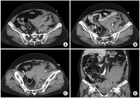
A 72-year-old female patient complaining of abdominal and back pain was admitted to the emergency room. She had no history of hypertension, diabetes mellitus, or other diseases and no medication history. In addition, she had no history of trauma. She suffered from sudden left abdominal and back pain without no specific causes 1 hour prior with abdominal distension and dizziness, directly. On arrival, her blood pressure was 70/40 mmHg and her pulse rate was 110 beats per minute. On abdominal palpation, we found abdominal distension and slight abdominal tenderness and rigidity. There was no leg swelling or leg pain. Blood tests showed anemia, with a hemoglobin of 7.3 g/dL and no other abnormal data except complete blood count. An abdominopelvic computed tomography (CT) showed large hematoma in the lower abdominal cavity and no evidence of extravasation of contrast (Fig. 1).
Fig. 1.
Initial abdominopelvic computed tomography finding. (A) Non-enhancing finding, (B) enhancing finding, (C) delayed finding, and (D) enhancing coronal view.
We assumed a left ovarian cyst rupture as initial diagnosis and performed an emergency operation. When the abdominal cavity was open, a large hematoma and active bleeding were found but the ovary and the uterus showed normal findings. Rather, we found a left retroperitoneal bulging mass-like lesion and performed an incision for left retroperitoneal lesion.
There was a longitudinal laceration, about 1.5 cm in length with sharp and clean margin on the medial wall of the left external iliac vein. This laceration was irrelevant to the adjoining site of the left internal iliac vein. The laceration was closed using continuous 4-0 Prolene sutures. She recovered quickly without complications.
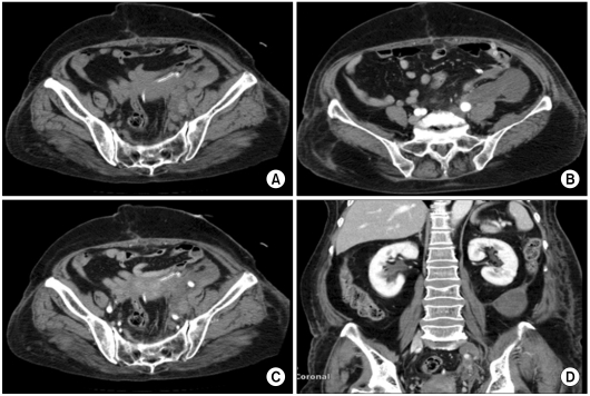
In period of repair, this patient suffered from left leg swelling similar to deep vein thrombosis after postoperative day 7. The patient showed no findings of deep vein thrombosis in postoperative follow-up (POD7) CT (Fig. 2). We administered low molecular weight heparin, changed later too warfarin and applied compressive stocking. Leg edema resolved gradually and she had stopped taking warfarin at 2 months. We followed up 6 months after.
Fig. 2.
Postoperative follow-up (POD7) abdominopelvic computed tomography finding. (A) Non-enhancing finding, (B) enhancing finding, (C) delayed finding, and (D) enhancing coronal view.
DISCUSSION
Reports of the spontaneous rupture of the iliac vein are very rare. The cause of this condition is not yet known in spite of several proposed etiologies including venous hypertension and constipation [1-6]. We assume that the cause of this case was the result of abdominal pressure rise owing to obesity and sudden position change.
This lethal condition was instigated by hypovolemic shock related symtoms and signs including syncope and hypotension in almost all cases. In the emergency room, this condition was mistaken for traumatic or gynecological emergency surgical cases because of the accompanying abdominal distension and syncope-related trauma. In fact, findings of leaked dye suggested vessel rupture were not easily found in abdominopelvic CT on account of heavily compressed hematoma.
Initial resuscitation and prompt surgical management should be operated, but the surgeon's appropriate decision was disturbed by the above reasons. So, if the patient's vital signs are stable, the surgical team should have help from preoperative radiologic examinations including the abdominopelvic CT in spite of dubitable effectiveness [4].
The principle of surgical management is maintaining continuity of ruptured iliac vein achieved by direct suture or bypass reconstruction [4-6]. In our case, we chose a direct suture method owing to her unstable vital signs. But because of a diminished vein diameter, she suffered from disturbed venous flow, as in the leg swelling. It is thought that postoperative anticoagulation will play an important role in maintenance of venous flow and patient's recovery.
The spontaneous rupture of the iliac vein is very rare and a lethal condition. Only prompt surgical management and postoperative anticoagulation therapy can prevent extreme results.
ACKNOWLEDGEMENTS
This work was supported by Konkuk University in 2011.
Footnotes
No potential conflict of interest relevant to this article was reported.
References
- 1.Gaschignard N, Le Paul Y, Maouni T, Le Priol PD. Spontaneous rupture of the left common iliac vein. Ann Vasc Surg. 2000;14:517–518. doi: 10.1007/s100169910096. [DOI] [PubMed] [Google Scholar]
- 2.Lin BC, Chen RJ, Fang JF, Lin KE, Wong YC. Spontaneous rupture of left external iliac vein: case report and review of the literature. J Vasc Surg. 1996;24:284–287. doi: 10.1016/s0741-5214(96)70106-4. [DOI] [PubMed] [Google Scholar]
- 3.Jazayeri S, Tatou E, Cheynel N, Becker F, Brenot R, David M. A spontaneous rupture of the external iliac vein revealed as a phlegmasia cerulea dolens with acute lower limb ischemia: case report and review of the literature. J Vasc Surg. 2002;35:999–1002. doi: 10.1067/mva.2002.121569. [DOI] [PubMed] [Google Scholar]
- 4.Kwon TW, Yang SM, Kim DK, Kim GE. Spontaneous rupture of the left external iliac vein. Yonsei Med J. 2004;45:174–176. doi: 10.3349/ymj.2004.45.1.174. [DOI] [PubMed] [Google Scholar]
- 5.Bracale G, Porcellini M, D'Armiento FP, Baldassarre M. Spontaneous rupture of the iliac vein. J Cardiovasc Surg (Torino) 1999;40:871–875. [PubMed] [Google Scholar]
- 6.Yamada M, Nonaka M, Murai N, Hanada H, Aiba M, Funami M, et al. Spontaneous rupture of the iliac vein: report of a case. Surg Today. 1995;25:465–467. doi: 10.1007/BF00311830. [DOI] [PubMed] [Google Scholar]


