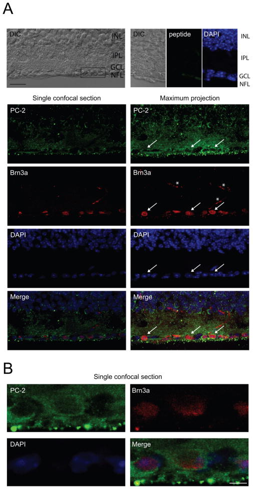
Figure 1. Polycystin-2 is localized to the cytosol of adult mouse RGCs.
A. Immunohistochemical analysis of vertical cryostat sections showing polycystin-2 immunoreactivity in adult mouse RGCs. Brn3a was used as nuclear RGC marker. Representative images are shown. A single confocal section acquired with differential interference contrast (DIC) is shown, indicating the retinal layers (INL, inner nuclear layer; GCL, ganglion cell layer; NFL, nerve fiber layer). The box shows the location of the high-magnification image shown in B. No significant immunoreactivity was obtained when pre-incubating with 100-fold excess of blocking peptide, confirming the specificity of the antibody. A single confocal section (252 nm thickness) and a maximum projection of a z-stack (8.82 μm thickness) are shown. Strong immunoreactivity was found in the cytosol of RGCs (indicated by white arrows) and throughout the inner plexiform layer (IPL). Scale bar, 20 μm. White asterisks indicate non-specific blood vessel staining by the AlexaFluor™ 594 goat anti-mouse secondary antibody. B. Single high magnification confocal section through the RGC layer. Polycystin-2 staining is confined to the cytosol, whereas Brn3a and DAPI are nuclear markers. Scale bar, 5 μm.

