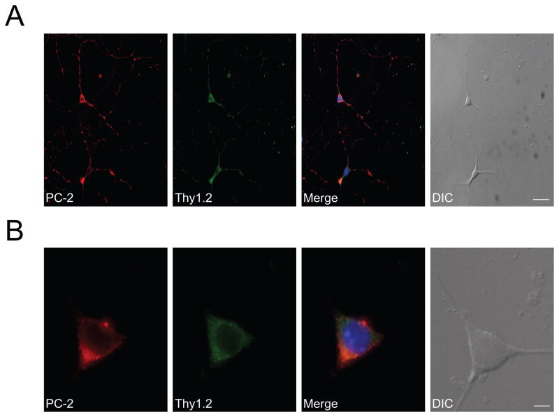
Figure 3. Polycystin-2 localizes to the cytosol of primary cultured RGCs.
A. Polycystin-2 is expressed in primary cultured RGCs. Representative images are shown. Identity of cultured cells was confirmed with the RGC marker Thy 1.2. In Thy 1.2-positive cells, polycystin-2 immunoreactivity was punctate and cytosolic, indicative of intracellular localization. Prominent polycystin-2 immunoreactivity was also observed in neurites. Thy1.2 was used as a cellular marker for positively identifying RGCs. The merged image also shows DAPI staining, positively identifying the nucleus. Scale bar, 25 μm. B. High magnification images showing that polycystin-2 immunoreactivity is confined to the cytosol. Scale bar, 5 μm.

