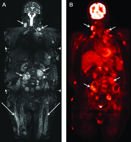
A 23-year-old man with schwannomatosis1 was imaged with whole-body MRI and 18F FDG-PET to assess the extent of disease prior to initiating systemic therapy for new and growing tumors. The patient had undergone multiple prior resections of schwannomas from peripheral nerves. He had ongoing pain, diffuse dysesthesias, and bilateral upper extremity weakness. Assessment of disease burden was similar on both modalities (small arrows). MRI (Figure, A) has superior demonstration of tumor localization and muscle denervation changes (large arrows) due to better contrast and spatial resolution than FDG-PET (Figure, B).2 FDG-PET demonstrated avid uptake in these benign lesions.
Figure. Imaging.
Coronal whole-body MRI (A) showing numerous mass lesions (short arrows) in a patient with schwannomatosis. FDG-PET (B) also shows the lesions, but MRI better depicts anatomy and displays lesions of any etiology regardless of FDG avidity. Muscle denervation (large arrows) is seen as edema-like T2 hyperintensity without perifascial edema on MRI.
Footnotes
Disclosure: Dr. Chhabra is a GE-AUR (GERRAF) fellow and has received research grants for MR neurography scans from Siemens Medical Solutions and Integra Life Sciences. Dr. Blakeley has served on a scientific advisory board for Novartis; receives research support from GlaxoSmithKline, the NIH/NCI, the Children's Tumor Foundation, Brain Tumor Funders Collaborative, and the American Society of Clinical Oncology; and has participated in medico-legal cases.
References
- 1. Gonzalvo A, Fowler A, Cook RJ, et al. Schwannomatosis, sporadic schwannomatosis, and familial schwannomatosis: a surgical series with long-term follow-up. J Neurosurg Epub 2010. October 8 [DOI] [PubMed] [Google Scholar]
- 2. Cai W, Kassarjian A, Bredella MA, et al. Tumor burden in patients with neurofibromatosis types 1 and 2 and schwannomatosis: determination on whole-body MR images. Radiology 2009;250:665–673 [DOI] [PubMed] [Google Scholar]


