Abstract
Background
CD33 is a well-known stem cell target in acute myeloid leukemia. So far, however, little is known about expression of CD33 on leukemic stem cells in chronic leukemias.
Design and Methods
We analyzed expression of CD33 in leukemic progenitors in chronic myeloid leukemia by multi-color flow cytometry and quantitative polymerase chain reaction. In addition, the effects of a CD33-targeting drug, gemtuzumab/ozogamicin, were examined.
Results
As assessed by flow cytometry, stem cell-enriched CD34+/CD38−/CD123+ leukemic cells expressed significantly higher levels of CD33 compared to normal CD34+/CD38− stem cells. Moreover, highly enriched leukemic CD34+/CD38− cells (>98% purity) displayed higher levels of CD33 mRNA. In chronic phase patients, CD33 was found to be expressed invariably on most or all stem cells, whereas in accelerated or blast phase of the disease, the levels of CD33 on stem cells varied from donor to donor. The MDR1 antigen, supposedly involved in resistance against ozogamicin, was not detectable on leukemic CD34+/CD38− cells. Correspondingly, gemtuzumab/ozogamicin produced growth inhibition in leukemic progenitor cells in all patients tested. The effects of gemtuzumab/ozogamicin were dose-dependent, occurred at low concentrations, and were accompanied by apoptosis in suspension culture. Moreover, the drug was found to inhibit growth of leukemic cells in a colony assay and long-term culture-initiating cell assay. Finally, gemtuzumab/ozogamicin was found to synergize with nilotinib and bosutinib in inducing growth inhibition in leukemic cells.
Conclusions
CD33 is expressed abundantly on immature CD34+/CD38− stem cells and may serve as a stem cell target in chronic myeloid leukemia.
Keywords: chronic myeloid leukemia, imatinib resistance, siglec-3, gemtuzumab/ozogamicin
Introduction
Chronic myeloid leukemia (CML) is a stem cell neoplasm characterized by the reciprocal translocation t(9;22).1–3 The resulting fusion gene-product, BCR/ABL, is an oncogenic kinase considered essential to disease evolution.1–3 This concept is supported by the impressive anti-leukemic effects of the BCR/ABL tyrosine kinase inhibitor (TKI) imatinib.4–6 Notably, imatinib produces complete cytogenetic responses (CCyR) and major molecular responses (MMR) in a majority of all patients with freshly diagnosed CML.4–6 Nevertheless, resistance against imatinib can occur in CML patients, and represents a major challenge in clinical practice.7–10 Imatinib-resistant patients may respond to a second generation BCR/ABL TKI such as nilotinib or dasatinib.9–15 However, not all CML patients are long-term responders, which can be explained by multidrug resistance.9–15
A generally accepted concept is that CML is organized hierarchically, and that only a smaller cell-fraction, the so-called leukemic stem cells (LSC), have the capacity of self-renewal, leukemia-initiation, and thus maintenance of CML, whereas more mature CML cells undergo apoptosis after a variable number of cell divisions.10,16,17 This concept predicts that anti-CML therapy is curative only when eliminating all CML LSC. A widely accepted hypothesis is that CML LSC reside within the CD34+/CD38− fraction of the leukemic clone. Unfortunately, however, CML LSC appear to be largely resistant against TKI. LSC-resistance may be due to intrinsic or/and acquired resistance.10,16–20 Whereas intrinsic resistance may result from abnormal expression of drug-transporters and LSC-quiescence, acquired resistance is mostly due to BCR/ABL mutations.10,16–20
To overcome LSC resistance in CML, several different pharmacological approaches have been proposed.10,16–20 One strategy is to apply novel targeted drugs. The ‘optimal’ target should be expressed in all CML LSC (all sub-clones), should be expressed preferentially in leukemic (but not in normal) stem cells, and should provide a BCR/ABL-independent mechanism of killing LSC.
Siglec-3 (CD33) is a cell surface antigen expressed on normal myeloid cells and CD34+ blasts in acute myeloid leukemia (AML).21–23 The antigen serves as a target of gemtuzumab/ozogamicin (GO), which exerts anti-leukemic effects in refractory AML.21–23 Recent data suggest that CD33 is expressed on NOD/SCID mouse-repopulating AML stem cells.24,25 In the present study, we provide evidence that CD34+/CD38−/CD123+ CML stem cells express high levels of CD33, and that CD33 may serve as a therapeutic target in CML.
Design and Methods
Patients
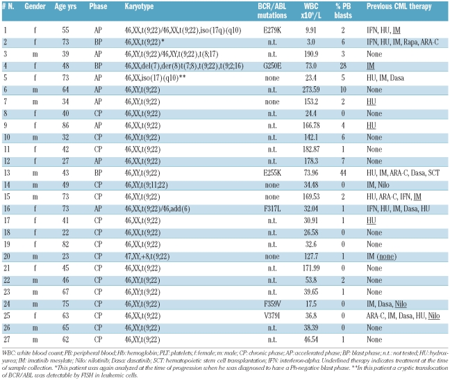
Twenty-seven patients with CML (14 females, 13 males) were examined. The median age was 49 years (range 22–86 years). Diagnoses were established according to WHO-criteria.26 Of the 27 patients, 16 were in chronic phase (CP) CML, 8 had accelerated phase (AP) CML, and 3 were in blast phase (BP) CML. Patients’ characteristics are shown in Table 1. Bone marrow (BM) was obtained from the iliac crest or sternum. Control BM cells were obtained from 7 patients with Hodgkin’s or non-Hodgkin’s lymphomas (pre-therapy staging) or idiopathic thrombocytopenia. All donors gave written informed consent. The study was approved by the ethics committee of the Medical University of Vienna.
Table 1.
Patients’ characteristics.
Flow cytometry and characterization of leukemic stem cells
Various commercial monoclonal antibodies (mAb) were used to characterize and isolate LSC, including mAb against CD33, CD34, CD38, CD45, CD123, and CD243 (MDR1). A specification of mAb used in this study is shown in the Online Supplementary Table S1. LSC were defined as CD34+/CD38−/CD123+ cells. Heparinized BM or peripheral blood (PB) cells (106/tube) were incubated with combinations of mAb (Online Supplementary Table S2) for 15 min. After erythrocyte-lysis (FACS-Lysing-Solution, BD Biosciences, San José, CA, USA), expression of cell surface antigens on LSC was examined by multicolor flow cytometry on a FACSCalibur (BD Biosciences) using FlowJo software (TreeStar, Ashland, OR, USA). Antibody-reactivity was controlled by isotype-matched control antibodies. In select experiments, CD34+/CD38− CML LSC were examined for signs of apoptosis by combined staining for surface antigens and AnnexinV-FITC after exposure to control medium or GO (0.1 μg/mL) for 48 h. AnnexinV staining was performed according to the manufacturer’s instructions (Bender MedSystems, Vienna, Austria).
Purification of leukemic stem cells
In 3 patients with CML AP, CD34+/CD38− cells and CD34+/CD38+ cells were purified from Ficoll-separated PB mononuclear cells (MNC) by cell sorting using a PE-labeled CD34 mAb and APC-conjugated CD38 mAb (Online Supplementary Table S1). CD34+ subfractions were separated using a high-speed sorter (FACSAria, BD Biosciences). After sorting, the purity of CD34+/CD38− LSC was more than 98% in 2 patients, and 80% in the third patient. In 3 CP patients, Lin-negative BM cells were isolated by using the EasySep human progenitor cell enrichment kit following the manufacturer’s recommendation (StemCell Technologies, Vancouver, BC, Canada). The presence of BCR/ABL and thus the leukemic origin of LSC were confirmed by fluorescence in situ hybridization (FISH) performed as described27 using BCR/ABL triple color dual-fusion probe (Kreatech Diagnostics, Amsterdam, The Netherlands).
Quantitative PCR (qPCR)
Total RNA was extracted from highly enriched CD34+ subfractions of CML cells using RNeasy Mini-Kit (Qiagen, Hilden, Germany). CD33 and ABL mRNA levels in CD34+ fractions were quantified by qPCR as reported28,29 using the following primers: CD33: forward 5′-CCCAGCTCTCTGTGCATGTGA-3′, reverse 5′-GAGTGCCAGGGATGAGGATTT-3′; ABL: forward 5′-TGTATGATTTTGTGGCCAGTGGAG-3′, reverse 5′-GCCTAAGACCCGGAGCTTTTCA-3′; MDR1: forward 5′-GCAGCAA AGGAGGCCAACAT-3′, reverse 5′-TCTGGCCACCAGAGAGCTGA-3′. BCR/ABL levels were determined by qPCR following published techniques.28 The Rneasy Micro-Kit (Qiagen) was used to isolate RNA from colony-derived cells.
Evaluation of effects of gemtuzumab/ozogamicin on proliferation of leukemic cells
Primary CML cells (MNC n=16; CP n=13; AP n=3) and lineage-depleted MNC (LSC-enriched n=3) were incubated with increasing concentrations of GO (0.5–1,000 ng/mL) at 37°C for 48 h. In a separate set of experiments, nilotinib or bosutinib (Chemietek, Indianapolis, IN, USA) were applied alone or in combination with GO at various concentrations (fixed ratio of drug concentrations). After 48 h, 3H-thymidine uptake was analyzed as described.30
Clonogenic assay and evaluation of long-term growth of leukemic cells
In 4 patients with CML (CP), MNC were incubated in RPMI 1640 medium plus 10% FCS in the absence or presence of GO (0.1–5 μg/mL) for 2 h. Thereafter, viability was confirmed by try-pan blue exclusion. Cells were washed, and were then grown in a methylcellulose culture essentially as described.31 In brief, cells were seeded in 35 mm culture plates (2–5×10⁵ per well in triplicates) at 37°C (5% CO2) in 0.8% methylcellulose with 30% FCS, 10% bovine serum albumin (Gibco, Carlsbad, CA, USA), alpha-thioglycerol, GM-CSF (10 ng/mL) (R&D Systems, Minneapolis, MN, USA), IL-3 (5 ng/mL) (R&D), and erythropoietin (2 U/mL) (Roche, Basel, Switzerland). After 14 days, the numbers of CFU-GM, BFU-E, and CFU-GEMM were counted under an inverted microscope (Olympus, Tokyo, Japan). Individual CFU-GM (up to 30 per culture-condition) were examined for the presence (percentage) of BCR/ABL mRNA levels (relative to ABL) by qPCR. Growth of long-term culture-initiating cells (LTCIC) was analyzed using irradiated (60 Gy) feeder cells (M2-10B4) and CML MNC (n=3). The LTCICs were performed with Myelocult H5100 Medium (StemCell Technologies, Vancouver, Canada) according to the manufacturer’s protocol. In 3 patients, CML MNC were pre-incubated with GO (5 μg/mL) or control medium at 37°C for 2 h, washed, and then transferred to LTCIC cultures (1–2×10⁵ MNC/well). In 2 of the 3 patients, CML MNC (1–2×10⁵/well) were also cultured on M2-10B4 cells in the absence or presence of GO (10 or 100 ng/ml) at 37°C. After three weeks, cells were recovered and transferred to methylcellulose cultures. Pooled CFU-GM (5–15 per condition) were examined for BCR/ABL by qPCR.
Statistical analyses
Statistical tests were applied to define the level of significance in analyses comparing CD33 levels in CML cells and normal cells (Mann-Whitney U test) and drug effects on cell growth (Student’s t-test). In drug combination experiments, the median effect equation was applied following published guidelines.32 Drug interactions were determined by calculating combination index (CI) values using Calcusyn software (Calcusyn; Biosoft, Ferguson, MO, USA).30,31 A CI of less than 1 indicates synergy, a CI of 1 indicates additive effects, and a CI of more than 1 is indicative of antagonistic effects.
Results
Sorted CD34+/CD38− LSC express CD33 mRNA
As assessed by FISH analysis, the vast majority of highly enriched CD34+/CD38− CML LSC or Lin-negative CML cells were found to display BCR/ABL and thus the Ph-chromosome (Online Supplementary Figure S1A). Expression of BCR/ABL in these cells was confirmed by qPCR analysis (range 61–83% according to the international scale, IS). Moreover, CD34+/CD38− cells strongly expressed CD123 confirming their leukemic origin. As assessed by qPCR, these highly purified CML LSC were found to express CD33 mRNA (Online Supplementary Figure S1B). The levels of CD33 mRNA in these LSC were found to be almost identical compared to CD33 mRNA levels expressed in more mature CD34+/CD38+ cells (Online Supplementary Figure S1B).
CML LSC express high levels of surface CD33
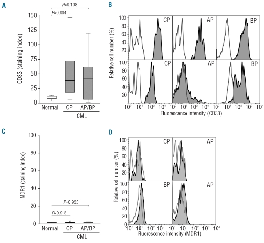
Compared to normal stem cells, CD34+/CD38− CML LSC expressed up to 10-fold higher levels of CD33 on their surface (Figure 1A). In CP patients, CD33 was found to be expressed invariably and homogeneously on most or all CD34+/CD38− cells. In patients with AP and BP, CML LSC also co-expressed CD33, but the expression levels varied from donor to donor, and in one AP patient, most LSC appeared to be CD33-negative cells (Figure 1B). Strong surface expression of CD33 was not only detectable on CML LSC in imatinib-responsive patients, but was also demonstrable in CML LSC obtained from patients with imatinib-resistant disease (Figure 1B). In one patient, we were able to analyze expression of CD33 on CML LSC before and at the time of progression to a Ph-negative blast phase. In this patient (#2 in Table 1), CD34+/CD38− cells were found to express CD33 at both time points (data not shown).
Figure 1.
CD34+/CD38− LSC in CML express high levels of CD33. CD34+/CD38− bone marrow (BM) stem cells in patients with CML in chronic phase (CP), accelerated phase (AP) or blast phase (BP, imatinib-resistant), as well as normal BM cells were analyzed for expression of CD33 (A, B) and MDR1 (C, D) by multicolor flow cytometry. (A and C) Comparison of CML cells (CD33: n=27 donors; MDR1: n=23 donors) to normal BM SC (n=7 donors) regarding expression of CD33 and MDR1 calculated as staining index (MFI test antibody: MFI control antibody). (B and D) Expression of CD33 and MDR1 (dark histograms) on LSC in individual CML donors. The isotype-matched control antibodies are also shown (open histograms).
Failure to detect surface MDR1 on CD34+/CD38− leukemic stem cells
Since the MDR1 antigen has been implicated in LSC resistance against various drugs, including the CD33-targeting drug GO, we also examined the expression of MDR1 (CD243) on CML stem cells. However, in the present study, we were unable to detect substantial amounts of MDR1 on the surface of CD34+/CD38−/CD123+ LSC in our CML patients (Figure 1D) which seems to contrast with previous studies.19 However, in most previous studies, expression of MDR1 in CML LSC was determined by PCR analysis but not by flow cytometry. In our study, we were also able to confirm expression of low levels of MDR1 mRNA in purified CML LSC (data not shown), but surface expression was not detectable.
Effects of gemtuzumab/ozogamicin on proliferation of leukemic cells
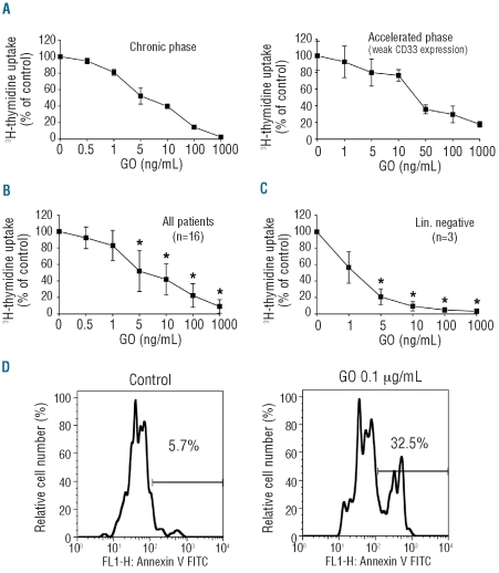
We next examined the effects of the CD33-targeting drug GO on in vitro growth of CML cells and LSC-enriched CML cells. In these experiments, GO was found to induce growth inhibition in primary CML cells (MNC) in all donors tested (Figure 2A–C). The effects of GO on CML cells were dose-dependent and occurred at relatively low and thus clinically relevant drug concentrations (IC50 ranging from 1–100 ng/mL). No differences in responses to GO (IC50) were seen when comparing bulk CML cells with LSC-enriched (Lin-negative) CML cells (Figure 2A–C). Finally, as determined by combined surface and AnnexinV staining, we were able to show that GO induces apoptosis in CML LSC (Figure 2D).
Figure 2.
Inhibition of proliferation and induction of apoptosis in CML cells by gemtuzumab/ozogamicin (GO). (A–C) Primary CML cells. (A) Individual donors. (B) All donors, n=16. (C) LSC-enriched bone marrow Lin- cells (n=3) were cultured in the presence or absence (0) of GO (1–1,000 ng/mL) at 37°C for 48 h. After incubation, 3H-thymidine uptake was measured. Results are expressed as percentage of control and represent (A) the mean±S.D. of triplicates, (B) the mean±S.D. of 16 donors, and (C) the mean±S.D. in 3 donors. (D) Mononuclear cells of a CML patient in blast phase were cultured in the presence or absence (control) of GO (0.1 μg/mL) at 37°C for 48 h. After incubation, the percentage of apoptotic cells within the CD34+/CD38− fraction was determined by combined staining with AnnexinV and cell surface markers. *P<0.05.
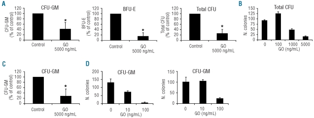
In a next step, we examined the effects of GO on colony formation of CML progenitor cells. In these experiments, preincubation with GO (5 μg/mL) for 2 h resulted in inhibition of growth of CFU-GM and BFU-E in all donors tested (n=4) (Figure 3A). The effect of GO on colony-formation was dose-dependent as exemplified for one donor in Figure 3B. As assessed by qPCR, more than 95% of the Day 14 CFU-GM colonies grown in the presence of GO (n=90 of 93) and 100% of the CFU-GM colonies grown in the absence of GO (n=41 of 41) were found to contain BCR/ABL. We then examined the effects of GO in an LTCIC assay. Pre-incubation of CML MNC with GO (5 μg/mL) for 2 h resulted in a significant decrease in the numbers of CFU-GM in the LTCIC (Figure 3C). In 2 donors, MNC were cultured on M2-10B4 cells in the presence of GO (10 or 100 ng/mL). Again, GO was found to reduce the number of colony-forming CFU-GM in both donors examined (Figure 3D). As assessed by qPCR, pooled CFU-GM-derived colonies, cultured in GO or in control medium, contained comparable amounts of BCR/ABL (control: 36.6±18.3% vs. GO, 100 ng/mL: 38.4±20.2%), suggesting that the growth-inhibitory effect of GO did not spare normal CFU-GM in the LTCIC. Together, these data suggest that GO inhibits the growth and survival of immature CML progenitor cells and CML LSC.
Figure 3.
Gemtuzumab/ozogamicin (GO) impairs CML colony formation. (A) Mononuclear cells (MNC) from 4 CML donors were preincubated in control medium or in GO (5 μg/mL) for 2 h. Thereafter, cells were cultured in methylcellulose in the presence of cytokines for 14 days. The numbers of CFU-GM, BFU-E, and CFU-GEMM were counted under an inverted microscope. Results show the numbers of CFU-GM, BFU-E, and all colonies (total CFU: CFU-GM+BFU-E+CFU-GEMM). Data are expressed as mean±S.D. from 4 donors. *P<0.05 compared to control. (B) Dose-dependent effect of GO (2-hour pre-incubation prior to seeding) on colony-formation of CML cells in one donor. Results show the numbers of total CFU and are expressed as mean±S.D. from triplicates. (C) Numbers of CFU-GM in a long term co-culture-initiating cell assay (LTCIC). MNC of 3 donors were pre-incubated in control medium or GO (5 μg/mL) for 2 h, washed, and transferred to a feeder layer of M2-10B4 cells for three weeks. Thereafter, CFU growth was determined. Data are expressed as mean±S.D. from 3 donors. *P<0.05 compared to control. (D) Numbers of CFU-GM in a LTCIC assay. Cells were maintained in the LTCIC assay in the presence or absence (0) of GO (10 or 100 ng/mL) for three weeks. Then, CFU growth was determined. Results show the numbers (N.) of CFU-GM in 2 donors and are expressed as mean±S.D. from triplicates.
Gemtuzumab/ozogamicin cooperates with nilotinib and bosutinib in producing growth inhibition in leukemic cells in chronic myeloid leukemia
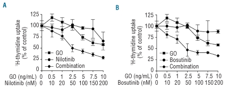
Finally, we asked whether GO would cooperate with BCR/ABL TKI in producing growth inhibition in CML cells. To address this question, we exposed primary CML cells to combinations of GO and nilotinib, and GO and bosutinib. As shown in Figure 4, GO was found to synergize with nilotinib (Figure 4A) as well as with bosutinib (Figure 4B) in producing growth inhibition in CML cells. Evaluation of drug combination effects by the median effect equation30,32 confirmed synergistic effects of these drug combinations (combination index values <1).
Figure 4.
Synergistic effects of gemtuzumab/ozogamicin (GO) and BCR/ABL tyrosine kinase inhibitors on growth of primary CML cells. CML cells were cultured in the presence of various concentrations of GO and nilotinib (A) or GO and bosutinib (B) alone or in combination (fixed ratio of drug concentrations) as indicated. Thereafter, 3H-thymidine uptake was measured. Results show typical experiments and represent the mean±S.D. of triplicates. In each case, the combination index value (CI) calculated by Calcusyn software was found to be <1, thereby indicating synergistic drug interactions.
Discussion
Although in most patients with CML the disease can be kept under control by BCR/ABL TKI, resistance may occur during therapy and this remains a challenge in the treatment of CML.7–10 Several different mechanisms of resistance have been described, including BCR/ABL mutations and intrinsic resistance of CML stem cells.7–10 A number of targeting concepts have recently been proposed with the aim of eradicating these cells.10,16,18 CML stem cells supposedly reside within the CD34+/CD38− fraction of the malignant clone. Results of this study show that CD34+/CD38− stem cells in CML express high levels of CD33 and that the CD33-targeting drug gemtuzumab/ozogamicin is a potent inhibitor of growth and survival of CML stem cells. Since drug effects occurred in the nanomolar range, and expression of CD33 on CML stem cells exceeded by far CD33 levels detectable on normal stem cells, these observations may have clinical implications.
To confirm that the CD34+/CD38− stem cell population in the CML patients analyzed were indeed of leukemic origin, we performed FISH analysis and qPCR. With both techniques, we were able to demonstrate that the majority of the CD34+/CD38− cells carried the BCR/ABL oncoprotein. Furthermore, we were able to show that these CD34+/CD38− cells express high levels of the IL-3 receptor alpha chain (CD123), supporting the conclusion that these cells were indeed leukemic stem cells.
We and others have recently shown that leukemic stem cells in AML express substantial amounts of CD33 whereas normal CD34+/CD38− stem cells express only low amounts of or even lack CD33.24,25 In the present study, we found that leukemic stem cells in CML patients express significantly higher levels of surface CD33 compared to normal stem cells. The mechanisms underlying the increased expression of CD33 on leukemic stem cells remains unknown. Since the phenomenon was also observed in AML,24,25 it is tempting to speculate that leukemic progenitors in general express higher levels of CD33 compared to normal stem cells. Another possibility would be that surface expression of CD33 is associated with cell cycle-specific or proliferative features of leukemic stem cells. The possibility that expression of CD33 is up-regulated by specific oncoproteins such as BCR/ABL would be another alternative. However, other clonal cells in CML such as monocytes or granulocytes displayed similar levels of CD33 compared to normal leukocytes.
Since CD33 may serve as a potential target for therapy,21–23 we asked ourselves whether CD33 is expressed invariably on all CML stem cells in all patients. In these experiments, we first noted that expression of CD33 on CML stem cells (CD34+/CD38−) depends on the phase of disease. Notably, whereas in CP virtually all CD34+/CD38− cells expressed high levels of CD33 in all patients examined, this was not the case in patients with advanced CML, i.e. AP or BP. Specifically, we found that in AP and BP, distinct subpopulations of CD34+/CD38− cells (presumably subclones) did not express detectable CD33 on their surface. In one patient, virtually all CD34+/CD38− CML cells were found to lack CD33. These observations suggest that CML progression is associated with evolution in subclones and that subclone formation may be associated with loss of CD33. Such a hypothesis would fit with the notion that CML transformation is a complex process that often involves multiple lineages, including even non-myeloid and thus CD33-negative cells.2,10,26 As far as a target for therapy is concerned, this observation may lead to the conclusion that CD33 is a suitable stem cell target in early rather than in advanced CML, and that drug combinations are still required to suppress all relevant subclones in these patients.
Recent data suggest that MDR1 is expressed in leukemic stem cells in CML and may serve as important gene mediating multi-drug resistance in these cells.19,34–36 With regard to gemtuzumab/ozogamicin, it has also been reported that MDR1 is involved in the mechanism of drug-efflux, and thus resistance, in AML and CML cell lines.37,38 Therefore, we asked ourselves whether CML LSC (CD34+/CD38−) express MDR1. However, in this study, we were unable to detect substantial amounts of the MDR1 antigen on the surface of CD34+/CD38− cells in our CML patients. In the light of the previously published literature,19,34–36 this result was unexpected and may have several explanations. The most likely is that in previous studies, MDR1 was examined in CML cells by PCR but not by surface staining techniques.19,34–36 In line with these previous data, we were also able to detect MDR1 mRNA in highly purified CML stem cells in this study. The failure to detect MDR1 on the surface of these cells may be explained by selective cytoplasmic expression or by very low expression of MDR1 on these cells. Alternatively, MDR1 is expressed on the surface of CML stem cells, but was not detectable by the antibody because the binding site was ‘masked’ by gangliosides or other antigens. Another explanation for the discrepant results obtained may be that in previous studies cell lines, but not primary CML stem cells, were examined. In fact, we were able to detect surface expression of MDR1 on the CML cell line KU812 which is resistant to gemtuzumab/ozogamicin (Online Supplementary Figures S2 and S3). All in all, our data strongly suggest that primary CD34+/CD38− CML stem cells do not express substantial amounts of surface MDR1 which is important when considering targeted drug therapies involving CD33.
Based on this observation, we asked ourselves whether the CD33-immunotoxin conjugate gemtuzumab/ozogamicin (GO) would exert growth-inhibitory effects on primary CML cells. The results of our study show that GO is a potent inhibitor of growth of primary CML cells, and also inhibits the growth of CD34+/CD38− or Lin-negative CML cells. Drug effects and IC50 were observed at a low nanomolar and thus pharmacological range. Moreover, the effect of GO was seen in CP and AP, and even in patients with imatinib-resistant disease; although, as expected, the effects of GO were less pronounced in CD33-negative subclones. Notably, in patients with AP and BP, CML LSC often expressed varying amounts of CD33, and in several cases subfractions of LSC were found to lack CD33. All in all there was a good correlation between GO effects and expression of CD33 on leukemic cells. However, it is still not clear whether all the GO effects on CML cells were mediated through CD33. Alternatively, some of the GO effects could have been mediated via non-specific drug-uptake (endocytosis) which has been described for leukemic cells.39
A number of previous and more recent data suggest that CML stem cells display multiple mechanisms of drug resistance and that drug combinations may be required to achieve stem cell-eradicating activity. We were, therefore, interested to learn whether combinations of BCR/ABL blockers and GO would produce cooperative or even synergistic drug combination effects on CML cells. Indeed, GO was found to cooperate with nilotinib and bosutinib in producing growth inhibition in primary CML cells. These observations may have clinical implications, even if GO has recently been withdrawn from the market in the US and in Europe. In particular, in the light of the LSC-targeting and ‘TKI-combination’ effects seen with GO, it seems reasonable to consider such drug combinations for eradication of advanced CML in clinical trials. For the moment, it is not known whether such studies should be performed with GO or with other CD33-targeting antibody constructs.
In conclusion, our data show that CD34+/CD38− CML LSC express high levels of CD33, and that GO is highly effective in producing growth arrest in CML stem cells. Whether the concept of targeting CD33 may also lead to growth inhibition or even eradication of CML stem cells in vivo remains to be clarified.
Acknowledgments
We would like to thank Günther Hofbauer and Andreas Spittler (both at the Cell Sorting Core Unit of the Medical University of Vienna), as well as Edith Pfeiffer and Gregor Hörmann for excellent technical assistance.
Footnotes
The online version of this article has a Supplementary Appendix.
Authorship and Disclosures
The information provided by the authors about contributions from persons listed as authors and in acknowledgments is available with the full text of this paper at www.haematologica.org.
Financial and other disclosures provided by the authors using the ICMJE (www.icmje.org) Uniform Format for Disclosure of Competing Interests are also available at www.haematologica.org.
References
- 1.Rowley JD. A new consistent chromosomal abnormality in chronic myelogenous leukaemia identified by quinacrine flourescence and Giemsa staining. Nature. 1973;243(5405):290–3. doi: 10.1038/243290a0. [DOI] [PubMed] [Google Scholar]
- 2.Melo JV, Deininger MW. Biology of chronic myelogenous leukemia--signaling pathways of initiation and transformation. Hematol Oncol Clin North Am. 2004;18(3):545–68. doi: 10.1016/j.hoc.2004.03.008. [DOI] [PubMed] [Google Scholar]
- 3.Arlinghaus R, Sun T. Signal transduction pathways in Bcr-Abl transformed cells. Cancer Treat Res. 2004;119:239–70. doi: 10.1007/1-4020-7847-1_12. [DOI] [PubMed] [Google Scholar]
- 4.Druker BJ, Talpaz M, Resta DJ, Peng B, Buchdunger E, Ford JM, et al. Efficacy and safety of a specific inhibitor of the BCR-ABL tyrosine kinase in chronic myeloid leukemia. N Engl J Med. 2001;344(14):1031–7. doi: 10.1056/NEJM200104053441401. [DOI] [PubMed] [Google Scholar]
- 5.O’Brien SG, Guilhot F, Larson RA, Gathmann I, Baccarani M, Cervantes F, et al. Imatinib compared with interferon and low-dose cytarabine for newly diagnosed chronic-phase chronic myeloid leukemia. N Engl J Med. 2003;348(11):994–1004. doi: 10.1056/NEJMoa022457. [DOI] [PubMed] [Google Scholar]
- 6.Druker BJ, Guilhot F, O’Brien SG, Gathmann I, Kantarjian H, Gattermann N, et al. Five-year follow-up of patients receiving imatinib for chronic myeloid leukemia. N Engl J Med. 2006;355(23):2408–17. doi: 10.1056/NEJMoa062867. [DOI] [PubMed] [Google Scholar]
- 7.Gorre ME, Mohammed M, Ellwood K, Hsu N, Paquette R, Rao PN, et al. Clinical resistance to STI-571 cancer therapy caused by BCR-ABL gene mutation or amplification. Science. 2001;293(5531):876–80. doi: 10.1126/science.1062538. [DOI] [PubMed] [Google Scholar]
- 8.Shah NP, Nicoll JM, Nagar B, Gorre ME, Paquette RL, Kuriyan J, et al. Multiple BCR-ABL kinase domain mutations confer poly-clonal resistance to the tyrosine kinase inhibitor imatinib (STI571) in chronic phase and blast crisis chronic myeloid leukemia. Cancer Cell. 2002;2(2):117–25. doi: 10.1016/s1535-6108(02)00096-x. [DOI] [PubMed] [Google Scholar]
- 9.Deininger M. Resistance and relapse with imatinib in CML: causes and consequences. J Natl Compr Canc Netw. 2008;6(S2):S11–S21. [PubMed] [Google Scholar]
- 10.Valent P. Emerging stem cell concepts for imatinib-resistant chronic myeloid leukaemia: implications for the biology, management, and therapy of the disease. Br J Haematol. 2008;142(3):361–78. doi: 10.1111/j.1365-2141.2008.07197.x. [DOI] [PubMed] [Google Scholar]
- 11.Kantarjian H, Giles F, Wunderle L, Bhalla K, O’Brien S, Wassmann B, et al. Nilotinib in imatinib-resistant CML and Philadelphia chromosome-positive ALL. N Engl J Med. 2006;354(24):2542–51. doi: 10.1056/NEJMoa055104. [DOI] [PubMed] [Google Scholar]
- 12.Talpaz M, Shah NP, Kantarjian H, Donato N, Nicoll J, Paquette R, et al. Dasatinib in imatinib-resistant Philadelphia chromosome-positive leukemias. N Engl J Med. 2006;354(24):2531–41. doi: 10.1056/NEJMoa055229. [DOI] [PubMed] [Google Scholar]
- 13.Weisberg E, Manley PW, Cowan-Jacob SW, Hochhaus A, Griffin JD. Second generation inhibitors of BCR-ABL for the treatment of imatinib-resistant chronic myeloid leukaemia. Nat Rev Cancer. 2007;7(5):345–56. doi: 10.1038/nrc2126. [DOI] [PubMed] [Google Scholar]
- 14.Martinelli G, Soverini S, Rosti G, Cilloni D, Baccarani M. New tyrosine kinase inhibitors in chronic myeloid leukemia. Haematologica. 2005;90(4):534–41. [PubMed] [Google Scholar]
- 15.Giles FJ, O’Dwyer M, Swords R. Class effects of tyrosine kinase inhibitors in the treatment of chronic myeloid leukemia. Leukemia. 2009;23(10):1698–707. doi: 10.1038/leu.2009.111. [DOI] [PubMed] [Google Scholar]
- 16.Elrick LJ, Jorgensen HG, Mountford JC, Holyoake TL. Punish the parent not the progeny. Blood. 2005;105(5):1862–6. doi: 10.1182/blood-2004-08-3373. [DOI] [PubMed] [Google Scholar]
- 17.Barnes DJ, Melo JV. Primitive, quiescent and difficult to kill: the role of non-proliferating stem cells in chronic myeloid leukemia. Cell Cycle. 2006;5(24):2862–6. doi: 10.4161/cc.5.24.3573. [DOI] [PubMed] [Google Scholar]
- 18.Copland M, Fraser AR, Harrison SJ, Holyoake TL. Targeting the silent minority: emerging immunotherapeutic strategies for eradication of malignant stem cells in chronic myeloid leukaemia. Cancer Immunol Immunother. 2005;54(4):297–306. doi: 10.1007/s00262-004-0573-1. [DOI] [PMC free article] [PubMed] [Google Scholar]
- 19.Jiang X, Zhao Y, Smith C, Gasparetto M, Turhan A, Eaves A, et al. Chronic myeloid leukemia stem cells possess multiple unique features of resistance to BCR-ABL targeted therapies. Leukemia. 2007;21(5):926–35. doi: 10.1038/sj.leu.2404609. [DOI] [PubMed] [Google Scholar]
- 20.Sullivan C, Peng C, Chen Y, Li D, Li S. Targeted therapy of chronic myeloid leukemia. Biochem Pharmacol. 2010;80(5):584–91. doi: 10.1016/j.bcp.2010.05.001. [DOI] [PMC free article] [PubMed] [Google Scholar]
- 21.Sievers EL, Appelbaum FR, Spielberger RT, Forman SJ, Flowers D, Smith FO, et al. Selective ablation of acute myeloid leukemia using antibody-targeted chemotherapy: a phase I study of an anti-CD33 calicheamicin immunoconjugate. Blood. 1999;93(11):3678–84. [PubMed] [Google Scholar]
- 22.van Der Velden VH, te Marvelde JG, Hoogeveen PG, Bernstein ID, Houtsmuller AB, Berger MS, et al. Targeting of the CD33-calicheamicin immunoconjugate Mylotarg (CMA-676) in acute myeloid leukemia: in vivo and in vitro saturation and internalization by leukemic and normal myeloid cells. Blood. 2001;97(10):3197–204. doi: 10.1182/blood.v97.10.3197. [DOI] [PubMed] [Google Scholar]
- 23.Sievers EL, Larson RA, Stadtmauer EA, Estey E, Löwenberg B, Dombret H, et al. Efficacy and safety of gemtuzumab ozogamicin in patients with CD33-positive acute myeloid leukemia in first relapse. J Clin Oncol. 2001;19(13):3244–54. doi: 10.1200/JCO.2001.19.13.3244. [DOI] [PubMed] [Google Scholar]
- 24.Taussig DC, Pearce DJ, Simpson C, Rohatiner AZ, Lister TA, Kelly G, et al. Hematopoietic stem cells express multiple myeloid markers: implications for the origin and targeted therapy of acute myeloid leukemia. Blood. 2005;106(13):4086–92. doi: 10.1182/blood-2005-03-1072. [DOI] [PMC free article] [PubMed] [Google Scholar]
- 25.Hauswirth AW, Florian S, Printz D, Sotlar K, Krauth MT, Fritsch G, et al. Expression of the target receptor CD33 in CD34+/CD38−/CD123+ AML stem cells. Eur J Clin Invest. 2007;37(1):73–82. doi: 10.1111/j.1365-2362.2007.01746.x. [DOI] [PubMed] [Google Scholar]
- 26.Vardiman JW, Melo J, Baccarani M, Thiele J. Chronic myelogenous leukemia, BCR/ABL1 positive. In: Swerdlow SH, Campo E, Harris NL, Jaffe ES, Pileri SA, Stein H, et al., editors. WHO Classification of Tumours of Haematopoietic and Lymphoid Tissues. 4th ed. IARC Press; Lyon, France: 2008. pp. 32–7. [Google Scholar]
- 27.Wöhrer S, Rabitsch W, Shehata M, Kondo R, Esterbauer H, Streubel B, et al. Mesenchymal stem cells in patients with chronic myelogenous leukaemia or bi-phenotypic Ph+ acute leukaemia are not related to the leukaemic clone. Anticancer Res. 2007;27(6B):3837–41. [PubMed] [Google Scholar]
- 28.Müller MC, Cross NC, Erben P, Schenk T, Hanfstein B, Ernst T, et al. Harmonization of molecular monitoring of CML therapy in Europe. Leukemia. 2009;23(11):1957–63. doi: 10.1038/leu.2009.168. [DOI] [PubMed] [Google Scholar]
- 29.Aichberger KJ, Gleixner KV, Mirkina I, Cerny-Reiterer S, Peter B, Ferenc V, et al. Identification of proapoptotic Bim as a tumor suppressor in neoplastic mast cells: role of KIT D816V and effects of various targeted drugs. Blood. 2009;114(26):5342–51. doi: 10.1182/blood-2008-08-175190. [DOI] [PubMed] [Google Scholar]
- 30.Gleixner KV, Ferenc V, Peter B, Gruze A, Meyer RA, Hadzijusufovic E, et al. Polo-like kinase 1 (Plk1) as a novel drug target in chronic myeloid leukemia: overriding imatinib resistance with the Plk1 inhibitor BI 2536. Cancer Res. 2010;70(4):1513–23. doi: 10.1158/0008-5472.CAN-09-2181. [DOI] [PubMed] [Google Scholar]
- 31.Agis H, Jager E, Doninger B, Sillaber C, Marosi C, Drach J, et al. In vivo effects of imatinib mesylate on human haematopoietic progenitor cells. Eur J Clin Invest. 2006;36(6):402–8. doi: 10.1111/j.1365-2362.2006.01645.x. [DOI] [PubMed] [Google Scholar]
- 32.Chou TC, Talalay P. Quantitative analysis of dose-effect relationships: the combined effects of multiple drugs or enzyme inhibitors. Adv Enzyme Regul. 1984;22:27–55. doi: 10.1016/0065-2571(84)90007-4. [DOI] [PubMed] [Google Scholar]
- 33.Florian S, Sonneck K, Hauswirth AW, Krauth MT, Schernthaner GH, Sperr WR, et al. Detection of molecular targets on the surface of CD34+/CD38−-stem cells in various myeloid malignancies. Leuk Lymphoma. 2006;47(2):207–22. doi: 10.1080/10428190500272507. [DOI] [PubMed] [Google Scholar]
- 34.Giles FJ, Kantarjian HM, Cortes J, Thomas DA, Talpaz M, Manshouri T, et al. Multidrug resistance protein expression in chronic myeloid leukemia: associations and significance. Cancer. 1999;86(5):805–13. [PubMed] [Google Scholar]
- 35.Mahon FX, Belloc F, Lagarde V, Chollet C, Moreau-Gaudry F, Reiffers J, et al. MDR1 gene overexpression confers resistance to imatinib mesylate in leukemia cell line models. Blood. 2003;101(6):2368–73. doi: 10.1182/blood.V101.6.2368. [DOI] [PubMed] [Google Scholar]
- 36.Illmer T, Schaich M, Platzbecker U, Freiberg-Richter J, Oelschlägel U, von Bonin M, et al. P-glycoprotein-mediated drug efflux is a resistance mechanism of chronic myelogenous leukemia cells to treatment with imatinib mesylate. Leukemia. 2004;18(3):401–8. doi: 10.1038/sj.leu.2403257. [DOI] [PubMed] [Google Scholar]
- 37.Naito K, Takeshita A, Shigeno K, Nakamura S, Fujisawa S, Shinjo K, et al. Calicheamicin-conjugated humanized anti-CD33 monoclonal antibody (gemtuzumab zogamicin, CMA-676) shows cytocidal effect on CD33-positive leukemia cell lines, but is inactive on P-glycoprotein-expressing sublines. Leukemia. 2000;14(8):1436–43. doi: 10.1038/sj.leu.2401851. [DOI] [PubMed] [Google Scholar]
- 38.Jawad M, Seedhouse C, Mony U, Grundy M, Russell NH, Pallis M. Analysis of factors that affect in vitro chemosensitivity of leukaemic stem and progenitor cells to gemtuzumab ozogamicin (Mylotarg) in acute myeloid leukaemia. Leukemia. 2010;24(1):74–80. doi: 10.1038/leu.2009.199. [DOI] [PubMed] [Google Scholar]
- 39.Jedema I, Barge RM, van der Velden VH, Nijmeijer BA, van Dongen JJ, Willemze R, et al. Internalization and cell cycle-dependent killing of leukemic cells by Gemtuzumab Ozogamicin: rationale for efficacy in CD33-negative malignancies with endocytic capacity. Leukemia. 2004;18(2):316–25. doi: 10.1038/sj.leu.2403205. [DOI] [PubMed] [Google Scholar]


