

Abstract
Context:
Congenital heart disease (CHD) patients bear a higher risk of scoliosis during their lifetime compared to their normal counterparts. On the other hand, operation on chest wall has been shown to increase the risk of scoliosis. However, the data are inconclusive. The present retrospective analysis is undertaken to determine the frequency of post-thoracotomy/sternotomy scoliosis in children with CHD.
Materials and Methods:
One hundred and eighty children with CHD who underwent thoracotomy/sternotomy and had a minimum followup of 3 years in a teaching center from 1997 to 2010 were recruited. After operation, all the patients were regularly examined for the development of scoliosis. 102 patients underwent thoracotomy and 78 sternotomy. Student's t test, Chi-square test, Fisher's exact test were used for statistical analyses.
Results:
Eighty-eight males and 92 females with a mean age of 9.95 ± 2.31 (range: 5–15) years were enrolled. The mean age at operation was 2.59 ± 1.66 (range: 0–9) years and the mean follow-up period was 7.36 ± 2.12 (range: 5–13) years. Scoliosis was confirmed in two patients (1.1%): 1 (1%) in the thoracotomy group (a 12-year-old female operated 2 years earlier with a spinal 22° convexity to the right and 78° kyphosis) and another (1.1%) in the sternotomy group (an 8-year-old female operated during her neonatal period with a spinal 23° convexity to the left).
Conclusion:
Scoliosis is not a common finding among the operated children with CHD in our center.
Keywords: Congenital heart disease, scoliosis, sternotomy, thoracotomy
INTRODUCTION
The prevalence of congenital heart disease (CHD) is reported to be about 1% in liveborn infants.1,2 This condition is a multifactorial entity and majority of the patients are treated by open heart surgery. Amazing advances in pediatric cardiac intensive care have dramatically increased the survival rate after open heart operations, and so longer follow-ups are now possible.3 The main approaches in open heart surgeries are thoracotomy, sternotomy or both.4 It has been previously claimed that these operations may cause an increased risk of spinal deformities, particularly, scoliosis. This is seemingly apart from the inherited increased risk of scoliosis in patients with CHD, which is estimated to be between 2% and 31%.5–7 It is noteworthy that the incidence of adolescent idiopathic scoliosis is almost 2–3% in the general population.7 Some authors believe that this increased risk of scoliosis is due to the manipulation of the thoracic cage itself in the early life because these operations had also increased the incidence of scoliosis in patients who underwent thoracotomy/sternotomy with underlying noncardiac etiologies.8,9 However, the exact cause is still unknown and there is ongoing debate in this regard.9 Due to lack of homogeneity in the available data in the literature, scarce methodologically appropriate studies, a possible effect of racial differences and ethnicity,10,11 and no known study on the targeted population, we aimed to assess the prevalence of scoliosis in a group of Iranian children who underwent open heart surgery and to determine the role of available technical approaches (thoracotomy, sternotomy) in this regard.
MATERIALS AND METHODS
In this retrospective, study, 196 neonates, infants and children with CHD, who were recruited in a Teaching Center during a period of 13 years (1997–2010), were studied. These patients underwent open heart surgery with the minimum follow-up of 3 years. Sixteen patients were lost during the follow-up period, and hence the study was completed in 180 cases: 102 patients underwent thoracotomy and 78 patients underwent sternotomy. The inclusion criterion was history of first-ever sternotomy or thoracotomy for open heart surgery due to CHD. The exclusion criterion was pre-operative history of any spinal deformity. During the follow-up period, Adam's Forward Bending (AFB) test was performed by a skilled pediatric orthopedics surgeon with over 25 years experience, in every session of physical examination, with or without extra tests. For this test, the patient was asked to lean forward with his or her feet together and bend 90° at the waist. Then, the examiner documented any asymmetry of the trunk or any abnormal spinal curvatures.12 Chest and spinal radiographs were taken if there was any indication determined by the corresponding physician, such as presence of shoulder and breast asymmetry, unequal scapular prominence, elevated or prominent hip, greater space between the arm and body on one side, head not centered over the pelvis, or flank crease asymmetry;13 and in the cases with positive AFB test as determined by the pediatric orthopedics surgeon. The radiographs were standing posteroanterior and lateral chest films. With no tilt of L2, the lumbar scoliosis was reported. The Cobb's method was used to measure the spinal curvature on both anteroposterior and lateral radiographs.14 Scoliosis was defined as curvature above 10°. On lateral films (T2–T12), normal thoracic kyphosis was defined between 21° and 40°.15 All the films were interpreted by a radiologist and an orthopedics surgeon. The deviation was quantified by a standard scoliometer (Medline Industries, Mundelein, IL, USA). Patients’ characteristics as well as the frequency of scoliosis were compared between the two groups (thoracotomy and sternotomy). Statistical analysis was performed using SPSS version 19.0 (IBM, USA). The Student's t test, Chi-square test or Fisher's exact test were used where appropriate. A P value of less than 0.05 was considered statistically significant. This study was approved by the Ethics Committee of Tabriz University of Medical Sciences.
RESULTS
Patients’ demographic and general data are summarized in Table 1. At the time of followup examination, the mean age of the patients in the sternotomy group was significantly higher than the age of patients in the thoracotomy group (P=0.02) [Figure 1]. Patients in the thoracotomy group were operated significantly earlier than the patients in the sternotomy group (P=0.001) [Figure 2]. Percentage of male patients was significantly higher in the sternotomy group (P =0.02).
Table 1.
Patients’ demographics and characteristics

Figure 1.

Bar diagram showing age of the patients at followup
Figure 2.

Bar diagram showing age of the patients at the time of heart surgery
The mean postoperative follow-up time was comparable between the two groups (P=0.89).
The AFB test was reported as positive by the pediatric orthopedics surgeon in two patients (1.1%), one case in the thoracotomy group and one case in the sternotomy group. Radiological assessments in both the patients confirmed the presence of scoliosis [Figures 3 and 4]. There was no significant difference between the two groups in this regard (P=0.68) [Figure 5]. Percentage of cases with scoliosis was comparable between the male (n=1, 1.1%) and the female (n=1, 1.1%) groups of patients (p=0.74)
Figure 3.

Anteroposterior and lateral radiographs of a 12-year-old male patient with ventricular septal defect as the primary underlying cause of thoracotomy at the age of 9 years with scoliosis (22° to right) and hyperkyphosis (78°)
Figure 4.
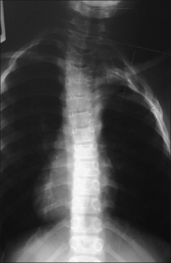
Anteroposterior radiograph of an 8-year-old female patient with patent ductus arteriosus as the primary underlying cause of sternotomy at the age of 1 month with scoliosis (23° to left)
Figure 5.

Bar diagram showing percentage of cases with and without scoliosis in the two studied groups
DISCUSSION
In the current study, prevalence of post-thoracotomy/sternotomy scoliosis after open heart surgery for CHD was assessed in a pediatric population. With an average follow-up time of 7.4 (range: 5–13) years, scoliosis was documented in 1.1% of the studied patients (1.3% of the sternotomy group vs. 1% of the thoracotomy group; P=0.68). It is assumed that the incidence of scoliosis among CHD patients is higher than the normal counterparts even without open heart surgery. This incidence ranged between 0.2% and 31% in the CHD patients versus 2% and 3% in age- and sex-matched unaffected group. In other studies, however, it is proposed that the scoliosis is associated only with thoracotomy or any open heart surgery irrelevant to the approaching technique.5,8,9,16–18 Ruiz-Iban et al. studied 128 children with CHD after median sternotomy. Scoliosis (deviation >10°) was documented in 34.4% of the patients.9 In another series by Herrea-Soto et al., 68 children with CHD first underwent thoracotomy and then sternotomy. In a mean follow-up time of 14.9 (range: 5–20) years, the incidence of scoliosis was about 26%.13 In another study by Herrea-Soto et al., the incidence of scoliosis was about 28% in 108 children with CHD who underwent medial sternotomy. The mean follow-up period was 13 years.7 It is obvious that although the rate of scoliosis after open heart surgery varies significantly between studies, our result is far less than the reported range. Many justifications may be proposed in this regard, such as different definitions of scoliosis, heterogeneous underlying CHD, type of open heart surgery, age at operation and length of follow-up.9 We used the AFB test as the primary screening method in our population. This test is the only standard method for detecting even mild scoliosis in a target population. In addition, this was not the sole screening modality, and different criteria such as alerting findings during examination and other non-standard tests proposed in these patients had been employed by an experienced pediatric orthopedics surgeon, if needed. Likewise, the radiological diagnosis was also based on an accepted method in the literature.19 The severity of scoliosis was moderate in our patients according to the criteria of scoliosis severity classification.8 It had been previously shown that the majority of patients with scoliosis after open heart surgery suffer from mild to moderate scoliosis.5,8,16–18 So, our results are in conformity with the available data in this regard. There is not yet an agreement on the influence of underlying CHD on the incidence of scoliosis. Whereas some investigations have found an association between the cyanotic cardiac disease and scoliosis, others did not confirm this.8 The two patients with scoliosis in our series were operated due to ventricular septal defect (VSD) and patent ductus arteriosus (PDA), both classified as non cyanotic conditions.1,2 Due to limited sample size, we could not elucidate this association in the present study. To the best of our knowledge, the current study is the first one that compares the frequency of scoliosis after thoracotomy and sternotomy in CHD patients. Apparently, further studies are required for definite conclusion. It seems that the age at operation and length of follow-up are the two key factors in this regard. Ruiz-Iban et al. concluded that the risk of scoliosis will increase significantly if the patients undergo open heart surgery under the age of 18 months.9 Herrea-Soto et al. believe that for a better conclusion, the following up should be at least by the time of bony maturation.7 The mean age of our patients at the time of operation was 2.22 ± 1.45 years in the thoracotomy group, 3.07 ± 1.81 years in the sternotomy group, and 2.59 ± 1.66 years overall. On the other hand, the mean follow-up time was 7.36 ± 2.12 years in this series. Longitudinal growth of the spine has been estimated to be 2 cm per year within the first 5 years of life, 0.9 cm per year between 5 and 10 years of life, and 1.8 cm per year thereafter until puberty.13 So, we assume that a considerable amount of spinal bony maturation has been achieved by the time of follow-up in our subjects. Further we propose that tethering is at least one of the major underlying mechanisms of scoliosis after thoracic surgeries. Although the maximum deformity is expected to be developed by puberty, it will be apparent to some degree within the first 5 years after operation.19 Thus, the follow-up period seems to be sufficient in the present study. However, the patients will be followed up until after puberty and the consequences will be compared with the present data.
CONCLUSION
This study showed a very low incidence of scoliosis after open heart surgery in pediatric patients with CHD. Further studies with larger samples sizes might be needed to determine possible contributing factors in this regard.
Footnotes
Source of Support: Nil
Conflict of Interest: None.
REFERENCES
- 1.Rudolph C, Rudolph A, Hostetter M, Lister G, Siegel N. Rudolph's Pediatrics. 21st ed. USA: McGraw-Hill Professional; 2002. [Google Scholar]
- 2.Kliegman RM, Stanton BM, Geme J, St, Schor N, Behrman RE. Nelson Textbook of Pediatrics. 19th ed. USA: Saunders; 2011. [Google Scholar]
- 3.Martin RJ, Fanaroff AA, Michele MC, Walsh C. Fanaroff and Martin's Neonatal-Perinatal Medicine: Diseases of the fetus and infant. 9th ed. USA: Mosby; 2010. [Google Scholar]
- 4.Mavroudis C, Backer C. Pediatric Cardiac Surgery. 3rd ed. USA: Mosby; 2003. [DOI] [PubMed] [Google Scholar]
- 5.Kawakami N, Mimatsu K, Deguchi M, Kato F, Maki S. Scoliosis and congenital heart disease. Spine (Phila Pa 1976) 1995;20:1252–5. doi: 10.1097/00007632-199506000-00008. [DOI] [PubMed] [Google Scholar]
- 6.Bal S, Elshershari H, Celiker R, Celiker A. Thoracic sequels after thoracotomies in children with congenital cardiac disease. Cardiol Young. 2003;13:264–7. [PubMed] [Google Scholar]
- 7.Herrera-Soto JA, Vander Have KL, Barry-Lane P, Myers JL. Retrospective study on the development of spinal deformities following sternotomy for congenital heart disease. Spine (Phila Pa 1976) 2007;32:1998–2004. doi: 10.1097/BRS.0b013e318131b225. [DOI] [PubMed] [Google Scholar]
- 8.Van Biezen FC, Bakx PA, De Villeneuve VH, Hop WC. Scoliosis in children after thoracotomy for aortic coarctation. J Bone Joint Surg Am. 1993;75:514–8. doi: 10.2106/00004623-199304000-00006. [DOI] [PubMed] [Google Scholar]
- 9.Ruiz-Iban MA, Burgos J, Aguado HJ, Diaz-Heredia J, Roger I, Muriel A, et al. Scoliosis after median sternotomy in children with congenital heart disease. Spine (Phila Pa 1976) 2005;30:E214–8. doi: 10.1097/01.brs.0000158959.91925.43. [DOI] [PubMed] [Google Scholar]
- 10.Verma K, Lonner B, Hoashi JS, Lafage V, Dean L, Engel I, et al. Demographic factors affect Scoliosis Research Society-22 performance in healthy adolescents: A comparative baseline for adolescents with idiopathic scoliosis. Spine (Phila Pa 1976) 2010;35:2134–9. doi: 10.1097/BRS.0b013e3181cb474f. [DOI] [PubMed] [Google Scholar]
- 11.Fei Q, Wu ZH, Zhou X, Wang H, Wang NG, Yin RF, et al. Association study of SIM2 gene polymorphisms with susceptibility to congenital scoliosis in a Chinese Han population. Zhonghua Yi Xue Za Zhi. 2009;89:2888–93. [PubMed] [Google Scholar]
- 12.Bonagamba GH, Coelho DM, Oliveira AS. Inter and intra-rater reliability of the scoliometer. Rev Bras Fisioter. 2010;14:432–8. [PubMed] [Google Scholar]
- 13.Canale ST, Beaty JH. Campbell's Operative Orthopaedics. 11th ed. SA: Mosby; 2008. [Google Scholar]
- 14.Cobb JR. Outline for the study of scoliosis: Surgeons. Instr Course Lect. 1948;5:261–75. [Google Scholar]
- 15.Lowe T. Scoliosis Research Society Meeting. Vancouver, Canada: 1987. Mortality-morbidity committee report. [Google Scholar]
- 16.Herrera-Soto JA, Vander Have KL, Barry-Lane P, Woo A. Spinal deformity after combined thoracotomy and sternotomy for congenital heart disease. J Pediatr Orthop. 2006;26:211–5. doi: 10.1097/01.bpo.0000218527.36362.76. [DOI] [PubMed] [Google Scholar]
- 17.Coran DL, Rodgers WB, Keane JF, Hall JE, Emans JB. Spinal fusion in patients with congenital heart disease. Predictors of outcome. Clin Orthop Relat Res. 1999;364:99–107. doi: 10.1097/00003086-199907000-00014. [DOI] [PubMed] [Google Scholar]
- 18.Wong-Chung J, France J, Gillespie R. Scoliosis caused by rib fusion after thoracotomy for esophageal atresia. Report of a case and review of the literature. Spine (Phila Pa 1976) 1992;17:851–4. doi: 10.1097/00007632-199207000-00024. [DOI] [PubMed] [Google Scholar]
- 19.Herring JA. Tachdjian's Pediatric Orthopaedics. 4th ed. USA: Saunders; 2007. [Google Scholar]