Abstract
A glucosyltransferase (GT) of Phytolacca americana (PaGT3) was expressed in Escherichia coli and purified for the synthesis of two O-β-glucoside products of trans-resveratrol. The reaction was moderately regioselective with a ratio of 4′-O-β-glucoside: 3-O-β-glucoside at 10:3. We used not only the purified enzyme but also the E. coli cells containing the PaGT3 gene for the synthesis of glycoconjugates. E. coli cell cultures also have other advantages, such as a shorter incubation time compared with cultured plant cells, no need for the addition of exogenous glucosyl donor compounds such as UDP-glucose, and almost complete conversion of the aglycone to the glucoside products. Furthermore, a homology model of PaGT3 and mutagenesis studies suggested that His-20 would be a catalytically important residue.
Keywords: Glucosyltransferase, Glycoconjugates, Phytolacca americana, Resveratrol
Introduction
Resveratrol, which is a naturally occurring hydroxystilbene, has a broad range of biological activities and can be used in treatments for the metabolic syndrome (Cherniack 2011; Xianfeng-Huang and Zhu 2011). It can lower the weight, blood pressure, glucose, and insulin resistance in rodents, and a human trial with it is currently underway. However, the water-insolubility of resveratrol limits its pharmacological exploitation but its water-soluble derivatives, resveratrol 3-O-β-D-glucoside and resveratrol 4′-O-β-D-glucoside, show pharmaceutical properties such as cancer prevention (Ingram et al. 1998), anti-oxidant activity (Tikkanen et al. 1998) and estrogenic activity (Gehm et al. 1997). The glucosidation of trans-resveratrol is therefore of considerable importance the further exploitation of trans-resveratrol.
Cultured plant cells can be used to convert organic molecules to more useful compounds (Suga and Hirata 1990) by carrying out hydrolysis, oxidation, reduction, esterification, isomerization, and glycosylation reactions (Ishihara et al. 2003; Suga and Hirata 1990). Glycosylation of bioactive compounds can enhance water solubility, physicochemical stability, intestinal absorption, and biological half-life (Vogt and Jones 2000). Many secondary metabolites accumulate in the form of glycosides in plants suggesting that suspension of such cells would contain glucosyltransferases (GTs), which catalyze the conjugation of an aglycone and a glucosyl donor molecule (Furuya et al. 1989).
We previously reported that the plant cell cultures from Phytolacca americana (Pa) glycosylated exogenous compounds (Noguchi et al. 2009). Three glucosyltransferase cDNAs (PaGT1-3) were isolated from P. americana. In this study, we report that PaGT3, which accepts a broad range of substrates, could catalyze regio- and stereo-specific glucosidation of resveratrol using a purified enzyme as well as the recombinant E. coli cells bearing the PaGT3 gene. Furthermore, we constructed the PaGT3 model structure and performed mutagenesis studies in order to examine amino acid residues involved in the substrate recognition and the catalytic process.
Methods and materials
General
All chemicals were purchased from Wako, Nakalai, and Sigma-Aldrich, and enzymes for mutagenesis were obtained from Toyobo. HRFAB-MS spectra were measured on a JEOL MS station JMS-700 spectrometer. The 1H-and 13C-NMR spectra were recorded using Varian XL-400 spectrometer.
Glucosidation of trans-resveratrol with cultured plant cells
Callus tissues from Phytolacca americana L. were prepared (see Hamada et al. 2003). Callus tissue (20 g) was cultured under light for 4 weeks, transferred to an Erlenmeyer flask (300 ml) containing 100 ml Murashige and Skoog (MS) liquid medium, and cultured with continuous shaking at 25°C. Ten mg of trans-resveratrol was added to the flask and the cells were incubated for an additional 3 days at 25°C. Following incubation, the cells were isolated by filtration, and extracted with methanol (200 ml) for 24 h. The methanol fraction was concentrated and partitioned between water (100 ml) and ethyl acetate (300 ml); the ethyl acetate fraction was analyzed by HPLC. The substrate and glucosylated products were separated using YMC-Pack R&D ODS column and acetonitrile/water (15:85, v/v) at 1 ml/min, with detection at 280 nm. Product yields were calculated based on the peak area from the HPLC profile using the calibration curves prepared by similar HPLC analyses of known glycosides.
Glucosidation of trans-resveratrol by purified PaGT3 enzyme
PaGT3 cDNA was cloned into pQE30 containing 6X-His tag at the N-terminus, and the resulting pQE30-PaGT3 plasmid was transformed into E. coli M15 cells. The transformants were cultured with continuous shaking at 30°C in the Luria- Bertani (LB) broth containing 50 μg ampicillin/ml. After 16 h, 20 ml culture was transferred to 2 l fresh LB broth, and cultured with shaking at 30°C; at OD600 = 1, IPTG was added to 0.1 mM, followed by incubation at 30°C for an additional 16 h. To purify the PaGT3 enzyme, the harvested cells were collected by centrifugation for 10 min 5,000×g resuspended in buffer H (15 mM potassium phosphate, 1 mM EDTA, 2 mM 2-mercapotoethanol). Cells were lysed by sonication, and cellular debris were removed by centrifugation for 15 min 5,000×g, and the supernatant was applied to a His-accept column equilibrated with buffer H. The column was washed with buffer H to remove impurities, and the bound PaGT3 enzyme was eluted with buffer H supplemented with 200 mM imidazole. The purified PaGT3 enzyme solution was dialyzed for 16 h at 4°C with 50 mM potassium phosphate buffer (pH 7.2).
The His-20 PaGT3 mutants were constructed from pQE30-PaGT3 by polymerase chain reaction (PCR) using primers 5′-GCCCATGGGGCCATGATCCCG-3′ and 5′- CGGGATCATGGCCCCATGGGC-3′ for H20A PaGT3, and 5′-GCCCATGGGGATATGATCCCG-3′ and 5′- CGGGATCATATCCCCATGGGC-3′ for H20D PaGT3. The plasmids with His-20 mutations were transformed into E. coli M15 cells and characterized by sequencing.
Glucosidation reactions were performed at 30°C for 60 min in 0.5 ml 50 mM potassium phosphate buffer (pH 7.2) supplemented with 50 μM trans-resveratrol, 100 μM UDP-glucose, and 1 μM PaGT3. The substrates and glucosylated products were separated using Insertsil ODS-3 column, and the standard gradient program, which was as follows: the ratio of water (A) to acetonitrile (B) was initially 80:20 at a time 0–15 min, then the A:B ratio was linearly increased from 80:20 (v/v) at t = 15 min to 20:80 (v/v) at t = 40 min followed by a final wash with 100% acetonitrile. The UV–visible detector was set at 280 nm.
Glucosidation of trans-resveratrol by the E. coli cell cultures
E. coli M15 cells containing pQE30-PaGT3 were cultured as described above in 10 ml LB medium containing 0.1 mg tans-resveratrol/l and 50 μg/ml ampicillin. After induction for 16 h with 0.1 mM IPTG, the cells and the medium were separated, and the products were extracted from the medium with 2 × 10 ml ethyl acetate. Ethyl acetate was evaporated using a rotary evaporator and the residue was analyzed by HPLC. Alternatively, the medium was applied to Diaion HP-20 (1 g), washed with water, and eluted with methanol. The methanol solution was analyzed by HPLC using the standard gradient program.
Analysis
The 1H-and 13C-NMR spectra were recorded in CD3OD. The chemical shifts were expressed in δ (ppm) referring to TMS. 1H-NMR spectra of trans-resveratrol 3-O-β-glucoside and 4′-O-β-glucoside were essentially identical to those of authentic spectra (Weis et al. 2006). The β-configuration was confirmed by anomeric proton signals at δ 4.80 (1H, d, J = 7.6 Hz) and δ 4.88 (1H, d, J = 7.6 Hz) for trans-resveratrol 3-O-β-glucoside and 4′-O-β-glucoside, respectively. 13C-NMR of trans-resveratrol 3-O-β-glucoside: δ139.3 (C-1), 107.1 (C-2), 158.0 (C-3), 102.7 (C-4), 158.8 (C-5), 104.7 (C-6), 125.1 (C-7), 128.5(C-8), 129.9 (C-1′), 128.0 (C-2′), 115.5 (C-3′), 157.2 (C-4′), 115.5 (C-5′), 127.8 (C-6′), 100.6 (C-1″), 73.3 (C-2″), 76.7 (C-3″), 69.8 (C-4″), 77.1 (C-5″), 60.8 (C-6″). 13C NMR of trans-resveratrol 4′-O-β-glucoside: δ138.3 (C-1), 104.4 (C-2), 158.4 (C-3), 102.0 (C-4), 158.2 (C-5), 104.4 (C-6), 127.1 (C-7), 127.5(C-8), 130.7 (C-1′), 127.3 (C-2′), 116.3 (C-3′), 156.8 (C-4′), 116.3 (C-5′), 127.3 (C-6′), 100.2 (C-1″), 73.2 (C-2″), 76.6 (C-3″), 69.7 (C-4″), 77.0 (C-5″), 60.7 (C-6″).
Construction of the homology modeling of PaGT3
The homology model of PaGT3 was constructed using the Swiss-model (http://swissmodel.expasy.org) with the crystal structure of GT from Vitis vinifera (pdb code: 2C1Z) as a template (Offen et al. 2006). The model and the template were compared using the Swiss-pdb viewer (http://spdbv.vital-it.ch/) and PyMol (http://www.pymol.org/) (Kiefer et al. 2009; Kopp and Schwede 2006; Peitsch 1995).
Results and discussion
The production of trans-resveratrol 3-O-β-glucoside and 4′-O-β-glucoside by PaGT3
Cell suspension cultures of P. americana could glucosylate trans-resveratrol to synthesize 3-O-β-glucoside and 4′-O-β-glucoside (Scheme 1). In this system, the addition of UDP-glucose was not required because glucosyl donor molecules were regenerated. Conjugation products were extracted from the cells and purified by reverse phase HPLC (Fig. 1a). Under the reverse phase HPLC conditions, 4′-O-β-glucoside eluted faster than 3-O-β-glucoside, as previously reported (Weis et al. 2006). The HRFABMS spectra of these isolated glucosides showed a pseudomolecular ion [M + H]+ peak at m/z 391, suggesting a molecular formula of C20O8H22 (calculated for 390). The comparison of NMR spectra with those of the known monoglucoside standards confirmed the synthesis of trans-resveratrol 3-O-β-glucoside and 4′-O-β-glucoside in the culture medium. Interestingly, the product distribution was considerably altered by the addition of 2,4-dichlorophenoxyacetic acid as a synthetic auxin to the culture medium (Fig. 1b). The synthesis of 4′-O-β-glucoside was notably enhanced following incubation with the plant hormone.
Scheme 1.
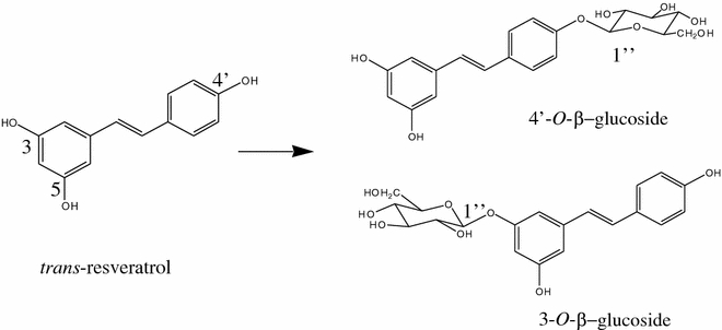
Glucosidation of trans-resveratrol
Fig. 1.
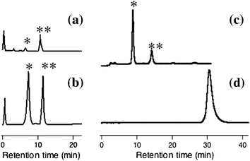
HPLC profiles of the glucosylated products obtained from incubation with a Phytolacca americana cells in the absence of auxin, b P. americana cells in the presence of 2,4-dichlorophenoxyacetic acid, and c the purified PaGT3 enzyme. * and ** indicate trans-resveratrol 4′-O-β-glucoside and trans-resveratrol 3-O-β-glucoside, respectively. d The starting material trans-resveratrol was eluted at 31 min
To investigate the changes in regiospecificity, we heterologously expressed PaGT3 in E. coli and purified the enzyme to synthesize the glucosides. When trans-resveratrol was incubated with UDP-glucose in the presence of PaGT3, 4′-O-β-glucoside was obtained as the major product (Fig. 1c). The ratio of peak area for 4′-O-β-glucoside to that for 3-O-β-glucoside in the HPLC profile was 10:3. The product distribution was not altered following the addition of 2,4-dichlorophenoxyacetic acid, phenyl acetic acid, or 1-naphthalene acetic acid, indicating that these hormones do not interact directly with the purified PaGT3 enzyme. From these observations, we believe that 2,4-dichlorophenoxyacetic acid may induce the expression of PaGT3 in the cultured cells, resulting in enhanced 4′-O-β-glucoside production. The expression and purification of PaGT1 and PaGT2 are now under way to examine the regiospecificity for the glucosidation reaction catalyzed by these isozymes.
Biotransformation of trans-resveratrol by E. coli cells
The results of this study show that purified PaGT3 catalyzes the glucosidation of trans-resveratrol. Next, we attempted to use E. coli cells expressing PaGT3 gene to promote the glucoside synthesis because E. coli cells grow faster than P. americana cells. When PaGT3-expressing E. coli cells were cultured in the presence of trans-resveratrol, glucoside products of trans-resveratrol were detected in the medium of overnight culture. Interestingly, endogenous glucosyl donor molecules were sufficient for the reaction, since the reaction occurred even when UDP-glucose was not added to the medium. The products were extracted from the medium using ethyl acetate and identified by reverse-phase HPLC as 3-O-β–glucoside and 4′-O-β–glucoside. The glucosidation of trans-resveratrol was almost 100% using this method, and a peak corresponding to resveratrol, starting material, was not observed in the HPLC profile (Fig. 2). This was a considerable improvement because only 39% of trans-resveratrol was glucosylated in P. americana cell cultures. Furthermore, the use of E. coli cells allowed the incubation time to be shortened from 3 days to 1. The glucoside products accumulated in the medium and could not be isolated from the cell extracts. Since PaGT3 is not secreted from the E. coli cells, trans-resveratrol and its glucoside products may pass through the cell membrane. These results indicate that PaGT3 expressing E. coli cells utilize endogenous UDP-glucose to convert trans-resveratrol completely into the glucosides.
Fig. 2.
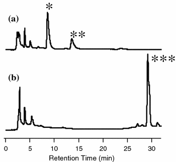
HPLC profiles of glucosylated products obtained from the incubation of E. coli expressing a wild type PaGT3 gene and b H20A PaGT3 gene. HPLC profile b was similar to that obtained in the incubation with E. coli expressing H20D PaGT3 gene. *, **, and *** indicate trans-resveratrol 4′-O-β-glucoside, trans-resveratrol 3-O-β-glucoside, and trans-resveratrol, respectively
We also found that Diaion HP20 could be used to isolate the glucoside products from the medium instead of extracting the glucosides with ethyl acetate. The aromatic moiety of trans-resveratrol glucosides appeared to interact with the non-polar hydrophobic surface of HP20 resin by van der Waals force, and the glucoside products bound on the resin were eluted with methanol. The use of HP20 is convenient for the isolation of glucosylated products from the medium obtained by a large scale incubation.
Molecular modeling and mutagenesis studies
The X-ray crystal structure of PaGT3 may yield important insights into the reaction mechanism; however, the structure of PaGT3 has not yet been determined. Therefore, we constructed a structural model of PaGT3 using the crystal structure of GT from V. vinifera (VvGT) as a template (Offen et al. 2006). The predicted PaGT3 structure was then superimposed upon the crystal structure of VvGT bearing UDP-2-deoxy-2-fluoro glucose (a nontransferable donor molecule) and kaempferol (a glucose acceptor molecule) at the active site for comparison (Fig. 3a). A putative model structure of PaGT3 implies that Gln-395 may play an important role in appropriate positioning of UDP-glucose, a glucose donor molecule. trans-Resveratrol, a glucose acceptor molecule, may bind PaGT3 in the hydrophobic pocket surrounded by Phe-126, Leu-155, and Phe-392. A histidine residue conserved in the active site of GTs normally accept a proton from the hydroxy group of an acceptor molecule and promote the nucleophilic attack on C1 of UDP-glucose (Fig. 3b) (Lairson and Withers 2004). The model structure of PaGT3 clearly suggests that His-20 rather than His-18 is the catalytic residue. Moreover, the hydrogen-bonding interaction between the carboxy side chain of Asp-124 and the Nδ atom of His-20 might increase the basicity of this imidazole to enhance the nucleophilicity.
Fig. 3.
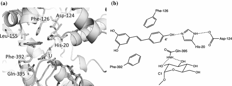
a The active site of PaGT3 model. The predicted PaGT3 structure was superimposed upon the crystal structure of VvGT bearing UDP-2-deoxy-2-fluoro glucose (nontransferable donor) and kaempferol (acceptor). UDP-2-deoxy-2-fluoro glucose (U), kaempferol (K), and some selected active site residues of PaGT3 are shown. b Schematic diagram of active site of PaGT3 model. The dashed lines indicate hydrogen bonds. A hydrogen bond between His-20 and the 4′-hydroxy group of trans-resveratrol may promote nucleophilic attack on C1 of UDP-glucose to produce 4′-β-O-glucoside
In order to determine if His-20 is a catalytically important residue, we decided to substitute His-20 with Ala and Asp and measure the GT activity of these mutants. The expression levels of the mutated enzymes were comparable to the wild type enzyme. We could successfully purify the mutant enzymes using nickel-nitrilotriacetic acid affinity columns and analyzed their GT activity (Fig. 4). We were unable to detect glucosidation products in the reaction mixture containing trans-resveratrol (acceptor), UDP-glucose (donor), and the H20A and H20D PaGT3 mutant enzymes. The incubation of E. coli cells expressing the H20A and H20D mutant PaGT3 enzymes in the medium containing trans-resveratrol did not produce the glucosides either (Fig. 2). These results provided the experimental evidence that His-20 in the active site was critical for the GT reaction. The mutagenesis of residues interacting with an acceptor molecule might allow us to construct a novel enzyme glucosylating a broad range of biologically active molecules.
Fig. 4.
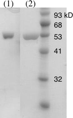
SDS-PAGE of the purified 1 H20A and 2 H20D PaGT3 enzymes
Conclusions
Purified PaGT3 glucosylates trans-resveratrol to 4′-O-β-glucoside (a major product) and 3-O-β-glucoside (a minor product). Based on homology modeling, resveratrol may bind PaGT3 in a hydrophobic pocket similar to the GT from V. vinifera (VvGT). The predicted structure of PaGT3 indicated that His-20 probably was a catalytically important residue for the nucleophilic attack of the aglycone on the C1 of UDP-glucose. This was confirmed by mutational analysis because H20A and H20D PaGT3 mutant enzymes lacked GT activity. Furthermore, E. coli expressing the PaGT3 gene was used for the glycoconjugation of trans-resveratrol in the culture medium. The advantages of using E. coli cell cultures are a shorter incubation time compared with cultured plant cells, no need for the addition of exogenous glucosyl donor compounds, such as UDP-glucose, and the almost complete conversion of the aglycone to the glucoside products. The use of E. coli cell cultures combined with molecular engineering of PaGT3 may provide a new biocatalyst for the glucosidation of aromatic compounds.
Acknowledgments
We thank Regional Innovation Creation R&D Programs (Japan) for their financial support
Open Access
This article is distributed under the terms of the Creative Commons Attribution Noncommercial License which permits any noncommercial use, distribution, and reproduction in any medium, provided the original author(s) and source are credited.
References
- Cherniack EP. Polyphenols: planting the seeds of treatment for the metabolic syndrome. Nutrition. 2011;27:617–623. doi: 10.1016/j.nut.2010.10.013. [DOI] [PubMed] [Google Scholar]
- Furuya T, Ushiyama M, Asada Y, Yoshikawa T. Biotransformation of 2-phenylpropionic acid in root culture of Panax ginseng. Phytochemistry. 1989;28:483–487. doi: 10.1016/0031-9422(89)80036-6. [DOI] [Google Scholar]
- Gehm BD, McAndrews JM, Chien PY, Jameson JL. Resveratrol, a polyphenolic compound found in grapes and wine, is an agonist for the estrogen receptor. Proc Natl Acad Sci USA. 1997;94:14138–14143. doi: 10.1073/pnas.94.25.14138. [DOI] [PMC free article] [PubMed] [Google Scholar]
- Hamada H, Ohiwa S, Nishida T, Katsuragi H, Takeda T, Hamada H, Nakajima N, Ishihara K, Hamada H, Hirata T, Nakajima N. One-step glucosylation of capsaicinoide by cultured cells of Phytolacca americana. Plant Biotechnol. 2003;20:253–255. doi: 10.5511/plantbiotechnology.20.253. [DOI] [Google Scholar]
- Ingram D, Sanders K, Kolybaba M, Lopez D. Case-control study of phyto-oestrogens and breast cancer. Lancet. 1998;350:990–994. doi: 10.1016/S0140-6736(97)01339-1. [DOI] [PubMed] [Google Scholar]
- Ishihara K, Hamada H, Hirata T, Nakajima N. Biotransformation using plant cultured cells. J Mol Catal B Enz. 2003;23:145–170. doi: 10.1016/S1381-1177(03)00080-8. [DOI] [Google Scholar]
- Kiefer F, Arnold K, Künzli M, Bordoli L, Schwede T. The SWISS-MODEL repository and associated resources. Nucleic Acids Res. 2009;37:D387–D392. doi: 10.1093/nar/gkn750. [DOI] [PMC free article] [PubMed] [Google Scholar]
- Kopp J, Schwede T. The SWISS-MODEL repository: new features and functionalities. Nucleic Acids Res. 2006;34:D315–D318. doi: 10.1093/nar/gkj056. [DOI] [PMC free article] [PubMed] [Google Scholar]
- Lairson LL, Withers SG. Mechanistic analogies amongst carbohydrate modifying enzymes. Chem Commun (Camb, Engl) 2004;20:2243–2248. doi: 10.1039/b406490a. [DOI] [PubMed] [Google Scholar]
- Noguchi A, Kunikane S, Homma H, Liu W, Sekiya T, Hosoya M, Kwon S, Ohiwa S, Katsuragi H, Nishino T, Takahashi S, Hamada H, Nakayama T. Identification of an inducible glucosyltransferase from Phytolacca americana L. cells that are capable of glucosylating capsaicin. Plant Biotechnol. 2009;26:285–292. doi: 10.5511/plantbiotechnology.26.285. [DOI] [Google Scholar]
- Offen W, Martinez-Fleites C, Yang M, Kiat-Lim E, Davis BG, Tarling CA, Ford CM, Bowles DJ, Davies GJ. Structure of a flavonoid glucosyltransferase reveals the basis for plant natural product modification. EMBO J. 2006;25:1396–1405. doi: 10.1038/sj.emboj.7600970. [DOI] [PMC free article] [PubMed] [Google Scholar]
- Peitsch MC. Protein modeling by E-mail. Biotechnology. 1995;13:658–660. doi: 10.1038/nbt0795-658. [DOI] [Google Scholar]
- Suga T, Hirata T. Biotransformation of exogenous substrates by plant cell cultures. Phytochemistry. 1990;29:2393–2406. doi: 10.1016/0031-9422(90)85155-9. [DOI] [Google Scholar]
- Tikkanen MJ, Wahala K, Ojala S, Vihma V, Adlercreutz H. Effect of soybean phytoestrogen intake on low density lipoprotein oxidation resistance. Proc Natl Acad Sci USA. 1998;95:3106–3110. doi: 10.1073/pnas.95.6.3106. [DOI] [PMC free article] [PubMed] [Google Scholar]
- Vogt T, Jones P. Glycosyltransferases in plant-natural product synthesis: characterization of a super gene family. Trend Plant Sci. 2000;5:380–386. doi: 10.1016/S1360-1385(00)01720-9. [DOI] [PubMed] [Google Scholar]
- Weis M, Lim EK, Bruce N, Bowles D. Regioselective glucosylation of aromatic compounds: screening of a recombinant glycosyltransferase library to identify biocatalysts. Angew Chem Int Ed. 2006;45:3534–3538. doi: 10.1002/anie.200504505. [DOI] [PubMed] [Google Scholar]
- Xianfeng-Huang X-H, Zhu H-L. Resveratrol and its analogues: promising antitumor agents. Anticancer Agents Med Chem. 2011;11:479–490. doi: 10.2174/187152011795677427. [DOI] [PubMed] [Google Scholar]


