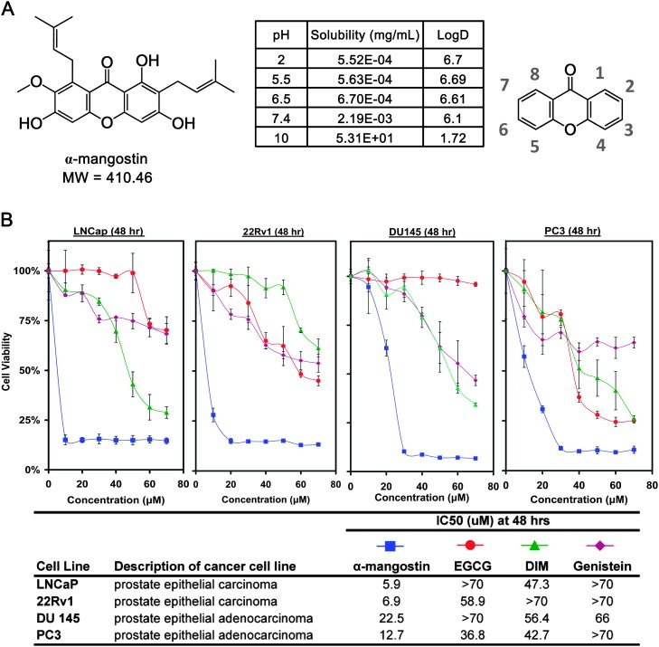
Fig. 1.
(A) Chemical structure of α-mangostin and nomenclature of a xanthone. (B) Cell viability assay was performed treating cells with α-mangostin, Epigallocatechin 3-gallate, Genistein and 3,3′-Diindolylmethane up to 70 μM against four PCa cell lines (LNCaP, 22Rv1, DU145 and PC3) for 48 h. Data points are represented by the average of three values with standard deviation and representative of two different experiments.

