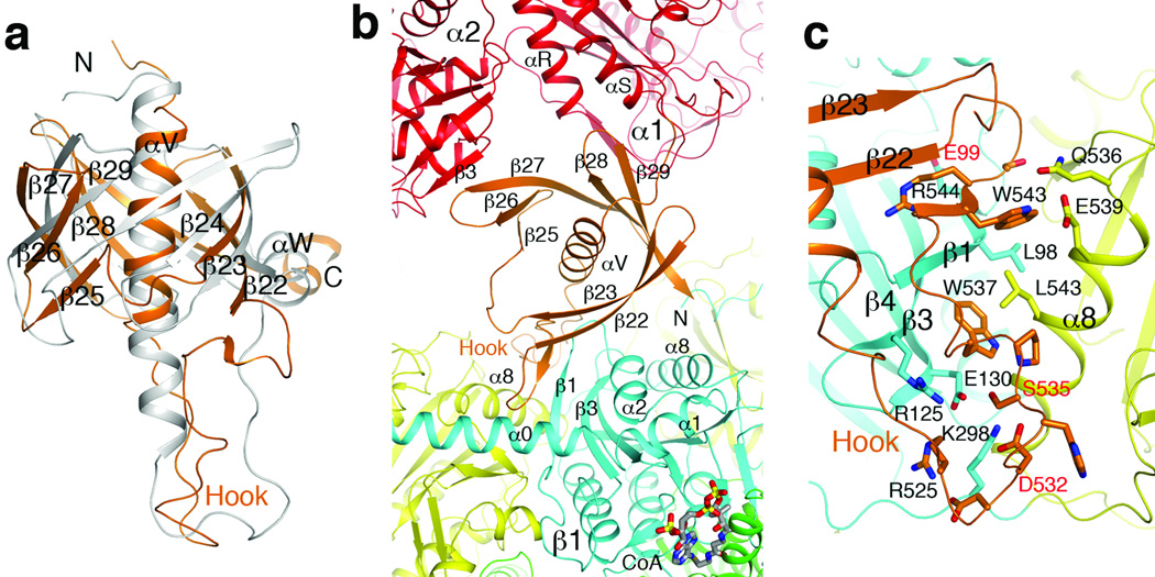
Figure 3. The BT domain mediates interactions in the MCC holoenzyme.
(a). Overlay of the structure of PaMCC BT domain (in orange) with that of PCC (gray). A large conformational difference for the hook is visible. The exact positions of many of the β-strands are different as well. (b). The BT domain (orange) contacts a β subunit (β1, N domain in cyan, C domain yellow) as well as a neighboring α subunit (α2, red) in the PaMCC holoenzyme. (c). Detailed interactions between the hook of the BT domain and the β subunit in PaMCC. Three disease-causing mutation sites near this interface are labeled in red. For stereo version of the panels, please see Supplementary Figs. 9, 10.

