Abstract
Visual loss in infectious posterior uveitis or panuveitis can occur if proper therapy is delayed because of diagnostic uncertainty. Some disorders, such as acute retinal necrosis and bacterial endophthalmitis, can be rapidly progressive, and therefore require prompt and accurate diagnosis to guide initial therapy. Other more slowly evolving infections, such as toxoplasmic chorioretinitis or fungal endophthalmitis, can be worsened by empiric use of corticosteroids without specific antimicrobial coverage. Key ocular diagnostic features are helpful but highly variable with overlap with both non-infectious uveitis and neoplastic masquerades, even for key signs such as hypopyon. Close examination of the fundus with attention to color, location, size, border, and opacity of lesions and associated arteriolitis or frosted branch angiitis is helpful in the diagnosis of chorioretinitis. Ultrasonography is an important tool in the evaluation of eyes with suspected endophthalmitis, especially those with intracapsular infection or focal infected deposits. Testing of intraocular fluid can be extremely useful but suffers from inaccessibility, poor sensitivity, and test selections dependent on a presumptive diagnosis, which may be wrong. The dilemma for clinician is to make the correct diagnosis of a rare, blinding, variegated disease quickly enough to intercede with specific therapy or to apply empiric therapy in a sufficiently skilled manner to avert disaster and confirm the diagnosis by response to treatment. When non-infectious uveitis is in the differential, empiric corticosteroids must sometimes be used, at great risk, if clinical examination, ancillary testing, and any available intraocular diagnostic tests have failed to confirm a diagnosis.
Keywords: chorioretinitis, endophthalmitis, acute retinal necrosis, cytomegalovirus retinitis, syphilis, PCR
Introduction
In 2008 the International Uveitis Study Group revised the original anatomic classification of uveitis to include only broad etiologic categories of infectious and non-infectious uveitis (as well as non-uveitic masquerades)1 recognizing an essential dichotomy in intraocular inflammation. Infections involving the posterior segment have tremendous visual impact in individual cases, especially if treatment is mistakenly begun with injectable or oral corticosteroids. The clinician preparing to manage a significant intermediate, posterior, or panuveitis is therefore faced with an urgent diagnostic challenge. First, is this infectious or non-infectious uveitis? If infectious, is a bacteria, fungus, virus or other agent more likely to produce these clinical features? Finally, what is the urgency of empiric anti-infective treatment before confirmation of the final diagnosis?
Diagnostic challenges
For some cases of infectious uveitis infection is sufficiently obvious that it presents no diagnostic challenge. For example, pain, hypopyon, and reduced vision within 1–2 weeks of cataract extraction is assumed infectious, probably bacterial, and treated with intravitreal antibiotics. A confluent, 360° peripheral necrotizing retinitis in one or both eyes with or without immunocompromise should at least initially be suspected to be acute retinal necrosis (ARN) due to herpes simplex or varicella zoster and potentially rapidly progressive, and receive aggressive intravitreal and systemic antiviral therapy. Even straightforward clinical scenarios such as these can present diagnostic dilemmas, as not all clinical endophthalmitis are confirmed to be infectious even with molecular techniques2 and other infections can mimic ARN.3 Neoplastic conditions can also mimic intraocular infections (Supplementary Figure 1).
Most posterior uveitis presents at least some difficulties in clinical diagnosis, in part because of variation introduced by the host response. Ipsilateral choroiditis in immunocompetent patients (‘thumbprint lesions') following herpes zoster ophthalmicus (Supplementary Figure 2) does not progress to ARN, but is clearly related to the prior infection, perhaps as an immunologic host response.4 Serpiginous choroidopathy is classified as a non-infectious uveitis, but there is a strong epidemiologic association with tuberculous infection and a serpiginous-like choroidopathy,5, 6 which could represent either direct choroidal infection or a hypersensitivity reaction to infection elsewhere. It has a different pattern of disease than classic serpiginous choroidopathy7 or the classic choroidal granulomas and dense periphlebitis that are pathognomonic for tuberculous uveitis (Supplementary Figure 3). The apparent requirement for both antituberculous treatment and corticosteroids in serpiginous-like choroidopathy as well as other cases of tuberculous uveitis supports a strong host–pathogen component in the clinical disease.8
Although some forms of uveitis, such as birdshot or sympathetic ophthalmia, are undoubtedly autoimmune, others, such as pars planitis and sarcoidosis, which are now classified as non-infectious, may ultimately be shown to be either direct infection or host response to infection.9 Multifocal choroiditis (MFC) and presumed ocular histoplasmosis syndrome (POHS) are distinguishable by subtle clinical appearance,10 but most reliably by the presence of vitreitis, which by definition is absent in POHS. If vitreitis is absent in MFC it is unlikely there is active choroidal inflammation requiring treatment.11 It is not clear whether it is reasonable to consider POHS an infectious choroiditis (or the sequela of one), but not MFC, when they are so similar in appearance and MFC is more likely to have active inflammation. Whether the chorioretinal lesions of POHS harbor organisms has not been proven. In immunocompromised individuals histoplasmosis produces an endophthalmitis with retinal involvement rather than a choroidal abscess.12, 13 Transient posterior uveitis such as acute posterior placoid pigment epitheliopathy or multiple evanescent white dot syndrome are also candidates for infections or at least the aftermath of a self-remitting infection.14, 15 The boundaries between non-infectious and infectious uveitis are thus somewhat blurred.
Most definite intraocular infections involve the vitreous cavity (endophthalmitis), in the case of extracellular organisms, or the retina (chorioretinitis), in the case of intracellular organisms such as viruses and protozoa. Different clinical appearances occur with different organisms. Candidal endophthalmitis is more likely to be located in the retina or vitreous than aspergillus endophthalmitis, which preferentially grows in the subretinal and sub-retinal pigment epithelial (RPE) spaces and viral invades vessels in the choroid.16 Other organisms, such as nocardia, also grow preferentially at the RPE level.17 It is possible that factors other than anatomy, such as oxygen levels or cell type influence the locus of initial infection. A limited spectrum of organisms cause choroidal abscess, possibly because of the redundant circulation.18 The initial focus of infection may be obscured by subsequent spread of the destructive process, for example, from retina to choroid in toxoplasmosis.19 Bacterial infections often progress rapidly so that the initial point of infection cannot be detected, but localized infections are recognized (Supplementary Figure 4). Screening for fungal endophthalmitis in hospitalized patients with fungemia was once widely performed. With the advent of effective preemptive treatment, most retinal lesions found on screening are actually microangiopathy rather than true infection.20
Diagnostic dilemmas for endophthalmitis arise when any of the key signs are missing, such as pain, redness, hypopyon, fibrin, or the vitreous is relatively clear. In chorioretinitis, problems in diagnosis mainly arise when preretinal opacities prevent adequate examination of the retina-choroid, or the pattern of infection is atypical for that expected, for example, when necrotizing herpetic retinitis is focal rather than diffuse (Supplementary Figure 5). The patient's history may lack clues to the source of a blood-borne infection causing an endogenous endophthalmitis. Similarly, immune-system impairment or residence in an endemic area for toxoplasmosis that could explain a susceptibility to an infectious chorioretinitis may be absent. Nonetheless, a comprehensive history in addition to physical examination of the eye remains an important first step in diagnosing in infectious uveitis.
Key ocular signs in endophthalmitis
Hypopyon is the classic diagnostic sign of endophthalmitis, yet is unreliable as a sole indicator. Flat-topped, layered, and shifting hypopyons are common in Behcet uveitis. They can also be manifestations of leukemic infiltration or diffuse retinoblastoma, and unfortunately for the clinician can be seen in very early and rapidly progressive infectious endophthalmitis in which sufficient fibrin has not formed to mold the upper edge of the hypopyon. Drug reactions, such as ‘sterile endophthalmitis' from intravitreal injection of triamcinolone acetonide or from oral administration of rifabutin also produce non-infectious hypopyon (Supplementary Figure 6).21 Except for Behcet, the hypopyon of non-infectious uveitis is more likely to have a curved upper border (like a fingernail or ‘onyx'). The eventual appearance of a hypopyon after waxing and waning postoperative inflammation with partial response to topical corticosteroids is almost always an indication to proceed with culture and intravitreal antibiotic treatment.
Key ocular signs in chorioretinitis
For chorioretinitis, the relevant clinical signs include presence or absence of necrosis of the retina; size, shape, and orientation of the lesions; degree of opacity; apparent thickness; and the confluency or focality of the lesions, along with their color and border characteristics. Associated inflammatory signs such as arteriolar or venular sheathing, vascular occlusion, frosted branch angiitis, (Supplementary Figure 7) and the intensity of vitreous and anterior chamber inflammation are also important. Pattern recognition is vastly assisted by experience because of the number of characteristics that must be recognized and the wide variation.
Syphilitic uveitis can mimic both endogenous endophthalmitis, with hypopyon and dense vitreous opacities, and viral chorioretinitis with retinal swelling and preretinal opacities (Supplementary Figure 8).22 After resolution, mild pigmentary changes can be seen with damage to the retinal pigment epithelium, but syphilis is rarely necrotizing.22 Conversion to a necrotizing chorioretinitis has been described after intravitreal injection of triamcinolone.23
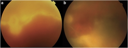
Table 1 summarizes the clinical features of the most common types of chorioretinitis and their variants. Figure 1 depicts diffuse toxoplasmosis, the infection which is most commonly confused with acute retinal necrosis, leading to delays in treatment.
Table 1. Key clinical features distinguishing different etiologies of infectious chorioretinitis.
| Location | Confluence | Border | Thickness | Vasculitis | |
|---|---|---|---|---|---|
| Acute retinal necrosis24 | Peripheral Multifocal or posterior25 | Confluent, Rapid centripetal spread | Smooth with large satellites | Necrotizing Full thickness, opaque, edematous | Occlusion in and outside lesions |
| Cytomegalovirus | Random Vasocentric | Unifocal or multifocal, Central healing, usually concentric spread | Granular small satellites | Necrotizing, Superficial | Occlusion in lesions FBA reported26 |
| Syphilis | Random Posterior polar predominance | Diffuse | Poorly defined | Non-necrotizing, Translucent, edematous | Vascular leakage Venous occlusion22 |
| Toxoplasmosis, focal | Random | Unifocal with border healing | Smooth | Thick, inner retina or full thickness | Arteriolar >venular sheathing, FBA reported |
| Toxoplasmosis, diffuse27, 28 | Random | Confluent, random spread | Smooth | Usually thick | Arteriolar >venular sheathing |
FBA, frosted branch angiitis: heavy deposits of inflammatory material along multiple arteriolar and venular branches.
Figure 1.
Diffuse toxoplasmosis. The chorioretinitis was initially misdiagnosed as acute retinal necrosis. (a) This elderly man may have acquired toxoplasmosis while he gardened at his new home; he was IgM and IgG antibody positive for toxoplasmosis. Note the focal lesion on the left that appears to have spread diffusely into a smooth-bordered chorioretinitis. There is vitreous haze. PCR from the vitreous was positive for toxoplasmosis. (b) This elderly woman developed what appeared to be a classic focal reactivation of toxoplasmic chorioretinitis after cataract extraction. She was treated with multiple courses of doxycycline, recurring each time the medication was stopped. The infection spread inferiorly and temporally. Vision was hand motions only.
Ancillary testing to confirm the working diagnosis
It is assumed that all patients with intraocular inflammation will have relevant histories performed, radiographs, and basic blood laboratory testing, including syphilis serology. Chorioretinitis is almost always an indication for HIV testing.
A complete blood count, chemistry panel, urinanalysis, C-reactive protein, herpes simplex, cytomegalovirus, Epstein-Barr virus, and toxoplasmosis infectious serologies can help assess prior exposures even if they do not directly confirm the etiology of the intraocular infection. Prior chickenpox infection is usually accepted in lieu of varicella zoster serology; prior varicella vaccination does not necessarily rule out varicella-related acute retinal necrosis.
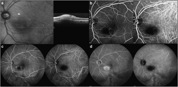
Angiography is most useful in toxoplasmic chorioretinitis, which has a distinctive early blockage in the lesion with a late hyperfluorescent border with fluorescein dye. On indocyanine green angiography, distinctive dark dots surround the lesion (Figure 2). Optical coherence tomography (OCT) also typically shows inner retinal hyperreflectivity. In thick, elevated, opaque lesions, OCT is very useful in ruling out choroidal or subretinal involvement.
Figure 2.
Imaging studies of toxoplasmic focal chorioretinitis. (a) OCT image through lesion showing inner retinal hyperreflectivity with shadowing of the outer retina and choroid. (b) Early angiogram, 50 s. There is hypofluorescence of the lesion in both the fluorescein (left) and the indocyanine angiogram (right). (c) Mid-phase of the angiogram, 2.33 min. Hyperfluorescence begins at the edge of the focal lesion. The ICG remains hypofluorescent. (d) Late angiogram, 15.11 min. The lesion is almost fully stained with fluorescein. In the ICG the lesion is hypofluorescent and surrounded by dark dots most visible in the temporal macula.
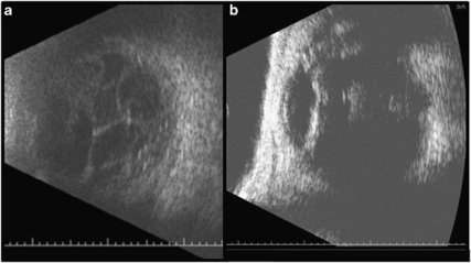
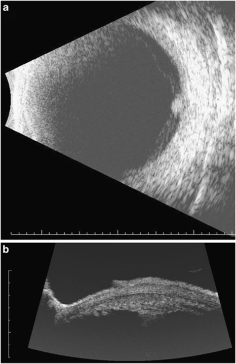
Ultrasonography is routinely used in the diagnosis of endophthalmitis when the posterior segment cannot be viewed. If the vitreous is not involved, it is less likely to be endophthalmitis unless there is limited exogenous endophthalmitis with an anterior entry or keratitis. Ultrasound is nonspecific, however: it can only indicate severity of the posterior involvement and whether retinal detachment or abscess is present. Vitreitis may be minimal in intracapsular, delayed endophthalmitis, leading to misdiagnosis, however, vitreitis is ordinarily the sine qua non of endophthalmitis (Figure 3). Features compatible with endophthalmitis include strands and membranes with reduced mobility. For delayed postoperative infection, the extent of intracapsular infection determines the amount of surgical debridement that will be required including whether the intraocular lens needs to be removed. Ultrasound can be used to predict findings before surgery. In persistent endophthalmitis, ultrasound may help locate infected foci (Figure 4).
Figure 3.
Ultrasonography in the diagnosis of postoperative endophthalmitis. (a) Classic appearance of vitreous stands and membranes on B-scan ultrasound. Variations in gain can alter the appearance of the vitreous opacities. (b) Capsular hyperreflectivity in a case of delayed onset endophthalmitis with dense intracapsular deposits. The vitreous contains some dense deposits but is not diffusely infiltrated. Absence of vitreous inflammation or opacities is suggestive that endophthalmitis is not present, except for limited anterior forms.
Figure 4.
Persistent fungal endophthalmitis following pars plana vitrectomy and removal of infected capsular bag. During a second surgery the entire bag and lens implant were removed, but inflammation persisted. Ultrasound confirmed focal deposits in the ciliary body region in the meridian where the infected capsular plaques had been noted. (a) Anterior to the equator at 3:00 (3EA) there is a focal deposit in the ciliary body region. (b) Anterior segment B scan confirms a ciliary body deposit at the 3:00 position (3T). Notice the ciliary processes to the left of the deposit. At surgery, a focal white deposit was found between 3 and 4:00 adherent to the ciliary processes. Removal of it with the vitreous cutter and picks followed by repeated injections of amphotericin enabled the infection to be cured.
PCR testing of vitreous specimens in suspected bacterial or fungal endophthalmitis is well established in certain centers.2, 29, 30, 31, 32, 33, 34 Concordance with culture is close to 100%, with greater sensitivity with PCR testing.29, 34 The presumption is that PCR will eventually replace culture and sensitivity testing (by amplifying loci known to determine resistance to antibiotics35) and enable the detection of unsuspected, fastidious, or previously unknown pathogens.9, 36, 37 Intraocular Whipple disease is diagnosable by PCR of aqueous humor or vitreous. Two positive results are considered a definitive diagnosis of uveitis related to Whipple disease.38
There are multiple case series summarizing the results of PCR testing of aqueous or vitreous humor in cases in which culture is inefficient or unavailable, mainly in the case of viral or protozoal chorioretinitis.39, 40, 41, 42, 43 Aqueous humor appears to be an adequate substrate for testing and vitreous tap is rarely needed although vitreous humor can also be used for testing. Culture of vitreous fluid for toxoplasmosis using viral media has been reported to be possible in cases of extensive, diffuse infections.44 Tuberculous chorioretinitis45 is potentially confirmable by PCR testing, although higher copy numbers seem to be required than exist in ocular specimens that have not been grown out in culture.46 PCR protocols optimized for the confirmation of Mycobacterium tuberculosis from cultures rather than from biologic fluids are not efficient diagnostic tools and yield false negatives when applied to ocular specimens. Intraocular syphilis antibody can be assayed from aqueous humor and PCR confirmation of syphilitic uveitis has also been reported.47
Table 2 summarizes the preferred diagnostic tests that can be performed on intraocular fluid. Culture of other body fluids can be helpful in endogenous uveitis and serology is helpful in syphilis. Delayed-type hypersensitivity reactions (Mantoux) and interferon-γ release assays are helpful in the diagnosis of tuberculous chorioretinitis.
Table 2. Preferred diagnostic testing of intraocular fluid in infectious posterior uveitis.
| Infectious uveitis | Culture | PCR | Antibodies |
|---|---|---|---|
| Viral—CMV | No | Yes, untreated | Yes, treated or healed |
| Viral—ARN, herpes simplex and herpes zoster | No | Yes, untreated | Yes, treated or healed |
| Bacterial | Yes | Available in some centers | No |
| Fungal | Yes | Available in some centers | No |
| Protozoal—focal toxoplasmosis | No | Large lesions or immunocompromised hosts | Yes |
| Protozoal—diffuse toxoplasmosis >1 clock hour in extent | Possible, not used routinely | Yes | Yes |
| Spirochetes | No | Available in some centers | Yes |
| Tuberculosis | Yes, with confirmatory PCR | Requires high copy numbers in biological specimens | No |
In some cases, retinal biopsy or aspiration is the next step after PCR or culture. Usually this is in cases in which lymphoma is in the differential, which would require histopathology for diagnosis. Retinal biopsy showing cytomegalic inclusions or toxoplasmic tachzoites can be diagnostic. Confirmation by immunohistochemical testing is advised in herpes simplex or varicella zoster. Histopathology has distinct advantages over selective molecular tests such as PCR when the clinical condition is a true unknown and testing is not just for confirmation. It enables a correct assignment of the case to non-infectious uveitis, infectious uveitis, or neoplasia. The slides acquired can be stained with iodine-containing preparations to identify organisms that would be Gram positive on conventional smears, stained with selective antibodies, or processed by in situ hybridization as a slide-based form of PCR.48
Response to empiric therapy as a diagnostic maneuver
The first goal in empiric therapy in suspected infectious uveitis is to reduce the risk of vision loss by treating potentially rapidly progressive and destructive infections before confirmatory testing is complete. The comparable success of vitreous tap and injection of antibiotics vs pars plana vitrectomy with injection of antibiotics may relate in part to the rapidity with which the antibiotics can be administered with the tap and inject protocol.49 It is possible that immediate tap for PCR with intravitreal antibiotics followed by pars plana vitrectomy when operating room time can be arranged would lead to better visual outcomes by removing inflammatory mediators and lytic enzymes from the eye.
For chorioretinitis, it is common to begin antiherpetic therapy at the time of presentation if acute retinal necrosis is suspected. A diagnostic aqueous tap for herpes simplex, herpes zoster, and cytomegalovirus can be stored until results of syphilis serology are known and then can be used to specify preferred antiviral (valganciclovir vs valacyclovir) and in the case of herpes simplex or zoster, the dose, as zoster usually requires higher antiviral doses.
The potential damage from high-dose empiric corticosteroids is extreme in undiagnosed infectious uveitis. Oral corticosteroids or intravenous corticosteroids are preferred to regional corticosteroids because they are more easily reversible. Nonetheless, corticosteroid administration without specific antimicrobial coverage can lead to vastly worsened prognosis. It is usually more helpful to try to elicit a response with specific anti-infective treatment first and then use corticosteroids as an adjunct to protect the eye against the secondary inflammatory reaction in infections. In endophthalmitis, it has been difficult to demonstrate that routine injection of intravitreal corticosteroids is beneficial.50, 51 They are avoided in fungal endophthalmitis, viral chorioretinitis in immunocompromised patients, and syphilitic uveitis except for topical agents, however, in diagnostic dilemmas a point is often reached at which a patient's failure to respond to empiric anti-infective treatment and negative diagnostic tests culminates in an aggressive therapeutic trial of corticosteroids for a presumption of autoimmune posterior or panuveitis. Establishing a timeframe at the initiation of empiric treatment in which a response is expected is helpful in interpreting results. In general, effective treatment for viral retinitis should lead to no progression after treatment with full healing within 4 to 6 weeks, bacterial infections should improve within 72 h, syphilis should improve within 1 week, and tuberculosis should improve within 3 to 6 weeks.
Summary
Ocular infections rare enough and variable enough that the first clinical impressions are often incorrect. Focused clinical skills and broad experience are helpful. Knowledgeable use of ancillary testing is essential. Treatment is urgent in some infections, especially acute endophthalmitis and acute retinal necrosis. The safe use of corticosteroids in equivocal cases and masquerades requires mastery.
Acknowledgments
This study was supported by Len-Ari Foundation and Research to Prevent Blindness.
The author declares no conflict of interest.
Footnotes
Supplementary Information accompanies the paper on Eye website (http://www.nature.com/eye)
Presented at the Cambridge Ophthalmological Symposium, 9 September 2011.
Supplementary Material
References
- Deschenes J, Murray PI, Rao NA, Nussenblatt RB, International Uveitis Study Group International Uveitis Study Group (IUSG): clinical classification of uveitis. Ocul Immunol Inflamm. 2008;16:1–2. doi: 10.1080/09273940801899822. [DOI] [PubMed] [Google Scholar]
- Chiquet C, Cornut PL, Benito Y, Thuret G, Maurin M, Lafontaine PO, French Institutional Endophthalmitis Study Group et al. Eubacterial PCR for bacterial detection and identification in 100 acute postcataract surgery endophthalmitis. Invest Ophthalmol Vis Sci. 2008;49:1971–1978. doi: 10.1167/iovs.07-1377. [DOI] [PubMed] [Google Scholar]
- Balansard B, Bodaghi B, Cassoux N, Fardeau C, Romand S, Rozenberg F, et al. Necrotising retinopathies simulating acute retinal necrosis syndrome. Br J Ophthalmol. 2005;89:96–101. doi: 10.1136/bjo.2004.042226. [DOI] [PMC free article] [PubMed] [Google Scholar]
- McKelvie PA, Francis IC, Watson S, Nuovo G. Multifocal chorioretinal atrophy associated with herpes zoster ophthalmicus. Clin Exp Ophthalmol. 2001;29:429–432. doi: 10.1046/j.1442-9071.2001.d01-30.x. [DOI] [PubMed] [Google Scholar]
- Gupta A, Bansal R, Gupta V, Sharma A, Bambery P. Ocular signs predictive of tubercular uveitis. Am J Ophthalmol. 2010;149:562–570. doi: 10.1016/j.ajo.2009.11.020. [DOI] [PubMed] [Google Scholar]
- Mackensen F, Becker MD, Wiehler U, Max R, Dalpke A, Zimmermann S. QuantiFERON TB-Gold--a new test strengthening long-suspected tuberculous involvement in serpiginous-like choroiditis. Am J Ophthalmol. 2008;146:761–766. doi: 10.1016/j.ajo.2008.06.012. [DOI] [PubMed] [Google Scholar]
- Vasconcelos-Santos DV, Rao PK, Davies JB, Sohn EH, Rao NA. Clinical features of tuberculous serpiginouslike choroiditis in contrast to classic serpiginous choroiditis. Arch Ophthalmol. 2010;128:853–858. doi: 10.1001/archophthalmol.2010.116. [DOI] [PubMed] [Google Scholar]
- Basu S, Das T. Pitfalls in the management of TB-associated uveitis. Eye (Lond) 2010;24:1681–1684. doi: 10.1038/eye.2010.110. [DOI] [PubMed] [Google Scholar]
- Drancourt M, Berger P, Terrada C, Bodaghi B, Conrath J, Raoult D, et al. High prevalence of fastidious bacteria in 1520 cases of uveitis of unknown etiology. Medicine (Baltimore) 2008;87:167–176. doi: 10.1097/MD.0b013e31817b0747. [DOI] [PubMed] [Google Scholar]
- Parnell JR, Jampol LM, Yannuzzi LA, Gass JD, Tittl MK. Differentiation between presumed ocular histoplasmosis syndrome and multifocal choroiditis with panuveitis based on morphology of photographed fundus lesions and fluorescein angiography. Arch Ophthalmol. 2001;119:208–212. [PubMed] [Google Scholar]
- Shimada H, Yuzawa M, Hirose T, Nakashizuka H, Hattori T, Kazato Y. Pathological findings of multifocal choroiditis with panuveitis and punctate inner choroidopathy. Jpn J Ophthalmol. 2008;52:282–288. doi: 10.1007/s10384-008-0566-2. [DOI] [PubMed] [Google Scholar]
- Goldstein BG, Buettner H. Histoplasmic endophthalmitis. A clinicopathologic correlation. Arch Ophthalmol. 1983;101:774–777. doi: 10.1001/archopht.1983.01040010774016. [DOI] [PubMed] [Google Scholar]
- Gonzales CA, Scott IU, Chaudhry NA, Luu KM, Miller D, Murray TG, et al. Endogenous endophthalmitis caused by Histoplasma capsulatum var. capsulatum: a case report and literature review. Ophthalmology. 2000;107:725–729. doi: 10.1016/s0161-6420(99)00179-7. [DOI] [PubMed] [Google Scholar]
- Schaal S, Schiff WM, Kaplan HJ, Tezel TH. Simultaneous appearance of multiple evanescent white dot syndrome and multifocal choroiditis indicate a common causal relationship. Ocul Immunol Inflamm. 2009;17:325–327. doi: 10.3109/09273940903043923. [DOI] [PubMed] [Google Scholar]
- Bryan RG, Freund KB, Yannuzzi LA, Spaide RF, Huang SJ, Costa DL. Multiple evanescent white dot syndrome in patients with multifocal choroiditis. Retina. 2002;22:317–322. doi: 10.1097/00006982-200206000-00010. [DOI] [PubMed] [Google Scholar]
- Rao NA, Hidayat AA. Endogenous mycotic endophthalmitis: variations in clinical and histopathologic changes in candidiasis compared with aspergillosis. Am J Ophthalmol. 2001;132:244–251. doi: 10.1016/s0002-9394(01)00968-0. [DOI] [PubMed] [Google Scholar]
- Chaudhry NA, Tabandeh H, Davis J. Successive intraocular nocardiosis and cytomegalovirus retinitis after cardiac transplantation. Arch Ophthalmol. 1998;116:960–961. doi: 10.1001/archopht.116.7.960. [DOI] [PubMed] [Google Scholar]
- Kaburaki T, Takamoto M, Araki F, Fujino Y, Nagahara M, Kawashima H, et al. Endogenous Candida albicans infection causing subretinal abscess. Int Ophthalmol. 2010;30:203–206. doi: 10.1007/s10792-009-9304-0. [DOI] [PubMed] [Google Scholar]
- Tabbara KF. Disruption of the choroidoretinal interface by toxoplasma. Eye (Lond) 1990;4:366–373. doi: 10.1038/eye.1990.49. [DOI] [PubMed] [Google Scholar]
- Dozier CC, Tarantola RM, Jiramongkolchai K, Donahue SP. Fungal eye disease at a tertiary care center: the utility of routine inpatient consultation. Ophthalmology. 2011;118:1671–1676. doi: 10.1016/j.ophtha.2011.01.038. [DOI] [PubMed] [Google Scholar]
- Saran BR, Maguire AM, Nichols C, Frank I, Hertle RW, Brucker AJ, et al. Hypopyon uveitis in patients with acquired immunodeficiency syndrome treated for systemic Mycobacterium avium complex infection with rifabutin. Arch Ophthalmol. 1994;112:1159–1165. doi: 10.1001/archopht.1994.01090210043015. [DOI] [PubMed] [Google Scholar]
- Fu EX, Geraets RL, Dodds EM, Echandi LV, Colombero D, McDonald HR, et al. Superficial retinal precipitates in patients with syphilitic retinitis. Retina. 2010;30:1135–1143. doi: 10.1097/IAE.0b013e3181cdf3ae. [DOI] [PubMed] [Google Scholar]
- Song JH, Hong YT, Kwon OW. Acute syphilitic posterior placoid chorioretinitis following intravitreal triamcinolone acetonide injection. Graefes Arch Clin Exp Ophthalmol. 2008;246:1775–1778. doi: 10.1007/s00417-008-0928-y. [DOI] [PubMed] [Google Scholar]
- Holland GN, Executive Committee of the American Uveitis Society Standard diagnostic criteria for the acute retinal necrosis syndrome (perspective) Am J Ophthalmol. 1994;117:663–667. doi: 10.1016/s0002-9394(14)70075-3. [DOI] [PubMed] [Google Scholar]
- Margolis R, Brasil OF, Lowder CY, Smith SD, Moshfeghi DM, Sears JE, et al. Multifocal posterior necrotizing retinitis. Am J Ophthalmol. 2007;143:1003–1008. doi: 10.1016/j.ajo.2007.02.033. [DOI] [PubMed] [Google Scholar]
- Walker S, Iguchi A, Jones NP. Frosted branch angiitis: a review. Eye (Lond) 2004;18:527–533. doi: 10.1038/sj.eye.6700712. [DOI] [PubMed] [Google Scholar]
- Moshfeghi DM, Dodds EM, Couto CA, Santos CI, Nicholson DH, Lowder CY, et al. Diagnostic approaches to severe, atypical toxoplasmosis mimicking acute retinal necrosis. Ophthalmology. 2004;111:716–725. doi: 10.1016/j.ophtha.2003.07.004. [DOI] [PubMed] [Google Scholar]
- Johnson MW, Greven GM, Jaffe GJ, Sudhalkar H, Vine AK. Atypical, severe toxoplasmic retinochoroiditis in elderly patients. Ophthalmology. 1997;104:48–57. doi: 10.1016/s0161-6420(97)30362-5. [DOI] [PubMed] [Google Scholar]
- Bispo PJ, de Melo GB, Hofling-Lima AL, Pignatari AC. Detection and gram discrimination of bacterial pathogens from aqueous and vitreous humor using real-time PCR assays. Invest Ophthalmol Vis Sci. 2011;52:873–881. doi: 10.1167/iovs.10-5712. [DOI] [PubMed] [Google Scholar]
- Sugita S, Kamoi K, Ogawa M, Watanabe K, Shimizu N, Mochizuki M.Detection of Candida and Aspergillus species DNA using broad-range real-time PCR for fungal endophthalmitis Graefes Arch Clin Exp Ophthalmol 2011. e-pub ahead of print 27 September 2011; DOI: 10.1007/s00417-011-1819-1 [DOI] [PubMed]
- Sugita S, Shimizu N, Watanabe K, Katayama M, Horie S, Ogawa M, et al. Diagnosis of bacterial endophthalmitis by broad-range quantitative PCR. Br J Ophthalmol. 2011;95:345–349. doi: 10.1136/bjo.2009.171504. [DOI] [PubMed] [Google Scholar]
- Sowmya P, Madhavan HN. Diagnostic utility of polymerase chain reaction on intraocular specimens to establish the etiology of infectious endophthalmitis. Eur J Ophthalmol. 2009;19:812–817. doi: 10.1177/112067210901900520. [DOI] [PubMed] [Google Scholar]
- Goldschmidt P, Degorge S, Benallaoua D, Basli E, Batellier L, Boutboul S, et al. New test for the diagnosis of bacterial endophthalmitis. Br J Ophthalmol. 2009;93:1089–1095. doi: 10.1136/bjo.2008.152181. [DOI] [PubMed] [Google Scholar]
- Seal D, Reischl U, Behr A, Ferrer C, Alivo J, Koerner RJ, et al. Laboratory diagnosis of endophthalmitis: comparison of microbiology and molecular methods in the European Society of Cataract & Refractive Surgeons multicenter study and susceptibility testing. J Cataract Refract Surg. 2008;34:1439–1450. doi: 10.1016/j.jcrs.2008.05.043. [DOI] [PubMed] [Google Scholar]
- Huletsky A, Giroux R, Rossbach V, Gagnon M, Vaillancourt M, Bernier M, et al. New real-time PCR assay for rapid detection of methicillin-resistant Staphylococcus aureus directly from specimens containing a mixture of staphylococci. J Clin Microbiol. 2004;42:1875–1884. doi: 10.1128/JCM.42.5.1875-1884.2004. [DOI] [PMC free article] [PubMed] [Google Scholar]
- Chiquet C, Bodaghi B, Mougin C, Najioullah F. Acute retinal necrosis diagnosed in a child with chronic panuveitis. Graefes Arch Clin Exp Ophthalmol. 2006;244:1206–1208. doi: 10.1007/s00417-005-0233-y. [DOI] [PubMed] [Google Scholar]
- Bodaghi B, Rozenberg F, Cassoux N, Fardeau C, LeHoang P. Nonnecrotizing herpetic retinopathies masquerading as severe posterior uveitis. Ophthalmology. 2003;110:1737–1743. doi: 10.1016/S0161-6420(03)00580-3. [DOI] [PubMed] [Google Scholar]
- Lagier JC, Lepidi H, Raoult D, Fenollar F. Systemic Tropheryma whipplei: clinical presentation of 142 patients with infections diagnosed or confirmed in a reference center. Medicine (Baltimore) 2010;89:337–345. doi: 10.1097/MD.0b013e3181f204a8. [DOI] [PubMed] [Google Scholar]
- Fekkar A, Bodaghi B, Touafek F, Le Hoang P, Mazier D, Paris L. Comparison of immunoblotting, calculation of the Goldmann-Witmer coefficient, and real-time PCR using aqueous humor samples for diagnosis of ocular toxoplasmosis. J Clin Microbiol. 2008;46:1965–1967. doi: 10.1128/JCM.01900-07. [DOI] [PMC free article] [PubMed] [Google Scholar]
- Harper TW, Miller D, Schiffman JC, Davis JL. Polymerase chain reaction analysis of aqueous and vitreous specimens in the diagnosis of posterior segment infectious uveitis. Am J Ophthalmol. 2009;147:140–147. doi: 10.1016/j.ajo.2008.07.043. [DOI] [PMC free article] [PubMed] [Google Scholar]
- Rothova A, de Boer JH, Ten Dam-van Loon NH, Postma G, de Visser L, Zuurveen SJ, et al. Usefulness of aqueous humor analysis for the diagnosis of posterior uveitis. Ophthalmology. 2008;115:306–311. doi: 10.1016/j.ophtha.2007.05.014. [DOI] [PubMed] [Google Scholar]
- Westeneng AC, Rothova A, de Boer JH, de Groot-Mijnes JD. Infectious uveitis in immunocompromised patients and the diagnostic value of polymerase chain reaction and Goldmann-Witmer coefficient in aqueous analysis. Am J Ophthalmol. 2007;144:781–785. doi: 10.1016/j.ajo.2007.06.034. [DOI] [PubMed] [Google Scholar]
- Errera MH, Goldschmidt P, Batellier L, Degorge S, Héron E, Laroche L, et al. Real-time polymerase chain reaction and intraocular antibody production for the diagnosis of viral vs toxoplasmic infectious posterior uveitis Graefes Arch Clin Exp Ophthalmol 2011. e-pub ahead of print 6 July 2011; DOI: 10.1007/s00417-011-1724-7 [DOI] [PubMed]
- Miller D, Davis J, Rosa R, Diaz M, Perez E. Utility of tissue culture for detection of Toxoplasma gondii in vitreous humor of patients diagnosed with toxoplasmic retinochoroiditis. J Clin Microbiol. 2000;38:3840–3842. doi: 10.1128/jcm.38.10.3840-3842.2000. [DOI] [PMC free article] [PubMed] [Google Scholar]
- Palani D, Kulandai LT, Naraharirao MH, Guruswami S, Ramendra B. Application of polymerase chain reaction-based restriction fragment length polymorphism in typing ocular rapid-growing nontuberculous mycobacterial isolates from three patients with postoperative endophthalmitis. Cornea. 2007;26:729–735. doi: 10.1097/ICO.0b013e318060ac3a. [DOI] [PubMed] [Google Scholar]
- Miller MB, Popowitch EB, Backlund MG, Ager EP. Performance of Xpert MTB/RIF RUO assay and IS6110 Real-Time PCR for Mycobacterium tuberculosis detection in clinical samples. J Clin Microbiol. 2011;49:3458–3462. doi: 10.1128/JCM.05212-11. [DOI] [PMC free article] [PubMed] [Google Scholar]
- Cornut PL, Sobas CR, Perard L, De Bats F, Salord H, Manificat HJ, et al. Detection of Treponema pallidum in aqueous humor by real-time polymerase chain reaction. Ocul Immunol Inflamm. 2011;19:127–128. doi: 10.3109/09273948.2010.531175. [DOI] [PubMed] [Google Scholar]
- Hayden RT, Qian X, Roberts GD, Lloyd RV. In situ hybridization for the identification of yeastlike organisms in tissue section. Diagn Mol Pathol. 2001;10:15–23. doi: 10.1097/00019606-200103000-00004. [DOI] [PubMed] [Google Scholar]
- Endophthalmitis Vitrectomy Study Group Results of the Endophthalmitis Vitrectomy Study. A randomized trial of immediate vitrectomy and of intravenous antibiotics for the treatment of postoperative bacterial endophthalmitis. Arch Ophthalmol. 1995;113:1479–1496. [PubMed] [Google Scholar]
- Gan IM, Ugahary LC, van Dissel JT, Feron E, Peperkamp E, Veckeneer M, et al. Intravitreal dexamethasone as adjuvant in the treatment of postoperative endophthalmitis: a prospective randomized trial. Graefes Arch Clin Exp Ophthalmol. 2005;243:1200–1205. doi: 10.1007/s00417-005-0133-1. [DOI] [PubMed] [Google Scholar]
- Albrecht E, Richards JC, Pollock T, Cook C, Myers L. Adjunctive use of intravitreal dexamethasone in presumed bacterial endophthalmitis: a randomised trial. Br J Ophthalmol. 2011;95:1385–1388. doi: 10.1136/bjo.2010.187963. [DOI] [PubMed] [Google Scholar]
Associated Data
This section collects any data citations, data availability statements, or supplementary materials included in this article.


