

Abstract
Objective
Despite evidence in support of anti-inflammatory and triglyceride-lowering effects of fenofibrate, little is known about genetic determinants of the observed heterogeneity in treatment response. This study provides the first genome-wide examination of fenofibrate effects on systemic inflammation.
Methods
Biomarkers of inflammation were measured in participants of the Genetics of Lipid Lowering Drugs and Diet Network (GOLDN, n=1092) before and after a 3-week daily treatment with 160 mg of fenofibrate. Two inflammatory patterns (hsCRP-IL6 and MCP1-TNF-α) were derived using principal component analysis. Associations between single nucleotide polymorphisms on the Affymetrix 6.0 chip and phenotypes were assessed using mixed linear models, adjusted for age, sex, study center, and ancestry as fixed effects and pedigree as a random effect.
Results
Before fenofibrate treatment, the strongest evidence for association was observed for polymorphisms near or within the IL2RA gene with the hsCRP-IL6 pattern (rs7911500, P=5×10⁻⁹ and rs12722605, P=5×10⁻⁸). Associations of the MCP1-TNF-α pattern with loci in several biologically plausible genes (CYP4F8 (rs3764563), APBB1IP (rs1775246), COL13A1 (rs2683572), and COMMD10 (rs1396485)) approached genome-wide significance (P=3×10⁻⁷, 5×10⁻⁷, 6×10⁻⁷, and 7×10⁻⁷ respectively) before fenofibrate treatment. After fenofibrate treatment, the rs12722605 locus in IL2RA was also associated with the MCP1-TNF-α pattern (P=3×10⁻⁷). The analyses of individual biomarker response to fenofibrate did not yield genome-wide significant results, but the rs6517147 locus near the immunologically relevant IFNAR2 gene was suggestively associated with IL6 (P=7×10⁻⁷).
Conclusions
We have identified several novel biologically relevant loci associated with systemic inflammation before and after fenofibrate treatment.
Keywords: fenofibrate, inflammation, genome-wide association study
Background
Fenofibrate, a peroxisome proliferator-activated receptor-α (PPAR-α) agonist, has been shown to improve the lipid profile and reduce systemic inflammation [1–3]. However, individual response to fenofibrate is highly heterogeneous. While several genes have been implicated in mediating its lipid-lowering effects [4–6], only a limited number of studies to date have investigated the role of genetic variation in the anti-inflammatory effects of PPAR-α agonist treatment. Specifically, a previous analysis of the Genetics of Lipid Lowering and Diet Network (GOLDN) study data showed robust associations of common C-reactive protein (CRP) gene variants with baseline CRP levels, as well as with CRP response to fenofibrate among participants with metabolic syndrome [7]. In contrast, GOLDN investigators reported that two transcription factor 7 like-2 (TCF7L2) gene polymorphisms that had been implicated in diabetes were not associated with plasma concentrations of inflammatory biomarkers before or after fenofibrate treatment [8].
A genome-wide approach provides a more comprehensive examination of genetic determinants of fenofibrate response, potentially revealing novel markers associated with systemic inflammation. If successful, a genome-wide approach will identify new biological pathways and thus potential drug targets, as well as more personalized clinical approaches. In this study, we performed association tests between single nucleotide polymorphisms (SNPs) and biomarkers of systemic inflammation before and after three weeks of daily treatment with fenofibrate in participants of European ancestry in the GOLDN study.
Methods
Study Population
The National Heart, Lung, and Blood Institute GOLDN study, described in detail in previous publications [9, 10], was designed to identify genetic determinants of lipid response to treatment with 160 mg of micronized fenofibrate once a day for three weeks. Briefly, Caucasian families with at least 2 siblings were recruited from the genetically homogeneous centers of the National Heart, Lung, and Blood Institute Family Heart study in Minneapolis, MN, and Salt Lake City, UT. Participants were asked to discontinue the use of lipid-lowering agents for at least 4 weeks, to fast for at least 8 hours prior to study visits, and to abstain from alcohol for at least 24 hours prior to study visits. The study protocol was approved by Institutional Review Boards at the University of Minnesota, University of Utah, and Tufts University/New England Medical Center.
Biochemical Measurements
All samples were centrifuged at 2000 × g for 15 minutes at 4 degrees C within 20 minutes of collection, stored frozen at −70 degrees C, and analyzed at the same time for each participant to eliminate inter-assay variability. High-sensitivity C-reactive protein (hsCRP) was measured on the Hitachi 911 using a latex particle enhanced immunoturbidimetric assay (Kamiya Biomedical Company, Seattle, WA). Interleukin-6 (IL6), interleukin-2 soluble receptor (IL2sR)-α, tumor necrosis factor (TNF)-α and monocyte chemoattractant protein-1 (MCP1) were measured using quantitative sandwich enzyme immunoassay techniques (ELISA kit assays, R&D Systems Inc., Minneapolis, MN) as described in previous publications [12, 13]. The reliability coefficients ranged from 0.76 for TNF-α to 0.99 for hsCRP [13].
Genotyping
DNA extraction and purification in the GOLDN study has been described in detail in Irvin et al [10]. A total of 906,600 SNPs were genotyped using the Affymetrix Genome-Wide Human 6.0 array and the Birdseed calling algorithm [14]. The samples were processed in two different batches by two different technicians. SNPs that were monomorphic (55,530) or had a call rate <96% (82,462) were removed from the analysis. Additionally, SNPs were excluded from the analysis based on the number of families with Mendelian errors as follows: for minor allele frequency (MAF) ≥ 20%, removed if errors were present in >3 families (1,486 SNPs); for 20%>MAF≥10%, removed if errors were present in >2 families (1,338 SNPs); for 10%>MAF≥5%, removed if errors were present in >1 family (1,767 SNPs); for MAF<5%, removed if any errors were present (9,592 SNPs). In families with remaining errors, SNPs that exhibited Mendelian error were set to missing (31,595 SNPs). Furthermore, 16 participants with call rates <96% were also removed from any subsequent analyses. Subsequently, 748 SNPs failing the Hardy-Weinberg equilibrium (HWE) test at P-value <10−6 were excluded from association analyses. Finally, after excluding markers with MAF < 1%, HWE P-value <10−6, missing strand information, or discrepancies with the mlinfo file, we used MACH software (Version 1.0.16) to impute untyped SNPs using Human Genome Build 36 as the reference. After the imputation we created a hybrid dataset that included 2,543,887 SNPs, of which 584,029 were initially genotyped in the GOLDN population. Missing typed data were kept as missing in the final genotype data set.
Statistical Methods
Participants were excluded from the analysis if they reported being sick with an infection or fever (n=29), or if they were missing outcome or covariate data, yielding n=1092 for the pre-fenofibrate and n=836 for the post-fenofibrate inflammatory pattern analyses, n=826 for IL2sR-α, IL6, and hsCRP, and n=825 for TNF-α and MCP1. Of those, participants that were missing genotype information at specific loci were excluded from the respective analyses.
Outcomes were defined using the individual inflammatory measures as ratios of pre- to post-fenofibrate treatment plasma concentrations of the biomarkers (hsCRP, IL2sR-α, IL6, MCP1, TNF-α), as well as inflammatory patterns pre- and post-fenofibrate treatment. Log- or square root- transformations were carried out for non-normally distributed ratios of individual inflammatory biomarkers. For the inflammatory biomarker with the strongest evidence of association (IL2sR-α), a sensitivity analysis was conducted with the outcome defined as the difference in post- and pre-fenofibrate treatment concentrations with the model adjusted for baseline concentrations. Patterns were derived as proposed by Kabagambe et al. [13] using principal component analysis (PROC FACTOR in SAS); all five inflammatory biomarkers, measured before fenofibrate treatment, were entered into the model, producing two inflammatory patterns based on evaluation of eigenvalues and the Scree plot. The patterns were rotated using the VARIMAX option to improve interpretability [15]. To derive post-fenofibrate inflammatory patterns, the procedure was repeated with the same markers measured in fasting serum collected at the end of the treatment period. Because none of the pre- or post-fenofibrate inflammatory principal components were normally distributed, PROC RANK was used to compute ranks of their values, which were subsequently used as the primary outcomes.
Population substructure was assessed using principal components generated using EIGENSOFT 3.0 and found to be limited in the GOLDN data. The first 10 principal components were retained and tested for association with dependent variables. If an association was found, a given principal component was included in the model as a covariate to adjust for population stratification. Of the 10 principal components initially retained to characterize population substructure, one (PC3) was found to be associated with the dependent variable in one of the models (IL6); therefore, only PC3 was retained as a covariate in the association model where the outcome was defined as pre- to post-fenofibrate treatment ratio of IL6 concentrations. The other models were not adjusted for ancestry as associations between the outcomes and the principal components were not statistically significant in the study population. Additionally, statistical adjustment for pedigree further removed heterogeneity due to population substructure in our data set. The associations of interest were assessed using linear mixed models, adjusted for sex, age, and center as fixed effects, and phenotypic dependence among family members as a function of their kinship (R software, kinship package [16]). The lmekin function fits a linear mixed effects model, which incorporates relatedness between individuals into the covariance structure of the random effects, while the fixed effects are used to test for associations and adjust for potential confounders. The additive assumption was used to model genotypes. P-values were adjusted for multiple testing using the Bonferroni approach based on 2,543,887 typed and imputed SNPs, yielding an α-level of 0.05/2,543,887= 1.97×10⁻⁸. Quantile-quantile (Q-Q) plots of p-values were constructed to evaluate deviations from the expected test statistic distribution. Genome-wide Manhattan plots were generated to visualize the results.
Results
General characteristics of the study population are summarized in Table 1. At baseline, the mean age of participants was 48±16 years; about half of the participants were female (51%) and about half were recruited at the Minnesota center (51%). All participants were of self-reported European ancestry. At baseline, 15% of all participants had triglyceride levels exceeding 150 mg/dL, 67% had high-density lipoprotein cholesterol levels lower than 50 mg/dL, 14% had low-density lipoprotein cholesterol levels higher than 150 mg/dL, and 31% had total cholesterol levels exceeding 200 mg/dL, indicating relatively low prevalence of dyslipidemia in the study population. Serum concentrations of all inflammatory biomarkers except hsCRP slightly increased from baseline to post-fenofibrate treatment, while dyslipidemia improved, as evidenced by decreases in triglycerides, low density lipoprotein- and total cholesterol, and a small increase in high density lipoprotein cholesterol. However, among subjects with baseline hypertriglyceridemia, defined as serum triglycerides >200 mg/dL, the levels of all inflammatory biomarkers except for IL2sR-α actually decreased (data not shown). The top two loci associated with the pre- to post-fenofibrate treatment ratio for each inflammatory biomarker are described in Table 2. Additionally, rs11206628, rs17471855, and rs11101115 were among the associated loci but are not included as they were in linkage disequilibrium (r-squared>0.8) with the top hits. None of the markers reached the threshold for genome-wide significance (P-value<1.97×10⁻⁸) for any of the individual inflammatory biomarkers. The strongest evidence for association (P-value=7×10⁻⁷) was observed between rs6517147 and IL2sR-α concentration. The top finding was validated (P-value=2×10⁻⁷) in a sensitivity analysis with the outcome defined as the difference between pre- and post-fenofibrate treatment concentrations, with the model adjusted for baseline IL2sR-α values (data not shown). Additionally, we observed associations between the pre- to post-fenofibrate treatment MCP1 concentration ratio and markers in the KIF6 gene on chromosome 6 (rs9394587, P-value=2×10⁻⁶ and rs721755, P-value=5×10⁻⁵). Although these associations did not reach genome-wide statistical significance, they may be biologically relevant as KIF6 polymorphisms have been implicated in mediating statin response and cardiovascular disease risk [17].
Table 1.
Characteristics of the GOLDN study participants (n= 1092 at baseline).
| Variable | Mean or Median1 ± SD or % |
|---|---|
| Age, years | 48 ± 16 |
| Sex, % female | 51 |
| Field center, % from Minnesota | 51 |
| High-sensitivity C-reactive protein, mg/dL | |
| Baseline | 0.12 ± 0.35 |
| After fenofibrate treatment | 0.12 ± 0.47 |
| Interleukin-2 soluble receptor-α, pg/mL | |
| Baseline | 943 ± 361 |
| After fenofibrate treatment | 1039 ± 530 |
| Interleukin-6, pg/mL | |
| Baseline | 1.40 ± 3.13 |
| After fenofibrate treatment | 1.47 ± 3.32 |
| Tumor necrosis factor-α, pg/mL | |
| Baseline | 2.88 ± 5.21 |
| After fenofibrate treatment | 3.13 ± 4.06 |
| Monocyte chemoattractant protein-1, pg/mL | |
| Baseline | 200 ± 16 |
| After fenofibrate treatment | 209 ± 75 |
| Triglycerides, mg/dL | |
| Baseline | 141.65 ± 119.47 |
| After fenofibrate treatment | 90.78 ± 55.22 |
|
| |
| High density lipoprotein cholesterol, mg/dL | |
| Baseline | 47.20 ± 13.15 |
| After fenofibrate treatment | 49.47 ± 13.41 |
|
| |
| Low density lipoprotein cholesterol, mg/dL | |
| Baseline | 121.63 ± 31.46 |
| After fenofibrate treatment | 104.30 ± 31.30 |
|
| |
| Total cholesterol, mg/dL | |
| Baseline | 191.20 ± 38.97 |
| After fenofibrate treatment | 166.83 ± 34.37 |
Medians were used only for the inflammatory biomarkers due to skewness in the data.
Table 2.
Top two loci associated with inflammatory response to fenofibrate treatment in GOLDN study participants.
| Marker | Chromosomal Position | Major/Minor Alleles | Minor Allele Frequency | Gene | P-value | n |
|---|---|---|---|---|---|---|
| C-reactive protein | ||||||
| rs13122273 | 4p15.33 | G/T | 0.23 | -- | 9×10⁻⁷ | 787 |
| rs17460823 | 14q13.1 | A/G | 0.05 | NPAS3 | 2×10⁻⁶ | 787 |
| Interleukin-6 | ||||||
| rs10888935 | 1p32.3 | A/T | 0.38 | -- | 7×10⁻⁷ | 787 |
| rs4513299 | 2q14.1 | C/G | 0.42 | -- | 4×10⁻⁶ | 775 |
| Interleukin-2 soluble receptor-α | ||||||
| rs6517147 | 21q22.11 | A/G | 0.23 | IFNAR21 | 7×10⁻⁷ | 788 |
| rs11661856 | 18q23 | A/G | 0.03 | -- | 1×10⁻⁶ | 788 |
| Tumor necrosis factor-α | ||||||
| rs17556665 | 11p15.2 | G/T | 0.11 | SPON1 | 1×10⁻⁶ | 786 |
| rs11979476 | 7q32.1 | G/T | 0.26 | AHCYL2 | 2×10⁻⁶ | 772 |
| Monocyte chemoattractant protein-1 | ||||||
| rs12220898 | 10q11.23 | C/T | 0.11 | DRGX | 1×10⁻⁶ | 786 |
| rs4909764 | 8q24.23 | C/T | 0.04 | FAM135B | 2×10⁻⁶ | 786 |
Within a 100 kb window
Consistent with findings by Kabagambe et al. [13], two inflammatory patterns were identified each before and after fenofibrate treatment: hsCRP-IL6 and MCP1-TNF-α. Scree plots characterizing each pattern are presented in Supplemental Digital Content 1 and 2 respectively. The hsCRP-IL6 pattern was dominant pre-, while the MCP1-TNF-α pattern was dominant post-fenofibrate treatment (factor loadings not shown). Table 3 presents genetic markers that were most significantly associated with the inflammatory patterns at both time points of the study. The top two loci for the hsCRP-IL6 pattern before fenofibrate, both of which reached the genome-wide significance level, were in or within 20 kb of the IL2RA (interleukin 2 receptor-α) gene. One of the two IL2RA SNPs (rs12722605) was also the locus that associated most significantly with the MCP1-TNF-α pattern after fenofibrate. Additionally, a number of biologically relevant yet borderline statistically significant markers in CYP4F8, AOBB1IP, COL13A1, and COMMD10 genes (P-values ranging from 3×10⁻⁷ to 7×10⁻⁷) were associated with the MCP1-TNF-α pattern. Similarly to the analysis of individual biomarkers, SNPs were excluded if they were in linkage disequilibrium (r-squared > 0.8) with the other top hits, leaving out rs10488183, rs4666349, and rs786870. The quantile-quantile (Q-Q) plots and Manhattan plots summarizing the results of the genome-wide analyses for each outcome are shown in Supplemental Digital Content 3–11 and Figure 1, respectively. The following lambda values were estimated for each model: 1.00 (hsCRP), 1.04 (IL2sR-α), 1.00 (IL6), 1.08 (MCP1), 1.02 (TNF-α), 1.12 (hsCRP-IL6 pattern at baseline), 1.11 (hsCRP-IL6 pattern after fenofibrate), 1.15 (MCP1-TNF-α pattern at baseline), 1.08 (MCP1-TNF-α after fenofibrate).
Table 3.
Top loci associated with inflammatory patterns pre- and post-fenofibrate treatment in GOLDN study participants.
| Marker | Chromosomal Position | Major/Minor Alleles | Minor Allele Frequency | Gene | P-value | n |
|---|---|---|---|---|---|---|
| The hsCRP-IL6 pattern pre-fenofibrate | ||||||
| rs7911500 | 10p15.1 | C/T | 0.07 | IL2RA | 5×10⁻⁹ | 796 |
| rs12532960 | 7p14.1 | G/T | 0.12 | -- | 3×10⁻⁷ | 796 |
| The hsCRP-IL6 pattern post-fenofibrate | ||||||
| rs6728440 | 2p42.1 | A/G | 0.15 | -- | 2×10⁻⁷ | 788 |
| The MCP1-TNF-α pattern pre-fenofibrate | ||||||
| rs3764563 | 19p13.12 | C/T | 0.08 | CYP4F8 | 3×10⁻⁷ | 785 |
| rs786870 | 10p12.1 | A/G | 0.28 | APBB1IP | 5×10⁻⁷ | 789 |
| rs17564315 | 2p24.1 | A/G | 0.17 | -- | 7×10⁻⁷ | 789 |
| rs1396485 | 5q23.1 | A/G | 0.17 | COMMD10 | 7×10⁻⁷ | 787 |
| The MCP1-TNF-α pattern post-fenofibrate | ||||||
| rs12722605 | 10p15.1 | A/T | 0.18 | IL2RA | 3×10⁻⁷ | 789 |
| rs391317 | 11p12 | A/T | 0.23 | -- | 5×10⁻⁷ | 789 |
Figure 1.


Manhattan plots of genome-wide results of testing for association between SNPs and inflammatory response to fenofibrate treatment (A–E) or with pre- (F,G) and post-fenofibrate (H,I) inflammatory patterns. The X-axes display the chromosome on which the SNP is located, the Y-axes display −log10(P-value).
A) High-sensitivity C-reactive protein
B) Interleukin-6
C) Interleukin-2 soluble receptor-α
D) Tumor necrosis factor-α
E) Monocyte chemoattractant protein-1
F) The hsCRP-IL6 pattern pre-fenofibrate treatment
G) The MCP1-TNF-α pattern pre-fenofibrate treatment
H) The hsCRP-IL6 pattern post-fenofibrate treatment I) The MCP1-TNF-α pattern post-fenofibrate treatment
Discussion
To our knowledge, we have conducted the first genome-wide study on the effect of fenofibrate on systemic inflammation and found several robust associations with loci in biologically plausible regions of the genome. Among individual inflammatory biomarkers’ responses to fenofibrate, the strongest association was observed between rs6517147 and the IL2sR-α concentration ratio. The rs6517147 marker is located on chromosome 21 within 100 kb of the IFNAR2 (interferon receptor 2) and within 150 kb of the IL10RB (interleukin 10 receptor β) genes; it has a minor allele frequency of 0.20 in the HapMap European (CEU) population. Despite not reaching genome-wide statistical significance, this finding is of biological relevance, as evidence from animal and human studies supports the role of both genes in the systemic inflammatory response. Specifically, IFNAR-knockout mice were shown to exhibit decreased resistance to inflammation-induced apoptotic stressors and impaired replenishment by precursors following infection with Pneumocystis [18]. Additionally, a family study in humans showed that mutations in the IL10RB gene impact secretion of pro-inflammatory cytokines from peripheral blood mononuclear cells in the setting of inflammatory bowel disease [19].
In the analysis of both inflammatory patterns, the top statistically significant markers varied from pre- to post-fenofibrate treatment, suggesting involvement of different biologic pathways. At baseline, the strongest and the only genome-wide statistically significant signal for the hsCRP-IL6 pattern was found with rs7911500, an intergenic marker between the IL15RA and the IL2RA genes, and with rs12722605, located in the 3′ region of the IL2RA gene. The association with rs12722605 also approached statistical significance post-fenofibrate treatment for the MCP1-TNF-α pattern. These results are consistent with previously published reports that have linked this region of chromosome 10 to a variety of immunologically relevant phenotypes including response to seasonal influenza vaccine [20], Graves’ disease [21], and type 1 diabetes [22]. Additionally, rs12722605 was found to be borderline associated with susceptibility to multiple sclerosis [23]. The IL2R-α/CD25 subunit encoded by the IL2RA gene is a component of the interleukin 2 receptor involved in control of T-cell response and autoimmunity [21, 24]. Studies have shown that regulatory T-cells play a role in atherogenesis [25], suggesting that variation in the IL2RA gene is a possible mediator of the relationship between fenofibrate treatment, systemic inflammation, and cardiovascular health.
Additionally, our findings for the MCP1-TNF-α pattern at baseline highlight several variants that warrant further examination. We have observed associations approaching genome-wide significance with markers in the following biologically pertinent genes: CYP4F8 (rs3764563), APBB1IP (rs1775246), COL13A1 (rs2683572), and COMMD10 (rs1396485). The CYP4F8 gene encodes a member of the arachidonic acid pathway that affects inflammation by oxygenating and hydroxylating COX-derived products to prostaglandin E2 [26]. Laboratory studies have shown that the protein encoded by APBB1IP interacts with Rap1, which in turn is an important modulator of T-cell responses and thus plays a crucial role in the context of inflammation [27, 28]. Collagen type XIII-α1, encoded by the COL13A1 gene, has been previously linked to inflammation in the liver [29] and intestine [30]. Finally, the protein encoded by COMMD10 associates with and inhibits NF-κB, a transcription factor involved in regulating innate and adaptive immunity, and has been implicated in autoimmune conditions such as multiple sclerosis [31, 32].
The results of this study must be interpreted in light of several limitations. First, as this is the first genome-wide study of inflammatory response to PPAR-α agonist treatment, the findings need to be replicated in independent cohorts to provide evidence of validity. However, due to the uniqueness of the GOLDN intervention and the limited availability of information on inflammatory phenotypes in other clinical trials, replication presents a serious challenge common to pharmacogenomics. Second, because the magnitude of effect of each marker on the complex phenotype of inflammatory response is likely to be small, our sample size may not have been sufficient to identify all relevant loci. Third, the use of principal component analyses to identify patterns from individual inflammatory biomarkers may not fully capture the state of systemic inflammation [13] and does not provide an intuitive interpretation of regression coefficients from the final models. However, prior work from our group showed that individuals with higher scores on either pattern also had higher concentrations of the biomarkers with high factor loadings (i.e. hsCRP and IL6 or MCP1 and TNF-α) [13], suggesting that the pattern scores have a distinct biological meaning. Fourth, as our study population is a priori at lower risk for cardiovascular disease than those with a true indication for fenofibrate, the generalizability of our findings may be limited. Finally, our statistical models do not take into account potential epistatic interactions that may impact the final estimates of association for each individual locus.
In summary, we have identified a number of biologically plausible loci for systemic inflammation both before and after fenofibrate treatment. The results of this genome-wide study can inform future investigations of interactions between genetic factors and fenofibrate in the treatment of cardiovascular disease.
Supplementary Material
Supplemental Digital Content 1. Scree plot for the hsCRP-IL6 pattern. The X-axes display the number of principal components, the Y-axes display the eigenvalue.
Supplemental Digital Content 10. Quantile-quantile plot for association of each SNP with the hsCRP-IL6 pattern post-fenofibrate treatment. The X-axes display the expected −log10(P-value), the Y-axes display the observed −log10(P-value).
Supplemental Digital Content 11. Quantile-quantile plot for association of each SNP with the MCP1-TNF-α pattern post-fenofibrate treatment. The X-axes display the expected −log10(P-value), the Y-axes display the observed −log10(P-value).
Supplemental Digital Content 2. Scree plot for the MCP1-TNF-α pattern. The X-axes display the number of principal components, the Y-axes display the eigenvalue.
Supplemental Digital Content 3. Quantile-quantile plot for association of each SNP with the response in high-sensitivity C-reactive protein to fenofibrate treatment. The X-axes display the expected −log10(P-value), the Y-axes display the observed −log10(P-value).
Supplemental Digital Content 4. Quantile-quantile plot for association of each SNP with the response in interleukin-6 to fenofibrate treatment. The X-axes display the expected −log10(P-value), the Y-axes display the observed −log10(P-value).
Supplemental Digital Content 5. Quantile-quantile plot for association of each SNP with the response in interleukin-2 soluble receptor-α to fenofibrate treatment. The X-axes display the expected −log10(P-value), the Y-axes display the observed −log10(P-value).
Supplemental Digital Content 6. Quantile-quantile plot for association of each SNP with the response in tumor necrosis factor-α to fenofibrate treatment. The X-axes display the expected −log10(P-value), the Y-axes display the observed −log10(P-value).
Supplemental Digital Content 7. Quantile-quantile plot for association of each SNP with the response in monocyte chemoattractant protein-1 to fenofibrate treatment. The X-axes display the expected −log10(P-value), the Y-axes display the observed −log10(P-value).
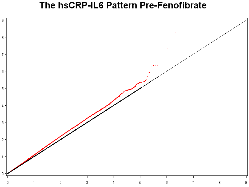
Supplemental Digital Content 8. Quantile-quantile plot for association of each SNP with the hsCRP-IL6 pattern pre-fenofibrate treatment. The X-axes display the expected −log10(P-value), the Y-axes display the observed −log10(P-value).
Supplemental Digital Content 9. Quantile-quantile plot for association of each SNP with the MCP1-TNF-α pattern pre-fenofibrate treatment. The X-axes display the expected −log10(P-value), the Y-axes display the observed −log10(P-value).
Acknowledgments
Source of funding: This work has been funded by the NHLBI grant U01HL072524-04.
Footnotes
Conflicts of interest: No authors declare conflict of interest.
References
- 1.Shen J, Ordovas JM. Impact of genetic and environmental factors on hsCRP concentrations and response to therapeutic agents. Clin Chem. 2009;55:256–264. doi: 10.1373/clinchem.2008.117754. [DOI] [PMC free article] [PubMed] [Google Scholar]
- 2.Libby P, Plutzky J. Inflammation in diabetes mellitus: role of peroxisome proliferator-activated receptor-alpha and peroxisome-activated receptor-gamma agonists. Am J Cardiol. 2007;99:27B–40B. doi: 10.1016/j.amjcard.2006.11.004. [DOI] [PubMed] [Google Scholar]
- 3.Bragt MCE, Mensink RP. Comparison of the effects of n-3 long chain polyunsaturated fatty acids and fenofibrate on markers of inflammation and vascular function, and on the serum lipoprotein profile in overweight and obese subjects. Nut Met Cardiovasc Dis. doi: 10.1016/j.numecd.2010.12.010. (in press) [DOI] [PubMed] [Google Scholar]
- 4.Brisson D, Ledoux K, Bosse Y, St-Pierre J, Julien P, Perron P, et al. Effect of apolipoprotein E, peroxisome proliferator-activated receptor alpha and lipoprotein lipase gene mutations on the ability of fenofibrate to improve lipid profiles and reach clinical guideline targets among hypertriglyceridemic patients. Pharmacogenetics. 2002;12:313–320. doi: 10.1097/00008571-200206000-00007. [DOI] [PubMed] [Google Scholar]
- 5.Schmitz G, Schmitz-Madry A, Ugocsai P. Pharmacogenetics and pharmacogenomics of cholesterol-lowering therapy. Curr Opin Lipidol. 2007;18:164–173. doi: 10.1097/MOL.0b013e3280555083. [DOI] [PubMed] [Google Scholar]
- 6.Wojczynski MK, Gao G, Borecki I, Hopkins PN, Parnell L, Lai CQ, et al. Apolipoprotein B genetic variants modify the response to fenofibrate: a GOLDN study. J Lipid Res. 2010;51:3316–3323. doi: 10.1194/jlr.P001834. [DOI] [PMC free article] [PubMed] [Google Scholar]
- 7.Shen J, Arnett DK, Parnell LD, Peacock JM, Lai CQ, Hixson JE, et al. Association of common C-reactive protein (CRP) gene polymorphisms with baseline plasma CRP levels and fenofibrate response: the GOLDN study. Diabetes Care. 2008;31:910–915. doi: 10.2337/dc07-1687. [DOI] [PMC free article] [PubMed] [Google Scholar]
- 8.Kabagambe EK, Glasser SP, Ordovas JM, Warodomwichit D, Tsai MY, Hopkins PN, et al. TCF7L2 polymorphisms and inflammatory markers before and after treatment with fenofibrate. Diabetol Metab Syndr. 2009;12:16. doi: 10.1186/1758-5996-1-16. [DOI] [PMC free article] [PubMed] [Google Scholar]
- 9.Corella D, Arnett DK, Tsai MY, Kabagambe EK, Peacock JM, Hixson JE, et al. The -256T>C polymorphism in the apolipoprotein A-II gene promoter is associated with body mass index and food intake in the genetics of lipid lowering drugs and diet network study. Clin Chem. 2007;53:1144–1152. doi: 10.1373/clinchem.2006.084863. [DOI] [PubMed] [Google Scholar]
- 10.Irvin MR, Kabagambe EK, Tiwari HK, Parnell LD, Straka RJ, Tsai M, et al. Apolipoprotein E polymorphisms and postprandial triglyceridemia before and after fenofibrate treatment in the Genetics of Lipid Lowering and Diet network (GOLDN) study. Circ Cardiovasc Genet. 2010;3:462–467. doi: 10.1161/CIRCGENETICS.110.950667. [DOI] [PMC free article] [PubMed] [Google Scholar]
- 11.Patsch JR, Miesenbock G, Hopferwieser T, Muhlberger V, Knapp E, Dunn JK, et al. Relation of triglyceride metabolism and coronary artery disease: studies in the postprandial state. Arterioscler Thromb. 1992;12:1336–1345. doi: 10.1161/01.atv.12.11.1336. [DOI] [PubMed] [Google Scholar]
- 12.Tsai MY, Hanson NQ, Straka RJ, Hoke TR, Ordovas JM, Peacock JM, et al. Effect of influenza vaccine on markers of inflammation and lipid profile. J Lab Clin Med. 2005;145:323–327. doi: 10.1016/j.lab.2005.03.009. [DOI] [PubMed] [Google Scholar]
- 13.Kabagambe EK, Ordovas JM, Tsai MY, Borecki IB, Hopkins PN, Glasser SP, et al. Smoking, inflammatory patterns and postprandial hypertriglyceridemia. Atherosclerosis. 2009;203:633–639. doi: 10.1016/j.atherosclerosis.2008.08.005. [DOI] [PMC free article] [PubMed] [Google Scholar]
- 14.Korn JM, Kuruvilla FG, McCarroll SA, Wysoker A, Nemesh J, Cawley S, et al. Integrated genotype calling and association analysis of SNPs, common copy number polymorphisms and rare CNVs. Nat Genet. 2008;40:1253–1260. doi: 10.1038/ng.237. [DOI] [PMC free article] [PubMed] [Google Scholar]
- 15.Hu FB, Rimm EB, Stampfer MJ, Ascherio A, Spiegelman D, Willett WC. Prospective study of major dietary patterns and risk of coronary heart disease in men. Am J Clin Nutr. 2000;72:912–921. doi: 10.1093/ajcn/72.4.912. [DOI] [PubMed] [Google Scholar]
- 16.Atkinson B, Therneau T. kinship: mixed-effects Cox models, sparse matrices, and modeling data from large pedigrees. R package version 1.1.0–17 2007 [Google Scholar]
- 17.Li Y, Sabatine MS, Tong CH, Ford I, Kirchgessner TG, Packard CJ, et al. Genetic variants in the KIF6 region and coronary event reduction from statin therapy. Hum Genet. 2011;129:17–23. doi: 10.1007/s00439-010-0892-6. [DOI] [PMC free article] [PubMed] [Google Scholar]
- 18.Taylor D, Wilkison M, Voyich J, Meissner N. Prevention of bone marrow cell apoptosis and regulation of hematopoiesis by type 1 IFNs during systemic responses to Pneumocystis lung infection. J Immunol. 2011;186:5956–5967. doi: 10.4049/jimmunol.1003558. [DOI] [PubMed] [Google Scholar]
- 19.Pfeifer D, Sykora KW, Sauer M, Kreipe H, Lacher M, Nustede R, et al. Inflammatory bowel disease and mutations affecting the interleukin-10 receptor. N Engl J Med. 2009;361:2033–2045. doi: 10.1056/NEJMoa0907206. [DOI] [PMC free article] [PubMed] [Google Scholar]
- 20.Poland GA, Ovsyannikova IG, Jacobson RM. Immunogenetics of seasonal influenza vaccine response. Vaccine. 2008;26:D35–D40. doi: 10.1016/j.vaccine.2008.07.065. [DOI] [PMC free article] [PubMed] [Google Scholar]
- 21.Brand OJ, Lowe CE, Heward JM, Franklin JA, Cooper JD, Todd JA, et al. Association of the interleukin-2 receptor alpha (IL-2Rα)/CD25 gene region with Graves’ disease using a multilocus test and tag SNPs. Clin Endocrinol. 2007;66:508–512. doi: 10.1111/j.1365-2265.2007.02762.x. [DOI] [PubMed] [Google Scholar]
- 22.Vella A, Cooper JD, Lowe CE, Walker N, Nutland S, Widmer B, et al. Localization of a type 1 diabetes locus in the IL2RA/CD25 region by use of tag single-nucleotide polymorphisms. Am J Hum Genet. 2005;76:773–779. doi: 10.1086/429843. [DOI] [PMC free article] [PubMed] [Google Scholar]
- 23.Perera D, Stankovich J, Butzkueven H, Taylor BV, Foote SJ, Kilpatrick TJ, et al. Fine mapping of multiple sclerosis susceptibility genes provides evidence of allelic heterogeneity at the IL2RA locus. J Neuroimmunol. 2009;211:105–109. doi: 10.1016/j.jneuroim.2009.03.010. [DOI] [PubMed] [Google Scholar]
- 24.O’Garra A, Vieira P. Regulatory T cells and mechanisms of immune system control. Nat Med. 2004;10:801–805. doi: 10.1038/nm0804-801. [DOI] [PubMed] [Google Scholar]
- 25.George J. Mechanisms of disease: the evolving role of regulatory T cells in atherosclerosis. Nat Clin Pract Cardiovasc Med. 2008;5:531–540. doi: 10.1038/ncpcardio1279. [DOI] [PubMed] [Google Scholar]
- 26.Vainio P, Gupta S, Ketola K, Mirtti T, Mpindi JP, Kohonen P, et al. Arachidonic acid pathway members PLA2G7, HPGD, EPHX2, and CYP4F8 identified as putative novel therapeutic targets in prostate cancer. Am J Pathol. 2011;178:525–536. doi: 10.1016/j.ajpath.2010.10.002. [DOI] [PMC free article] [PubMed] [Google Scholar]
- 27.Lafuente EM, van Puijenbroek AA, Krause M, Carman CV, Freeman GJ, Berezovskaya A, et al. RIAM, an Ena/VASP and Profilin ligand, interacts with Rap1-GTP and mediates RAP1-induced adhesion. Dev Cell. 2004;7:585–595. doi: 10.1016/j.devcel.2004.07.021. [DOI] [PubMed] [Google Scholar]
- 28.Katagiri K, Hattori M, Minato N, Kinashi T. Rap1 functions as a key regulator of T-cell and antigen-presenting cell interactions and modulates T-cell responses. Mol Cell Biol. 2002;22:1001–1015. doi: 10.1128/MCB.22.4.1001-1015.2002. [DOI] [PMC free article] [PubMed] [Google Scholar]
- 29.Chalasani N, Guo X, Loomba R, Goodarzi MO, Haritunians T, Kwon S, et al. Genome-wide association study identifies variants associated with histologic features of nonalcoholic fatty liver disease. Gastroenterol. 2010;139:1567–1576. doi: 10.1053/j.gastro.2010.07.057. [DOI] [PMC free article] [PubMed] [Google Scholar]
- 30.Tuomisto A, Sund M, Tahkola J, Latvanlehto A, Savolainen ER, Autio-Harmainen H, et al. A mutant collagen XIII alters intestinal expression of immune response genes and predisposes transgenic mice to develop B-cell lymphomas. Cancer Res. 2008;28:10324–10332. doi: 10.1158/0008-5472.CAN-08-2582. [DOI] [PMC free article] [PubMed] [Google Scholar]
- 31.Burstein E, Hoberg JE, Wilkinson AS, Rumble JM, Csomos RA, Komarck CM, et al. COMMD proteins, a novel family of structural and functional homologs of MURR1. J Biol Chem. 2005;28:22222–22232. doi: 10.1074/jbc.M501928200. [DOI] [PubMed] [Google Scholar]
- 32.Beck J, Urnovitz HB, Saresella M, Caputo D, Clerici M, Mitchell WM, et al. Serum DNA motifs predict disease and clinical status in multiple sclerosis. J Mol Diagn. 2010;12:312–319. doi: 10.2353/jmoldx.2010.090170. [DOI] [PMC free article] [PubMed] [Google Scholar]
Associated Data
This section collects any data citations, data availability statements, or supplementary materials included in this article.
Supplementary Materials
Supplemental Digital Content 1. Scree plot for the hsCRP-IL6 pattern. The X-axes display the number of principal components, the Y-axes display the eigenvalue.
Supplemental Digital Content 10. Quantile-quantile plot for association of each SNP with the hsCRP-IL6 pattern post-fenofibrate treatment. The X-axes display the expected −log10(P-value), the Y-axes display the observed −log10(P-value).
Supplemental Digital Content 11. Quantile-quantile plot for association of each SNP with the MCP1-TNF-α pattern post-fenofibrate treatment. The X-axes display the expected −log10(P-value), the Y-axes display the observed −log10(P-value).
Supplemental Digital Content 2. Scree plot for the MCP1-TNF-α pattern. The X-axes display the number of principal components, the Y-axes display the eigenvalue.
Supplemental Digital Content 3. Quantile-quantile plot for association of each SNP with the response in high-sensitivity C-reactive protein to fenofibrate treatment. The X-axes display the expected −log10(P-value), the Y-axes display the observed −log10(P-value).
Supplemental Digital Content 4. Quantile-quantile plot for association of each SNP with the response in interleukin-6 to fenofibrate treatment. The X-axes display the expected −log10(P-value), the Y-axes display the observed −log10(P-value).
Supplemental Digital Content 5. Quantile-quantile plot for association of each SNP with the response in interleukin-2 soluble receptor-α to fenofibrate treatment. The X-axes display the expected −log10(P-value), the Y-axes display the observed −log10(P-value).
Supplemental Digital Content 6. Quantile-quantile plot for association of each SNP with the response in tumor necrosis factor-α to fenofibrate treatment. The X-axes display the expected −log10(P-value), the Y-axes display the observed −log10(P-value).
Supplemental Digital Content 7. Quantile-quantile plot for association of each SNP with the response in monocyte chemoattractant protein-1 to fenofibrate treatment. The X-axes display the expected −log10(P-value), the Y-axes display the observed −log10(P-value).
Supplemental Digital Content 8. Quantile-quantile plot for association of each SNP with the hsCRP-IL6 pattern pre-fenofibrate treatment. The X-axes display the expected −log10(P-value), the Y-axes display the observed −log10(P-value).
Supplemental Digital Content 9. Quantile-quantile plot for association of each SNP with the MCP1-TNF-α pattern pre-fenofibrate treatment. The X-axes display the expected −log10(P-value), the Y-axes display the observed −log10(P-value).