Abstract
Background
Propofol exposure to neurons during synaptogenesis results in apoptosis leading to cognitive dysfunction in adulthood. Previous work from our laboratory showed that isoflurane neurotoxicity occurs through p75 neurotrophin receptor (p75NTR) and subsequent cytoskeleton depolymerization. Given that isoflurane and propofol both suppress neuronal activity, we hypothesized that propofol also induces apoptosis in developing neurons through p75NTR.
Methods
DIV5-7 neurons were exposed to propofol (3 µM) for 6 hr and apoptosis was assessed by cleaved caspase-3 (Cl-Csp3) immunoblot and immunofluorescence microscopy. Primary neurons from p75NTR−/− mice or wild-type neurons were treated with propofol, with or without pretreatment with TAT-Pep5 (10 µM, 15 min), a specific p75NTR inhibitor. P75NTR−/− neurons were transfected for 72 h with a lentiviral vector containing the synapsin driven p75NTR gene (Syn-p75NTR) or control vector (Syn-GFP) prior to propofol. To confirm our in vitro findings, wild type mice and p75NTR−/− mice (PND5) were pre-treated with either TAT-Pep5 or TAT-ctrl followed by propofol for 6 h.
Results
Neurons exposed to propofol showed a significant increase in Cl-Csp3, an effect attenuated by TAT-Pep5 and hydroxyfasudil. Apoptosis was significantly attenuated in p75NTR−/− neurons. In p75NTR−/− neurons transfected with Syn-p75NTR, propofol significantly increased Cl-Csp3 in comparison to Syn-GFP transfected p75NTR−/− neurons. Wild type mice exposed to propofol exhibited increased Cl-Csp3 in the hippocampus, an effect attenuated by TAT-Pep5. By contrast, propofol did not induce apoptosis in p75NTR−/− mice.
Conclusion
These results demonstrate that propofol induces apoptosis in developing neurons in vivo and in vitro and implicate a role for p75NTR and the downstream effector ROCK.
Introduction
During synaptogenesis (post-natal day 5–7, PND5-7), anesthetics lead to neurodegeneration.1–3 Many anesthetics cause neurotoxicity, which include midazolam and nitrous oxide, isoflurane, sevoflurane, propofol, thiopental and ketamine.4–11 In addition, isoflurane does not induce neuronal apoptosis PND 15, but does alter synaptic plasticity; these changes persist for at least 4 weeks postexposure.12 Of significant concern is that neonatal exposure to anesthetics results in neurocognitive and behavioral abnormalities during adolescence and adulthood.2,3,13,14 Although the mechanism by which this toxicity occurs is not clear, γ-aminobutyric acid (GABAA) agonism and N-methyl-D-aspartate receptor antagonism play a central role.
Recently we demonstrated that proBDNF-p75NTR signaling mediates isoflurane neurotoxicity in developing neurons in vivo and in vitro. Brain derived neurotrophic factor (BDNF) is important to both prosurvival and proapoptotic signaling pathways. BDNF is stored as a proneurotrophin (proBDNF) within synaptic vesicles and is proteolytically cleaved to mature BDNF (mBDNF) in the synaptic cleft by plasmin, a protease activated by tissue plasminogen activator (tPA).15–18 Prosurvival signaling is triggered by mBDNF agonism of tropomyosin receptor kinase B, which leads to neurite outgrowth and synapse maturation and stabilization.15,17,19 In contrast, non-cleaved proBDNF binds to the p75 neurotrophin receptor (p75NTR) and activates RhoA, a small GTPase that regulates actin cytoskeleton polymerization resulting in inhibition of axonal elongation, growth cone collapse, and apoptosis.16,20–22 Neuronal stimulation is important in this process since proBDNF is constitutively secreted while tPA release is regulated; without neuronal depolarization, conversion of proBDNF to mBDNF may be blunted, which then leads to preferential signaling through p75NTR. Upon neuronal excitation, tPA release results in plasmin production and subsequent generation of mBDNF-tropomyosin receptor kinase B activation leading to neuronal survival, neurite sprouting, and synaptogenesis.15–17,22
Head et al. showed that isoflurane induces apoptosis in DIV5 neurons, a finding that did not occur in DIV14 or DIV21 neurons.22 This effect was attenuated by TAT-Pep5, a p75NTR intracellular domain inhibitor, suggesting a role for p75NTR in isoflurane mediated neurotoxicity. Apoptosis was also attenuated by pretreatment with tPA or plasmin, suggesting that isoflurane mediated neurotoxicity is caused in part by suppressing tPA release and preventing proBDNF conversion to mBDNF, thus leading to preferential p75NTR signaling, RhoA activation, actin cytoskeleton depolymerization, and subsequent neuronal apoptosis. Lemkuil et al. extended these findings by showing that isoflurane exposure increases p75NTR-RhoA activation in parallel with apoptosis and that inhibition of RhoA activation or cytoskeleton stabilization attenuates the isoflurane-mediated neurotoxic effects.23
Although these data support the premise that proBDNF-p75NTR signaling plays a significant role in neonatal neurotoxicity, a number of questions remain. If this mechanism is important to anesthetic neurotoxicity, then it should also be involved in toxicity mediated by other anesthetics that activate GABAA receptor (e.g., propofol). In the studies of Head et al and Lemkuil et al, p75NTR inhibition was achieved pharmacologically. In addition to concerns about nonspecific effects of pharmacologic agents, complete suppression of RhoA activation was not achieved; residual RhoA activity may have influenced the results.24 To address these concerns, we investigated the role of p75NTR-RhoA-ROCK pathway in propofol-induced neurotoxicity.
Materials and Methods
Preparation of Neuronal Cell Cultures
All studies performed on animals were approved by Veteran Affairs San Diego Institutional Animal Care and Use Committee (San Diego, California) and conform to the guidelines of Public Health Service Policy on Human Care and Use of Laboratory Animals.
Neonatal mouse neurons (BALBc, The Jackson Laboratory, Bar Harbor, ME) were isolated using a papain dissociation kit (Worthington Biochemical, Lakewood, NJ) as previously described.22 p75NTR knockout BALBc mice were generously given to us by Don Pizzo, Ph.D. (Project Scientist, University of California, San Diego, Department of Pathology) at the Veterans Affairs San Diego Healthcare System (originally generated by Dr. Kuo-Fen Lee25). Neurons were isolated from 1–2 day old pups (PND1-2) and grown in culture for 4 to 7 days in vitro (DIV4-7). Neurons were cultured in Neuobasal A media supplemented with B27 (2%), 250 mM GLUTMax1, and penicillin/streptomycin (1%). Neurons were cultured on poly-D-lysine/laminin (2 g/cm2) coated plates or coverslips at 37°C in 5% CO2 for 4–7 days prior to experiments. Lentiviral (LV) vectors driven by neuronal specific synapsin promoters expressing p75NTR or green fluorescent protein (GFP) (LV-syn-p75NTR and LV-syn-GFP, respectively) were generated by and obtained from Atsushi Miyanohara, Ph.D. (Assistant Professor, Gene Therapy Program, University of California, San Diego, La Jolla, California) at the University of California, San Diego Viral Vector Core. Cleaved-caspase 3 (Cl-Csp3)(Cell Signaling, Danvers, MA) and drebrin (Abcam, Cambridge, MA) were used to detect apoptosis and the F-actin cytoskeleton respectively via immunoblot or immunofluorescence deconvolution or confocal microscopy. Cl-Csp3 and drebrin were normalized to the nuclear stain 4',6-diamidino-2-phenylindole (Molecular Probes/Invitrogen, Carlsbad, CA). Cl-Csp3 immunoblots were quantified by densitometry and normalized to total caspase-3 (T-Csp3). p75NTR antibody was obtained from Abcam. The cell-permeable peptide, TAT-Pep5 [H-YGRKKRRQRRR-CFFRGGFFNHNPRYC-OH], that blocks the intracellular association of p75NTR with RhoGDI, thus blocking its ability to activate RhoA and hydroxyfasudil (HA 1100 hydrochloride) were purchased from CalBiochem (Gibbstown, NJ) and Tocris (Ellsville MO), respectively.
Anesthetic Neurotoxicity Model
In vitro, primary neuronal cultures were placed within an incubator and exposed to propofol 3 µM for 6 h in a gas mixture of 5% CO2, 21% O2, balance nitrogen at a flow rate of 2 L/min. The temperature in the incubator was maintained at 37°C. Neurons were harvested for analysis 2 h postexposure. In vivo, neonatal mice (PND 5) were given a single intraperitoneal injection of propofol (100 mg/kg). Mice were sacrificed 6 hr post exposure and brains were prepared by perfusion fixation for immunofluorescence confocal microscopy. Mice were administered TAT-Pep5 intraperitoneal (10 µM) 15 min prior to propofol injections. All mice were kept in room air and on a warming pad maintained at 37°C.
Lentiviral vector transfection
Primary neuronal cultures were transfected with a lentiviral vector that expresses p75NTR driven by a neuronal specific synapsin-promoter (LV-syn-p75NTR) for 72 h and then exposed to propofol. A lentiviral vector that expresses GFP driven by a neuronal specific synapsin-promoter (LV-syn-GFP) served as control.
Protein extraction and Western blot Analysis
Proteins in cell lysates were separated by sodium dodecyl sulfate-polyacrylamide gel electrophoresis using 10% acrylamide gels (Invitrogen, Carlsbad, CA) and transferred to polyvinylidene difluoride membranes (Millipore, Billerica, MA) by electroelution. Membranes were blocked in 20 mM phosphate-buffered saline (PBS) Tween (1%) containing 4% bovine serum albumin and incubated with primary antibody overnight at 4°C as previously described.22,23 Primary antibodies were visualized using secondary antibodies conjugated to horseradish peroxidase (Santa Cruz Biotech, Santa Cruz, CA) and chemo luminescent reagent (Amersham Pharmacia Biotech, Piscataway, NJ). All displayed bands are expected to migrate to the appropriate size and were determined by comparison to molecular weight standards. We performed an oversaturation analysis with the UVP Imaging software. Red pixilation is assigned to the bands as an indicator of oversaturation. Image J (National Institutes of Health, Bethesda, MD) was used for densitometric analysis of immunoblots with normalization of cleaved caspase-3 to total caspase-3.
Immunofluorescence Confocal Microscopy
Neurons were prepared for immunofluorescence microscopy as previously described.22,23 Primary neurons were fixed with 4% paraformaldehyde in PBS for 10 min at room temperature, incubated with 100 mM glycine (pH. 7.4) for 10 min to quench aldehyde groups, permeabilized in buffered Triton X-100 (0.1%) for 10 min, blocked with 1% bovine serum albumin/PBS/Tween (0.05%) for 20 min and then incubated with primary antibodies in 1% bovine serum albumin/PBS/Tween (0.05%) for 24–48 h at 4°C. Excess antibody was removed by washing with PBS/Tween (0.1%) for 15 min followed by incubation with fluorescein isothiocyanate or Alexa-conjugated secondary antibody (1:250) for 1 h. To remove excess secondary antibody, tissue or cells were washed 6× at 5-min intervals with PBS/Tween (0.1%) and incubated for 20 min with the nuclear stain 4',6-diamidino-2-phenylindole (1:5000) diluted in PBS. Cells were washed for 10 min with PBS and mounted in gelvatol for microscopic imaging. Confocal images were captured with an Olympus confocal microscope system (Applied Precision, Inc., Issaquah, WA.) that included a Photometrics CCD (Photometrics, Tucson, AZ) mounted on a Nikon TE-200 (Nikon, Melville, NY) inverted epi-fluorescence microscope. Between 30 and 80 optical sections spaced by ~0.1–0.3 µm were captured. Exposure times were set such that the camera response was in the linear range for each fluorophore. Maximal projection volume views or single optical sections were visualized. Pixels were assessed quantitatively by CoLocalizer Pro 1.0 software (Colocalization Research Software, Japan and Switzerland). Statistical analysis was performed using Prism 4 (GraphPad Software, La Jolla, CA).
Apoptosis Quantification
The cleaved caspase-3 pixels (red, Alexa 594) were normalized to nuclear stained pixels (blue, 405).22,23 Cleaved caspase-3 is an executioner and marker of apoptosis. Ten visual fields at 40× magnification were counted per experimental condition.
Cytoskeletal Depolymerization Quantification
The drebrin pixels (green, Alexa 488) were normalized to nuclear stained pixels (blue, 405).22,23 Drebrin is a filamentous (F-)actin binding protein which stabilizes the actin cytoskeleton within neuritic processes. Pixel values were obtained after subtracting background through normalized threshold values in Co-localizer Pro as previously described.26,27 A reduction in neuritic processses is indicated by decreased drebrin protein expression. Sample size (n) equals number of neurons counted per experimental condition.
Statistical Analysis
All parametric data were analyzed by either a two-tailed unpaired t-tests (fig. 1–5) or by One-way ANOVA with Bonferroni’s Mutliple Camparison Test (fig. 6). Significance was set at p < 0.05. Statistical analysis was performed using Prism 4 (GraphPad Software). Sample size (n=) represents the amount of times the experiments were repeated on separate neuronal cell culture preparations derived from 12–20 PND1-3 pups.
Figure 1.
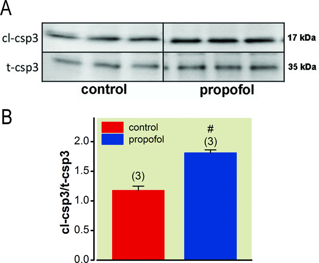
Primary neurons were isolated from neonatal rodent brains at postnatal day 1–3 (PND1-3) and grown in vitro for 5–7 days (DIV5-7). Neurons were then exposed to propofol 3.0 µM for 6 hr in 5% CO2 in air. Apoptosis was evaluated after propofol exposure by cleaved caspase-3 (Cl-Csp3) immunoblot. A, Immunoblot analysis shows an increase in apoptosis marker, Cl-Csp3, with propofol exposure. B, Quantitation of the data is represented in the graph (n=3; #p=0.002). Sample size is indicated above the error bars +/− SEM. These data demonstrate that, in vitro, primary neurons from neonatal rodents exposed to propofol exhibit increased apoptosis.
Figure 5.
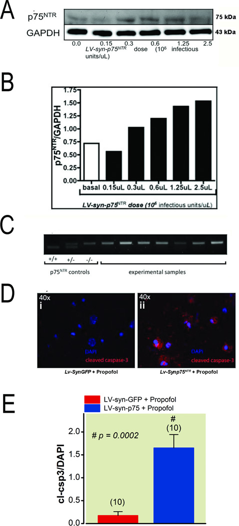
Primary neurons were isolated from neonatal rodent brains at postnatal day 1–3 (PND1-3) and grown in vitro for 5–7 days (DIV5-7). Neurons were then transfected for 72 hr with increasing doses of a lentiviral vector that expresses p75NTR driven by a neuronal specific synapsin-promoter (LV-syn-p75NTR). A, Immunoblot analysis shows an increase in p75NTR expression with increasing doses of LV-syn-p75NTR. B, Quantitation of the data is represented in the graph. These data demonstrate that, in vitro, primary neurons from neonatal rodents transfected for 72hr with LV-syn-p75NTR exhibits a dose dependent increase in p75NTR expression. C, PCR confirmed that rodents used for primary neuronal cultures were p75NTR knockout genotype (−/−). On DIV4, p75NTR knockout neurons were transfected with LV-syn-p75NTR for 72 hr LV-syn-GFP served as a control. On DIV7 neurons were exposed to propofol 3.0 µM for 6 hr and apoptosis was evaluated by cleaved caspase-3 (Cl-Csp3) immunofluorescence (IF) microscopy. D, IF microscopy shows an increase in Cl-Csp3 in p75NTR knockout neurons transfected with LV-syn-p75NTR and exposed to propofol versus LV-syn-GFP transfected neurons exposed to propofol. Quantitation of the data is represented in the graph (n=10; p=0.0002). Sample size is indicated above the error bars +/− SEM. These data demonstrate that, in vitro, re-expression of p75NTR in p75NTR knockout neurons reestablishes propofol mediated apoptosis. DAPI (4',6-diamidino-2-phenylindole), GAPDH (glyceraldehyde 3-phosphate dehydrogenase), LV (lentivirus), PCR (polymerase chain reactor), KO (knockout), GFP (green fluorescent protein), DIV (days in vitro).
Figure 6.
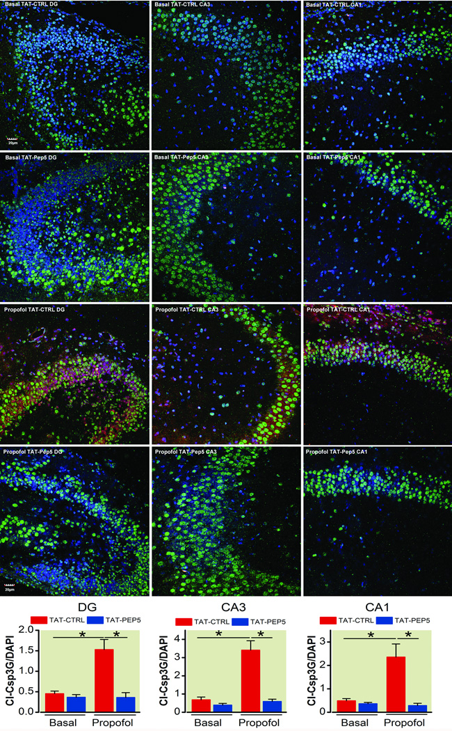
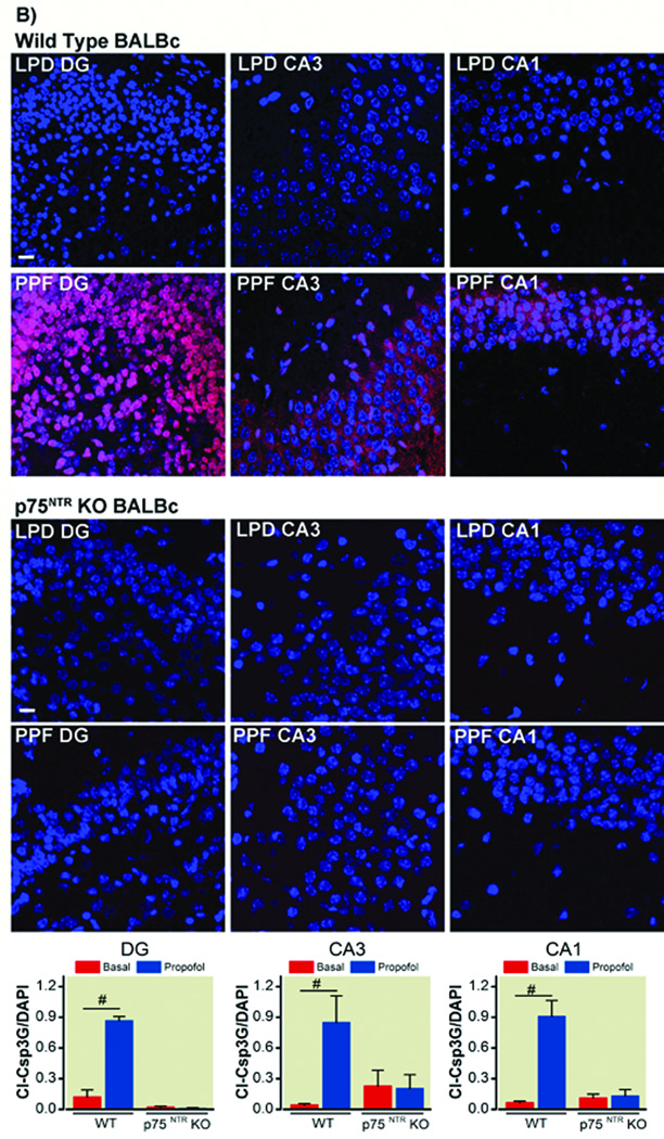
Postnatal day 5 (PND5) wild-type or p75NTR knockout mice were given an intraperitoneal (IP) injection of propofol (100 mg/kg) or intralipid for 6 hours and apoptosis was evaluated by cleaved caspase-3 (Cl-Csp3) immunofluorescence (IF). A, dentate gyrus, CA3, CA1 are indicated on image. Basal TAT-CTRL, Basal TAT-Pep5, propofol TAT-CTRL, propofol TAT-Pep5 are indicated on the image. Immunofluorescence microscopic analysis shows that wild-type mice exhibited a significant increase in Cl-Csp3 in the dentate gyrus (*p=0.002), CA3 (*p=0.008), and CA1 (*p=0.007) regions of the hippocampus compared to intralipid treated controls (n=4–5). TAT-Pep5 (10 µM, 15 min) significantly attenuated propofol-mediated apoptosis (*p=0.005, DG; *p=0.002, CA3; *p=0.004, dentate gyrus). B, Additional immunofluorescence microscopic analysis shows that p75NTR knockout mice exhibited no increase in Cl-Csp3 in the dentate gyrus, CA3, or CA1 following propofol exposure compared to wild-type (#p=0.0008, dentate gyrus; #p=0.03, CA3; #p=0.002, CA1). Quantitation of the data is represented in the graphs. Sample size is indicated above the error bars +/− SEM. Scale bar, 20 µm. DAPI (4',6-diamidino-2-phenylindole), KO (knockout), PND (postnatal day).
Results
Propofol exposure increases apoptosis in primary mouse neurons (DIV5-7)
Primary neurons were isolated from neonatal rodent brains at postnatal day 1–3 (PND1-3) and grown in vitro for 5–7 days (DIV5-7). Propofol (3 µM, 6 h) exposure resulted in a significantly increased (n = 3; p = 0.002) expression of Cl-Csp3 compared to control (fig. 1).
Propofol exposure decreases neuritic processes in primary mouse neurons (DIV5-7)
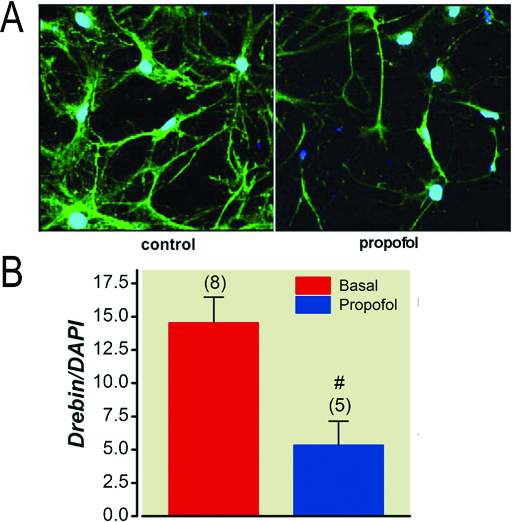
Based on our previous data on isoflurane-mediated neurotoxicity, a key mechanistic tenet of injury is preferential activation of p75NTR signaling pathway leading to actin cytoskeleton destabilization and subsequent apoptosis.22,23 Because we hypothesize that propofol-mediates neuronal apoptosis through a similar mechanism to that of isoflurane, we investigated the effects of propofol exposure on formation of neuritic processes as measured by drebrin, a neuronal F-actin binding protein and marker of dendritic filopodial spines.22,23 Primary neurons were isolated from neonatal rodent pup brains at postnatal day 1–3 (PND1-3) and grown in vitro for 5–7 days (DIV5-7). Propofol (3 µM, 6 h) exposure resulted in a significantly decreased (n = 5; p = 0.008) expression of drebrin compared to control (fig. 2).
Figure 2.
Primary neurons were isolated from neonatal rodent brains at postnatal day 1–3 (PND1-3) and grown in vitro for 5–7 days (DIV7). On DIV7 neurons were exposed to propofol (PPF) 3.0 µM for 6 hr in 5% CO2 in air. Neuritic processes were evaluated after propofol exposure by drebrin immunofluorescence (IF) microscopy. Nucleus was stained with DAPI (4',6-diamidino-2-phenylindole). A, IF microscopy shows a decrease in drebrin in neurons exposed to propofol versus control. B, Quantitation of the data is represented in the graph (n=5; #p=0.008). Sample size is indicated above the error bars +/− SEM. These data demonstrate that, in vitro, primary neurons from neonatal rodents exposed to propofol exhibit a reduction in neuritic processes
TAT-Pep5 attenuates propofol induced cleaved caspase-3 activation in primary mouse neurons (DIV5-7)
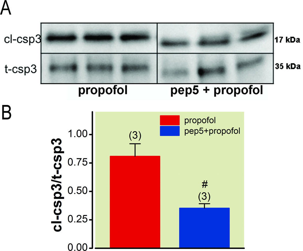
Previous work from our laboratory has demonstrated that isoflurane mediated neurotoxicity in developing primary neurons (DIV5-7) is mediated through neuronal suppression and subsequent proBDNF activation of p75NTR.22,23 Moreover, using TAT-Pep5, we showed that signaling from p75NTR to Rho was mediating by isoflurane neurotoxicity.23 We tested whether propofol exposure to DIV5-7 primary neurons also causes cellular injury in a pattern similar to isoflurane (i.e., propofol induces neuronal apoptosis through p75NTR activation). TAT-Pep5 (10 µM, 15 min) treatment of DIV5-7 primary mouse neurons prior to propofol exposure (3 µM, 6 hr) significantly attenuated Cl-Csp3 expression (n = 3; p = 0.046) compared to propofol exposure without TAT-Pep5 pretreatment (fig. 3).
Figure 3.
Primary neurons were isolated from neonatal rodent brains at postnatal day 1–3 (PND1-3) and grown in vitro for 5–7 days (DIV5-7). Neurons were pre-treated with TAT-Pep5 (10 µM; 15 min), a p75NTR intracellular domain antagonist, prior to propofol exposure. After pre-treatment, neurons were exposed to propofol 3.0 µM for 6 hr in 5% CO2 in air. Apoptosis was evaluated after propofol exposure by cleaved caspase-3 (Cl-Csp3) immunoblot. A, Immunoblot analysis shows a decrease in apoptosis marker, Cl-Csp3, with TAT-Pep5 pre-treatment. B, Quantitation of the data is represented in the graph (n=3; #p=0.046). Sample size is indicated above the error bars +/− SEM.
Hydroxyfasudil attenuates propofol induced cleaved caspase-3 activation in primary mouse neurons (DIV5-7)
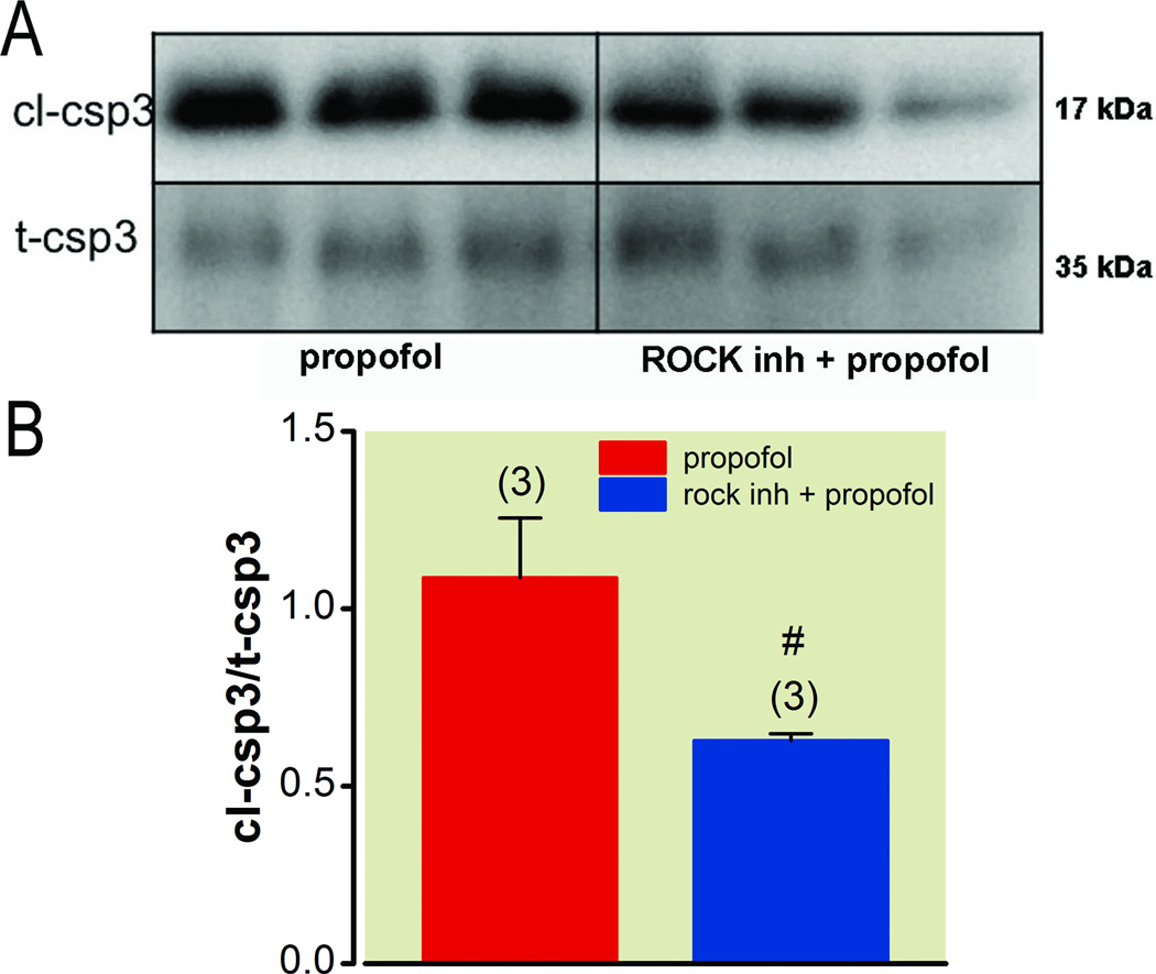
RhoA is known to regulate actin cytoskeleton dynamics in neurons and cause growth cone collapse, leading to apoptosis.28–30 RhoA is activated by p75NTR and mediates its effects through downstream activation of RhoA kinase (ROCK).31 Because ROCK is indirectly activated by p75NTR and inhibition of p75NTR with TAT-Pep5 attenuates propofol-mediated apoptosis in developing neurons (DIV5-7), we hypothesized that inhibition of ROCK would attenuate propofol-mediated apoptosis. DIV5-7 primary mouse neurons were pretreated with a ROCK inhibitor, hydroxyfasudil (10 µM, 15 min), prior to propofol exposure (3 µM, 6 h); hydroxyfasudil significantly attenuated Cl-Csp3 activation (n = 3; p = 0.007) compared to propofol exposure (with hydroxyfasudil vehicle) in the absence of hydroxyfasudil pre-treatment (fig. 4).
Figure 4.
Primary neurons were isolated from neonatal rodent brains at postnatal day 1–3 (PND1-3) and grown in vitro for 5–7 days (DIV5-7). Neurons were pre-treated with hydroxyfasudil (10 µM; 15 min), a Rho kinase (rock) inhibitor, prior to propofol exposure. After pre-treatment, neurons were exposed to propofol 3.0 µM for 6 hr in 5% CO2 in air. Apoptosis was evaluated after propofol exposure by cleaved caspase-3 (Cl-Csp3) immunoblot. A, Immunoblot analysis shows a decrease in apoptosis marker, Cl-Csp3, with hydroxyfasudil (rock inhibitor) pre-treatment. B, Quantitation of the data is represented in the graph (n=3; #p=0.007). Sample size is indicated above the error bars +/− SEM.
Primary neurons transfected with a lentiviral vector that expresses p75NTR driven by a neuronal specific synapsin-promoter (LV-syn-p75NTR) increases p75NTR expression
Because our results from figures 3 and 4 demonstrate that inhibition of p75NTR signaling attenuates propofol-mediated apoptosis in developing primary neurons, we hypothesized that primary neurons from p75NTR knockout (p75NTR−/−) mice would be less susceptible to propofol mediated apoptosis, and that reexpression of p75NTR in p75NTR−/− neurons would reestablish anesthetic vulnerability. Primary neurons were isolated from p75NTR−/− mice at postnatal day 1–3 (PND1-3) and grown in vitro for 5–7 days (DIV5-7). On DIV4, neurons were transfected with a lentiviral vector that expresses p75NTR driven by a neuron specific synapsin-promoter (LV-syn-p75NTR) or GFP control vector (LV-syn-GFP) for 72 h. Immunoblot analysis demonstrated a dose dependent increase in p75NTR protein expression after transfection with LV-syn-p75NTR (fig. 5 A and B). On DIV7 neurons were exposed to propofol (3 µM, 6 h) and subjected to immunofluroscence confocal microscopy. Fixed neurons were incubated with antibodies to drebrin, and Cl-Csp3, and the nuclear marker 4',6-diamidino-2-phenylindole. Propofol administration to p75NTR−/− neurons (as confirmed by polymerase chain reaction, fig. 5C) at DIV7 transfected with LV-syn-GFP did not result in neuronal apoptosis as indicated by Cl-Csp3 immunofluorescence (fig. 5Di), suggesting that p75NTR is necessary for propofol-induced neurotoxicity. By contrast, propofol administration to p75NTR−/− neurons at DIV7 transfected with LV-syn-p75NTR significantly increased (n = 10; p = 0.0002) levels of Cl-Csp3 (fig. 5Dii).
TAT-Pep5 attenuates propofol-mediated apoptosis in the hippocampus of wild type mice in vivo
To further confirm our in vitro findings, wild type mice were pretreated with either TAT-Pep5 (10 µM) or TAT-ctrl (10 µM) intraperitoneal followed by exposure to propofol with for 6 h in vivo. In addition, p75NTR−/− mice were exposed to propofol for 6 h. Wild-type mice exhibited a significant increase in Cl-Csp3 in the dentate gyrus (p < 0.0001), CA3 (p < 0.0001), and CA1 (p < 0.001) regions of the hippocampus compared to intralipid treated controls (n = 4–5, fig. 6A). TAT-Pep5 significantly attenuated propofol-mediated apoptosis (p < 0.001, dentate gyrus; p < 0.001, CA3; p < 0.001, dentate gyrus), suggesting that activation of Rho by p75NTR is involved in propofol neurotoxicity. There was no observed apoptosis in propofol-treated p75NTR−/− mice (n= 4, fig. 6B), suggesting that p75NTR expression mediates propofol-mediated neuronal cell death in vivo.
Discussion
Anesthetic agents inhibit neuronal activity in part by potentiating GABAA receptors, inhibiting N-methyl-D-aspartate channels, or activating two-pore potassium channels.32 We have recently shown that reduced neuronal activity during the critical period of synaptogenesis leads to neuronal apoptosis by preferential signaling of proBDNF via p75NTR.22,23 The mechanism by which p75NTR activation leads to neurodegeneration is mediated in part through RhoA activation, actin cytoskeleton destabilization, and subsequent apoptosis.23 Specifically, these findings pertained to studies conducted with isoflurane. Previous work has shown that propofol exposure during the neonatal period leads to a similar pattern of injury as seen with isoflurane.33 Because propofol and isoflurane depress neuronal activity and are GABAA receptor agonists, the present study was conducted to determine whether the mechanisms involved in isoflurane-mediated neurotoxicty are also the key mediators in propofol-induced neuronal cell death. The present study is the first to demonstrate that propofol exposure to developing neurons induces apoptosis through the p75NTR-RhoA-ROCK pathway.
Exposure of developing neurons (DIV5-7) to propofol results in decreased dendritic spines and increased apoptosis, and both these effects were attenuated by either p75NTR or RhoA kinase (ROCK) inhibition. Apoptosis was not observed in p75NTR−/− neurons in vitro or p75NTR−/− mice in vivo. Moreover, reexpression of p75NTR in p75NTR−/− neurons using a neuron-specific promoter (synapsin) reestablished the neurotoxic effects from propofol. These results strongly suggest a role for p75NTR in mediating anesthetic-induced neuronal apoptosis. While our data are in support of proBDNF-p75NTR signaling being involved in propofol-mediated neurotoxicity, it still remains to be answered whether this is the sole or dominant mechanism. Other studies have reported that GABAA activation contributes to anesthetic mediated neurotoxicity in immature neurons and that this toxicity might be mediated by GABA mediated excitation in neonatal neurons.34,35 However, our results show that propofol exposure does not induce apoptosis in p75NTR−/− neurons even though GABAA activation would be expected. This suggests that 1) GABAA signaling may be disrupted in p75NTR−/− neurons or 2) GABAA activation per se does not play a major role in propofol-mediated neurotoxicity. Although we did not directly measure GABAA activation, unpublished findings from our group show GABA protein expression in p75NTR−/− is similar to wild type, and that onset of, recovery from and sensitivity to propofol anesthesia in p75NTR−/− mice pups parallel that in wild type mice pups. Despite clinical evidence of deep anesthesia, neuronal apoptosis was not observed in the p75NTR−/−. Other work from our laboratory demonstrated that treatment of DIV4 neurons with 4-aminopyridine (4AP), an agent that induces synaptic release of neurotransmitters, prevented anesthetic-mediated neurotoxicity. Given that propofol and isoflurane are modulators of the GABA channel and increase Cl− current significantly only in the presence of GABA, one would have expected an increase in neurotoxicity with synaptic release of GABA. To the contrary, in our in vitro model, 4AP reduced death in neurons exposed to anesthetic. In addition, in our original paper in which we put forward the proBDNF-p75NTR hypothesis, we measured tPA levels with and without anesthetic administration in vitro and found that tPA levels were substantially reduced by isoflurane. If GABA mediated excitation had occurred, we would have expected increased tPA levels; what we observed was in fact the opposite. In aggregate, it is our belief that our data support the premise that a reduction in neuronal activity is one of the causes of anesthetic neurotoxicity; confirmation of this awaits results of electrophysiologic studies. Therefore, a reasonable argument can be made that in our in vitro and in vivo model system, propofol mediated neuronal death does not appear to be primarily mediated by GABAA activation but rather though activation of p75NTR.
Anesthetic agents have pleiotrophic effects and it is therefore not surprising that a number of other mechanisms by which they induce toxicity may be operative. Straiko et al. have shown that treatment of PND5 mice with lithium counteracted propofol and ketamine-mediated suppression of extracellular regulated kinase phosphorylation and subsequent neurapoptosis.36 Lithium has also been shown to be an inhibitor of GSK3β37; given the salutary effect of lithium, it is possible that GSK3β may also contribute to anesthetic neurotoxicity. Work by Wang et al. showed that antisense knockdown of N-methyl-D-aspartate receptor subunit NR1 or NR2A, but not NR2B, reduced phencyclidine-induced neurapoptosis.38 These authors suggested that blockade of N-methyl-D-aspartate receptors leads to an upregulation of N-methyl-D-aspartate receptor in neurons. These neurons may subsequently be more vulnerable to excitotoxic injury. A more recent finding demonstrated that ketamine exposure results in aberrant cell cycle reentry of neurons and subsequent apoptosis in the developing rat brain.39 What remains to be defined is whether anesthetic neurotoxicity is a function of a dominant mechanism or a combination of mechanisms. In addition, the possibility that the underlying mechanisms of toxicity might be dependent upon the class of anesthetic agents (predominantly GABAA agonists or N-methyl-D-aspartate receptor antagonists) remains to be explored.
Actin cytoskeleton organization and dynamics play a critical role for development and maturation of neurons.40 RhoA and its effector Rho-associated kinase (ROCK) are key mediators of actin rearrangement and formation. RhoA is a small GTPase that is activated by p75NTR signaling; RhoA activation leads to actin cytoskeleton depolymerization and subsequent apoptosis.28–31,41–46 The RhoA effects are through activation of ROCK, a serine/threonine kinase.47,48 In the present study pretreatment with a RhoA signaling pathway inhibitor prior to propofol exposure significantly attenuated apoptosis. This strongly supports proBDNF-p75NTR signaling as a common mechanism of isoflurane and propofol mediated neurotoxicity.22,23 A limitation to the present study is that we have not investigated the effects of anesthetics on other small GTPases, such as Rac1. Rac1 is a small GTPase known to be promote dendritic spine development through activation of N-methyl-D-aspartate receptors.49,50 Anesthetics antagonize N-methyl-D-aspartate receptors and therefore may promote actin cytoskeleton depolymerization through decreased Rac1 activity. This may contribute to the decrease in neuritic processes seen with propofol exposure in neonatal neurons (fig. 2), a notion worthy of further investigation. Although we investigated RhoA and actin depolymerization specifically in neurons, others have shown that anesthetics also affect RhoA/myosin light chain signaling cascade and leads to alterations in actin organization in astroglia, a finding that may shed light on how anesthetics affect the interplay between astroglia and neurons during development or following injury.51
Conclusion
Collectively, these results demonstrate that propofol induces apoptosis in developing neurons during the critical period of synaptogenesis and mechanistically links activation of p75NTR and the downstream effector ROCK in this neurotoxic process. As such, the results provide a mechanistic framework upon which novel therapeutic approaches for the prevention of anesthetic neurotoxicity can be developed.
Final Boxed Summary Statement.
What we already know about this topic
Isoflurane leads to reductions in the activity-dependent processing of brain-derived neurotrophic factor (proBDNF) to the survival-promoting mature form BDNF in immature brain
The authors hypothesized that this mechanism applies to other general anesthetics that reduce neuronal activity
What this article tells us that is new
Propofol-induced apoptosis in immature mouse neurons both in vitro and in vivo through alterations in BDNF signaling
This provides a potential therapeutic target for prevention of developmental neurotoxicity
ACKNOWLEDGMENTS
We are grateful for the assistance from the University of California, San Diego Cancer Center Digital Imaging Shared Resource, in particular James Feramisco, Ph.D. (Professor of Medicine, University of California, San Diego, La Jolla, California), Kersi Pestonjamasp, Ph.D. (Junior Faculty, University of California, San Diego, La Jolla, California). We are also grateful for the technical support from Michael Kidd, B.S. (Technician, University of California, San Diego, La Jolla, California), Ana Moreno, B.S. (Technician, University of California, San Diego, La Jolla, California), and Yue Hu (technician). In some cases, three-dimensional perspective views were made at the VisLab in the San Diego Supercomputer Center using National Partnership for Advanced Computational Infrastructure Scalable Visualization Tools.
FUNDING. This work is supported by National Institutes of Health, Bethesda, Maryland, RO1 GM085179 (P. M. Patel), National Institutes of Health, Bethesda, Maryland, RO1 HL091071-01 (H. H. Patel), National Institutes of Health, Bethesda, Maryland, R01NS051470 (K. Akassoglou), Veteran Affairs Career Development Award-2 (B.P. Head), and Merit Award from the Department of Veterans Affairs (D. M. Roth).
Footnotes
Publisher's Disclaimer: This is a PDF file of an unedited manuscript that has been accepted for publication. As a service to our customers we are providing this early version of the manuscript. The manuscript will undergo copyediting, typesetting, and review of the resulting proof before it is published in its final citable form. Please note that during the production process errors may be discovered which could affect the content, and all legal disclaimers that apply to the journal pertain.
SUMMARY STATEMENT: Exposure of developing mouse neurons (4–7 day postnatal) to propofol results in neuronal death. This neurotoxicity is mediated in part through p75 neurotrophin receptor-mediated activation of RhoA and subsequent RhoA kinase.
Work was received from the Department of Anesthesiology, Veterans Administration San Diego Healthcare System, University of San Diego, California
References
- 1.Jevtovic-Todorovic V, Wozniak DF, Benshoff ND, Olney JW. A comparative evaluation of the neurotoxic properties of ketamine and nitrous oxide. Brain Res. 2001;895:264–267. doi: 10.1016/s0006-8993(01)02079-0. [DOI] [PubMed] [Google Scholar]
- 2.Jevtovic-Todorovic V, Hartman RE, Izumi Y, Benshoff ND, Dikranian K, Zorumski CF, Olney JW, Wozniak DF. Early exposure to common anesthetic agents causes widespread neurodegeneration in the developing rat brain and persistent learning deficits. J Neurosci. 2003;23:876–882. doi: 10.1523/JNEUROSCI.23-03-00876.2003. [DOI] [PMC free article] [PubMed] [Google Scholar]
- 3.Fredriksson A, Ponten E, Gordh T, Eriksson P. Neonatal exposure to a combination of n-methyl-d-aspartate and γ-aminobutyric acid type A receptor anesthetic agents potentiates apoptotic neurodegeneration and persistent rehavioral deficits. Anesthesiology. 2007;107:427–436. doi: 10.1097/01.anes.0000278892.62305.9c. [DOI] [PubMed] [Google Scholar]
- 4.Honegger P, Matthieu JM. Selective toxicity of the general anesthetic propofol for GABAergic neurons in rat brain cell cultures. J Neurosci Res. 1996;45:631–636. doi: 10.1002/(SICI)1097-4547(19960901)45:5<631::AID-JNR12>3.0.CO;2-1. [DOI] [PubMed] [Google Scholar]
- 5.Olney JW, Ishimaru MJ, Bittigau P, Ikonomidou C. Ethanol-induced apoptotic neurodegeneration in the developing brain. Apoptosis. 2000;5:515–521. doi: 10.1023/a:1009685428847. [DOI] [PubMed] [Google Scholar]
- 6.Spahr-Schopfer I, Vutskits L, Toni N, Buchs PA, Parisi L, Muller D. Differential neurotoxic effects of propofol on dissociated cortical cells and organotypic hippocampal cultures. Anesthesiology. 2000;92:1408–1417. doi: 10.1097/00000542-200005000-00032. [DOI] [PubMed] [Google Scholar]
- 7.Young C, Jevtovic-Todorovic V, Qin YQ, Tenkova T, Wang H, Labruyere J, Olney JW. Potential of ketamine and midazolam, individually or in combination, to induce apoptotic neurodegeneration in the infant mouse brain. Br J Pharmacol. 2005;146:189–197. doi: 10.1038/sj.bjp.0706301. [DOI] [PMC free article] [PubMed] [Google Scholar]
- 8.Vutskits L, Gascon E, Tassonyi E, Kiss JZ. Effect of ketamine on dendritic arbor development and survival of immature GABAergic neurons in vitro. Toxicol Sci. 2006;91:540–549. doi: 10.1093/toxsci/kfj180. [DOI] [PubMed] [Google Scholar]
- 9.Slikker W, Jr, Zou X, Hotchkiss CE, Divine RL, Sadovova N, Twaddle NC, Doerge DR, Scallet AC, Patterson TA, Hanig JP, Paule MG, Wang C. Ketamine-induced neuronal cell death in the perinatal rhesus monkey. Toxicol Sci. 2007;98:145–158. doi: 10.1093/toxsci/kfm084. [DOI] [PubMed] [Google Scholar]
- 10.Wang C, Sadovova N, Patterson TA, Zou X, Fu X, Hanig JP, Paule MG, Ali SF, Zhang X, Slikker W., Jr Protective effects of 7-nitroindazole on ketamine-induced neurotoxicity in rat forebrain culture. Neurotoxicology. 2008;29:613–620. doi: 10.1016/j.neuro.2008.03.007. [DOI] [PubMed] [Google Scholar]
- 11.Shi Q, Guo L, Patterson TA, Dial S, Li Q, Sadovova N, Zhang X, Hanig JP, Paule MG, Slikker W, Jr, Wang C. Gene expression profiling in the developing rat brain exposed to ketamine. Neuroscience. 2010;166:852–863. doi: 10.1016/j.neuroscience.2010.01.007. [DOI] [PMC free article] [PubMed] [Google Scholar]
- 12.Briner A, De Roo M, Dayer A, Muller D, Habre W, Vutskits L. Volatile anesthetics rapidly increase dendritic spine density in the rat medial prefrontal cortex during synaptogenesis. Anesthesiology. 2010;112:546–556. doi: 10.1097/ALN.0b013e3181cd7942. [DOI] [PubMed] [Google Scholar]
- 13.Satomoto M, Satoh Y, Terui K, Miyao H, Takishima K, Ito M, Imaki J. Neonatal exposure to sevoflurane induces abnormal social behaviors and deficits in fear conditioning in mice. Anesthesiology. 2009;110:628–637. doi: 10.1097/ALN.0b013e3181974fa2. [DOI] [PubMed] [Google Scholar]
- 14.Paule MG, Li M, Allen RR, Liu F, Zou X, Hotchkiss C, Hanig JP, Patterson TA, Slikker W, Jr, Wang C. Ketamine anesthesia during the first week of life can cause long-lasting cognitive deficits in rhesus monkeys. Neurotoxicol Teratol. 2011;33:220–230. doi: 10.1016/j.ntt.2011.01.001. [DOI] [PMC free article] [PubMed] [Google Scholar]
- 15.Lee R, Kermani P, Teng KK, Hempstead BL. Regulation of cell survival by secreted proneurotrophins. Science. 2001;294:1945–1948. doi: 10.1126/science.1065057. [DOI] [PubMed] [Google Scholar]
- 16.Lu B. Pro-region of neurotrophins: Role in synaptic modulation. Neuron. 2003;39:735–738. doi: 10.1016/s0896-6273(03)00538-5. [DOI] [PubMed] [Google Scholar]
- 17.Pang PT, Teng HK, Zaitsev E, Woo NT, Sakata K, Zhen S, Teng KK, Yung WH, Hempstead BL, Lu B. Cleavage of proBDNF by tPA/plasmin is essential for long-term hippocampal plasticity. Science. 2004;306:487–491. doi: 10.1126/science.1100135. [DOI] [PubMed] [Google Scholar]
- 18.Lu LX, Yon JH, Carter LB, Jevtovic-Todorovic V. General anesthesia activates BDNF-dependent neuroapoptosis in the developing rat brain. Apoptosis. 2006;11:1603–1615. doi: 10.1007/s10495-006-8762-3. [DOI] [PubMed] [Google Scholar]
- 19.Lu B, Pang PT, Woo NH. The yin and yang of neurotrophin action. Nat Rev Neurosci. 2005;6:603–614. doi: 10.1038/nrn1726. [DOI] [PubMed] [Google Scholar]
- 20.Dubreuil CI, Winton MJ, McKerracher L. Rho activation patterns after spinal cord injury and the role of activated Rho in apoptosis in the central nervous system. J Cell Biol. 2003;162:233–243. doi: 10.1083/jcb.200301080. [DOI] [PMC free article] [PubMed] [Google Scholar]
- 21.Yamauchi J, Chan JR, Shooter EM. Neurotrophins regulate Schwann cell migration by activating divergent signaling pathways dependent on Rho GTPases. Proc Natl Acad Sci U S A. 2004;101:8774–8779. doi: 10.1073/pnas.0402795101. [DOI] [PMC free article] [PubMed] [Google Scholar]
- 22.Head BP, Patel HH, Niesman IR, Drummond JC, Roth DM, Patel PM. Inhibition of p75 neurotrophin receptor attenuates isoflurane-mediated neuronal apoptosis in the neonatal central nervous system. Anesthesiology. 2009;110:813–825. doi: 10.1097/ALN.0b013e31819b602b. [DOI] [PMC free article] [PubMed] [Google Scholar]
- 23.Lemkuil BP, Head BP, Pearn ML, Patel HH, Drummond JC, Patel PM. Isoflurane neurotoxicity is mediated by p75NTR-RhoA activation and actin depolymerization. Anesthesiology. 2011;114:49–57. doi: 10.1097/ALN.0b013e318201dcb3. [DOI] [PMC free article] [PubMed] [Google Scholar]
- 24.Morgan P, Sedensky M. A new phase in anesthetic-induced neurotoxicity research. Anesthesiology. 2011;114:49–57. doi: 10.1097/ALN.0b013e318201dda0. [DOI] [PubMed] [Google Scholar]
- 25.Lee KF, Li E, Huber LJ, Landis SC, Sharpe AH, Chao MV, Jaenisch R. Targeted mutation of the gene encoding the low affinity NGF receptor p75 leads to deficits in the peripheral sensory nervous system. Cell. 1992;69:737–749. doi: 10.1016/0092-8674(92)90286-l. [DOI] [PubMed] [Google Scholar]
- 26.Zinchuk O, Fukushima A, Hangstefer E. Dynamics of PAF-induced conjunctivitis reveals differential expression of PAF receptor by macrophages and eosinophils in the rat. Cell Tissue Res. 2004;317:265–277. doi: 10.1007/s00441-004-0919-1. [DOI] [PubMed] [Google Scholar]
- 27.Head BP, Patel HH, Roth DM, Murray F, Swaney JS, Niesman IR, Farquhar MG, Insel PA. Microtubules and actin microfilaments regulate lipid raft/caveolae localization of adenylyl cyclase signaling components. J Biol Chem. 2006;281:26391–26399. doi: 10.1074/jbc.M602577200. [DOI] [PubMed] [Google Scholar]
- 28.Schubert V, Dotti CG. Transmitting on actin: Synaptic control of dendritic architecture. J Cell Sci. 2007;120:205–212. doi: 10.1242/jcs.03337. [DOI] [PubMed] [Google Scholar]
- 29.Davies AM. Neurotrophins: neurotrophic modulation of neurite growth. Curr Biol. 2000;10:R198–R200. doi: 10.1016/s0960-9822(00)00351-1. [DOI] [PubMed] [Google Scholar]
- 30.Schmidt A, Hall A. Guanine nucleotide exchange factors for Rho GTPases: Turning on the switch. Genes Dev. 2002;16:1587–1609. doi: 10.1101/gad.1003302. [DOI] [PubMed] [Google Scholar]
- 31.Yamashita T, Tohyama M. The p75 receptor acts as a displacement factor that releases Rho from Rho-GDI. Nat Neurosci. 2003;6:461–467. doi: 10.1038/nn1045. [DOI] [PubMed] [Google Scholar]
- 32.Franks NP, Lieb WR. Molecular and cellular mechanisms of general anaesthesia. Nature. 1994;367:607–614. doi: 10.1038/367607a0. [DOI] [PubMed] [Google Scholar]
- 33.Cattano D, Young C, Straiko MM, Olney JW. Subanesthetic doses of propofol induce neuroapoptosis in the infant mouse brain. Anesth Analg. 2008;106:1712–1714. doi: 10.1213/ane.0b013e318172ba0a. [DOI] [PubMed] [Google Scholar]
- 34.Ben-Ari Y. Excitatory actions of gaba during development: The nature of the nurture. Nat Rev Neurosci. 2002;3:728–739. doi: 10.1038/nrn920. [DOI] [PubMed] [Google Scholar]
- 35.Kahraman S, Zup SL, McCarthy MM, Fiskum G. GABAergic mechanism of propofol toxicity in immature neurons. J Neurosurg Anesthesiol. 2008;20:233–240. doi: 10.1097/ANA.0b013e31817ec34d. [DOI] [PMC free article] [PubMed] [Google Scholar]
- 36.Straiko MM, Young C, Cattano D, Creeley CE, Wang H, Smith DJ, Johnson SA, Li ES, Olney JW. Lithium protects against anesthesia-induced developmental neuroapoptosis. Anesthesiology. 2009;110:862–868. doi: 10.1097/ALN.0b013e31819b5eab. [DOI] [PMC free article] [PubMed] [Google Scholar]
- 37.Xu CM, Wang J, Wu P, Xue YX, Zhu WL, Li QQ, Zhai HF, Shi J, Lu L. Glycogen synthase kinase 3β in the nucleus accumbens core is critical for methamphetamine-induced behavioral sensitization. J Neurochem. 2011;118:126–139. doi: 10.1111/j.1471-4159.2011.07281.x. [DOI] [PubMed] [Google Scholar]
- 38.Wang C, Fridley J, Johnson KM. The role of NMDA receptor upregulation in phencyclidine-induced cortical apoptosis in organotypic culture. Biochem Pharmacol. 2005;69:1373–1383. doi: 10.1016/j.bcp.2005.02.013. [DOI] [PubMed] [Google Scholar]
- 39.Soriano SG, Liu Q, Li J, Liu JR, Han XH, Kanter JL, Bajic D, Ibla JC. Ketamine activates cell cycle signaling and apoptosis in the neonatal rat brain. Anesthesiology. 2010;112:1155–1163. doi: 10.1097/ALN.0b013e3181d3e0c2. [DOI] [PubMed] [Google Scholar]
- 40.Sekino Y, Kojima N, Shirao T. Role of actin cytoskeleton in dendritic spine morphogenesis. Neurochem Int. 2007;51:92–104. doi: 10.1016/j.neuint.2007.04.029. [DOI] [PubMed] [Google Scholar]
- 41.Yamashita T, Tucker KL, Barde YA. Neurotrophin binding to the p75 receptor modulates Rho activity and axonal outgrowth. Neuron. 1999;24:585–593. doi: 10.1016/s0896-6273(00)81114-9. [DOI] [PubMed] [Google Scholar]
- 42.Luo L. Rho GTPases in neuronal morphogenesis. Nat Rev Neurosci. 2000;1:173–180. doi: 10.1038/35044547. [DOI] [PubMed] [Google Scholar]
- 43.Yamashita T, Higuchi H, Tohyama M. The p75 receptor transduces the signal from myelin-associated glycoprotein to Rho. J Cell Biol. 2002;157:565–570. doi: 10.1083/jcb.200202010. [DOI] [PMC free article] [PubMed] [Google Scholar]
- 44.Hable WE, Miller NR, Kropf DL. Polarity establishment requires dynamic actin in fucoid zygotes. Protoplasma. 2003;221:193–204. doi: 10.1007/s00709-002-0081-0. [DOI] [PubMed] [Google Scholar]
- 45.Vallotton P, Gupton SL, Waterman-Storer CM, Danuser G. Simultaneous mapping of filamentous actin flow and turnover in migrating cells by quantitative fluorescent speckle microscopy. Proc Natl Acad Sci U S A. 2004;101:9660–9665. doi: 10.1073/pnas.0300552101. [DOI] [PMC free article] [PubMed] [Google Scholar]
- 46.Ridley AJ. Rho GTPases and actin dynamics in membrane protrusions and vesicle trafficking. Trends Cell Biol. 2006;16:522–529. doi: 10.1016/j.tcb.2006.08.006. [DOI] [PubMed] [Google Scholar]
- 47.Coleman ML, Olson MF. Rho GTPase signalling pathways in the morphological changes associated with apoptosis. Cell Death Differ. 2002;9:493–504. doi: 10.1038/sj.cdd.4400987. [DOI] [PubMed] [Google Scholar]
- 48.Etienne-Manneville S, Hall A. Rho GTPases in cell biology. Nature. 2002;420:629–635. doi: 10.1038/nature01148. [DOI] [PubMed] [Google Scholar]
- 49.Tolias KF, Bikoff JB, Burette A, Paradis S, Harrar D, Tavazoie S, Weinberg RJ, Greenberg ME. The Rac1-GEF Tiam1 couples the NMDA receptor to the activity-dependent development of dendritic arbors and spines. Neuron. 2005;45:525–538. doi: 10.1016/j.neuron.2005.01.024. [DOI] [PubMed] [Google Scholar]
- 50.Tolias KF, Bikoff JB, Kane CG, Tolias CS, Hu L, Greenberg ME. The Rac1 guanine nucleotide exchange factor Tiam1 mediates EphB receptor-dependent dendritic spine development. Proc Natl Acad Sci U S A. 2007;104:7265–7270. doi: 10.1073/pnas.0702044104. [DOI] [PMC free article] [PubMed] [Google Scholar]
- 51.Lunardi N, Hucklenbruch C, Latham JR, Scarpa J, Jevtovic-Todorovic V. Isoflurane impairs immature astroglia development in vitro: The role of actin cytoskeleton. J Neuropathol Exp Neurol. 2011;70:281–291. doi: 10.1097/NEN.0b013e31821284e9. [DOI] [PMC free article] [PubMed] [Google Scholar]


