Abstract
Benign Masseteric Hypertrophy is a relatively uncommon condition that can occur unilaterally or bilaterally. Pain may be a symptom, but most frequently a clinician is consulted for cosmetic reasons. In some cases prominent Exostoses at the angle of the mandible are noted. Although it is tempting to point to Malocclusion, Bruxism, clenching, or Temporomandibular joint disorders, the etiology in the majority of cases is unclear. Diagnosis is based on awareness of the condition, clinical and radiographic findings, and exclusion of more serious Pathology such as Benign and Malignant Parotid Disease, Rhabdomyoma, and Lymphangioma. Treatment usually involves resection of a portion of the Masseter muscle with or without the underlying bone.
Introduction
Idiopathic masseter muscle hypertrophy (IMMH) was first described by Legg in 1880, reporting on the case of a 10-year-old girl with concurrent idiopathic temporalis muscle hypertrophy. The masseter muscle is essential for adequate mastication and is located laterally to the mandibular ramus, and thus plays an important role in facial esthetics. A hypertrophied masseter will alter facial lines, generating discomfort and negative cosmetic impacts for many patients (1, 2).
Benign masseteric hypertrophy is a relatively uncommon condition that can occur unilaterally or bilaterally. Unilateral- or bilateral hypertrophy of the masseter muscle is characterized by an increase in the volume of the muscle mass. This condition is benign, asymptomatic, and must be differentiated from parotid gland illnesses, Odontogenic problems, and rare neoplasms of muscular tissue. The reasons why patients request a medical consultation are predominantly related to aesthetics, especially if the hypertrophy is unilateral due to a noticeable asymmetry of the lower third of the face (3, 4).
Case Report
A 25 year-old patient came to Jimma University Dental Clinic with complain of unilateral increase of the angle of the mandible with the onset of two years prior to the visit.
The patient explained the area grew slowly which has been painless until now. Furthermore, he had no history of difficulty in opening his mouth and he has also been chewing on the affected side since childhood until he noticed the growth. Also he did not have facial trauma or Temporomandibular joint clicking and he has no family history of Masseter Muscle Hypertrophy.
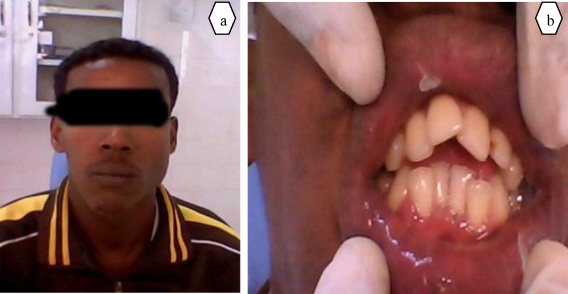
During Intra and Extra oral physical examination, inspection and palpation was done as a result ,firm unilateral tissue was detected over the angle of the mandible ,which became more prominent when the patient clenched his jaws (Fig. 1a) but the opening and closing of the jaws were normal. He has open bite, crowding and Malocclusion [class II division I] during Occlusion (Fig. 1b) and he has dental caries on 37.
Figure 1.
a) Frontal view showing Mandibular left angle prominence; b) Intra oral view showing crowding and open bite.
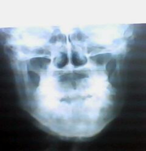
An antero-posterior and lateral oblique radiography was taken to rule out unilateral masseter muscle hypertrophy. The finding showed a compensatory hypertrophy in the area of muscle insertion due to the increase of the muscle size and tension. Prominence of the mandible angle and bone spur development was detected (Fig. 2).
Figure 2.
Antero-posterior radiograph showing spur development in the left angle of the mandible.
Then, extraction of the lower second molar was done and muscle relaxant was prescribed (Diazepam 5mg for five days) and patient reassurance was provided accordingly. Finally, the patient was referred to orthodontic sub-team for treatment of malocclusion. A written informed consent was obtained for case report and disclosure of photographs and radiography for scientific purposes.
Discussion
The masseter, a thick quadrate masticatory muscle, arises from the zygomatic arch and inserts into the inferior lateral aspect and angle area of the mandibular ramus. MH is an asymptomatic persistent enlargement of one or both masseter muscles resulting from a work hypertrophy, initiated by clenching, bruxing, or heavy gum chewing and this occurs primarily in younger patients. In older age groups with dental deterioration, there is an inability to fully activate the masseters and any pre-existing MH tends to recede. Anatomically, most of the masseteric thickness is along the inferior portion of the mandibular ramus, where the facial contour normally tapers. With MH, the patient's face takes on a characteristic rectangular configuration (5,6).
Idiopathic hypertrophy of the masseter muscle is a rare disorder of unknown cause. Some authors associate it with defective teeth, habit of chewing gum, temporo-mandibular joint disorder, congenital and functional hypertrophies, and emotional disorders (stress and nervousness). This case has been chewing on the left side (affected side) since his childhood because he believed that chewing on the right side is out of norm. Most patients complain of the cosmetic change caused by facial asymmetry, also called square face, however, symptoms such as trismus, protrusion and bruxism may also occur (5).
Diagnosis can be produced from clinical examination, directed interview, panoramic x-ray, and muscle palpation. This last diagnostic test consists of palpating the muscle with the fingers while the patient clenches his/her teeth so that the muscle is more prominent during contraction. With the muscle relaxed and the patient's mouth slightly open, extra-oral palpation using both hands will pinpoint the intramuscular location of the hypertrophy. In this case during physical examination the enlargement of masseter muscle was detected and the antro -posterior x-ray shows bone growth at the angle of mandible on the affected side. According to Seltzer and Wang (1987), CT and MRI scans produce excellent images for the diagnosis of various masseter muscle conditions. In our case, the diagnosis was made by palpating the fiber of masseter muscle at the insertion especially at the angle of mandible during clenching. Antro -posterior x-ray view of the patent shows bone growth at the angle of the mandible (2).
The bone spurs at the mandible angle are commonly associated findings and they can be observed in the anteroposterior radiograph (Fig. 3) in this case report. However, Bloem and Hoof stated that approximately 20% of normal people have this finding and that it cannot be considered as diagnostic aid. Guggenheim and Cohen reported that bone spurs are caused by periostal irritation and new bone deposition responding to increased forces exerted by the muscles bundles (7).Idiopathic masseter muscle hypertrophy must be accurately diagnosed, as it may be mistaken for other diseases. Among them are unilateral compensatory hypertrophy (due to hypotrophy or hypoplasia in the contralateral side), masseter tumor, salivary gland disease, parotid tumor, parotid inflammatory disease, and masseter muscle intrinsic myopathy (2,7). Therapy for masseteric enlargement is usually unnecessary, non-surgical modality of treatment include reassurance tranquilizer or muscle relaxant, psychiatric care and injection of very small dose of botulin toxin type A (8).
Excision of the internal layer of the masseter muscle and reduction of the thickened bone in the region of the mandibular angle via intraoral approach is the treatment of choice. Complication from surgical incision of master includes Hematoma, facial nerve paralysis, infection, trismus and sequelae from general anesthesia (9,10).
Injection of botulinum toxin type A in to the masseter muscle is also another alternative as a treatment. Botulinum toxin type A injection is reported to be safe and effective treatment modality in orofacial dystonies, sialorrhea, frey's syndrome, muscle hypertrophies, etc. Botulinum toxin type A, a power full neurotoxin, is produced by the anaerobic organism Clostridium botulinum .When injected in to muscle it causes interference with the neurotransmitter mechanism producing selective paralysis and subsequent atrophy of muscle (11–16).
In conclusion, MMH is a disease with unknown etiology and rare condition. Its diagnosis is mainly via clinical, accompanied with radiographic examination as it helps for the differential diagnosis against other conditions. Though it has no effect if left untreated, for dodging of cosmetic effect surgical treatment is applicable.
References
- 1.Arthur B K. masseter muscle hypertrophy. AMA Arch Derm Syphilol. 1954;69(5):558–562. doi: 10.1001/archderm.1954.01540170028004. [DOI] [PubMed] [Google Scholar]
- 2.Daniel Z R, Paulo MC, José L, Pires JR, Vinicius RF, Mandelli Karina K, Marcela AC. Benign masseter muscle hypertrophy. Rev Bras Otorrinolaringol. 2008;74(5):790–793. doi: 10.1016/S1808-8694(15)31393-8. [DOI] [PMC free article] [PubMed] [Google Scholar]
- 3.Rispoli DZ, Camargo PM, Pires JL, Fonseca VR, Mandelli KK, Pereira MA. Benign masseter muscle hypertrophy. Braz J Otorhinolaryngol. 2008;74(5):790–793. doi: 10.1016/S1808-8694(15)31393-8. [DOI] [PMC free article] [PubMed] [Google Scholar]
- 4.Rocco RA. Masseter muscle hypertrophy: Report of case and literature review. Journal of Oral and Maxillofacial Surgery. 1994;52(11):1199–1202. doi: 10.1016/0278-2391(94)90546-0. [DOI] [PubMed] [Google Scholar]
- 5.Waldhart E. benign hypertrophy of the masseter muscles and mandibular angles. AMA Arch Surg. 1971;102(2):115–118. doi: 10.1001/archsurg.1971.01350020025007. [DOI] [PubMed] [Google Scholar]
- 6.Yu C-C, et al. Botulinum Toxin A for Lower Facial Contouring: A Prospective Study. Aesth Plast Surg. 2007;31:445–451. doi: 10.1007/s00266-007-0081-8. [DOI] [PubMed] [Google Scholar]
- 7.Eduardo KS, Marcelo G, Marcelo PC. Masseter Muscle Hypertrophy - Case Report. Braz Dent J. 2006;17(4):347–350. doi: 10.1590/s0103-64402006000400015. [DOI] [PubMed] [Google Scholar]
- 8.Al-Ahmad HT, Al-Qudah MA. The treatment of masseter hypertrophy with botulinum toxin type A. Saudi Med J. 2006;27:397–400. [PubMed] [Google Scholar]
- 9.Ham JW. Masseter muscle reduction procedure with radiofrequency Coagulation. J Oral Maxillofac Surg. 2009;67:457–463. doi: 10.1016/j.joms.2006.04.012. [DOI] [PubMed] [Google Scholar]
- 10.Roncevic R. Masseter muscle hypertrophy: Aetiology and therapy. Journal of Maxillofacial Surgery. 1986;14:344–348. doi: 10.1016/s0301-0503(86)80322-8. [DOI] [PubMed] [Google Scholar]
- 11.Smyth AG. Botulinum toxin treatment of bilateral masseteric Hypertrophy. Br J Oral Maxillofac Surg. 1994;32:29–33. doi: 10.1016/0266-4356(94)90169-4. [DOI] [PubMed] [Google Scholar]
- 12.Moore AP, Wood GD. The medical management of masseteric hypertrophy with botulinum toxin types A. Br J Oral Maxillofac Surg. 1994;32:26–28. doi: 10.1016/0266-4356(94)90168-6. [DOI] [PubMed] [Google Scholar]
- 13.Maestre-Ferrín L, Burguera JA, Peñarrocha-Diago M, Peñarrocha Diago M. Oromandibular dystonia: a dental approach. Med Oral Patol Oral Cir Bucal. 2009;15:e25–e27. [PubMed] [Google Scholar]
- 14.Luna Ortiz K, Rascon Ortiz M, Sansón Riofrio JA, VillavicencioValencia V, Mosqueda Taylor A. Control of Frey's syndrome in patients treated with botulinum toxin type A. Med Oral Patol Oral Cir Bucal. 2007;12:E79–E84. [PubMed] [Google Scholar]
- 15.Kwon JS, Kim ST, Jeon YM, Choi JH. Effect of botulinum toxin type A injection into human masseter muscle on stimulated parotid saliva flow rate. Int J Oral Maxillofac Surg. 2009;38:316–320. doi: 10.1016/j.ijom.2009.01.008. [DOI] [PubMed] [Google Scholar]
- 16.Al-Muharraqi MA, Fedorowicz Z, Al Bareeq J, Al Bareeq R, Nasser M. Botulinum toxin for masseter hypertrophy. Cochrane Database Syst Rev. 2009;1:CD007510. doi: 10.1002/14651858.CD007510.pub2. [DOI] [PubMed] [Google Scholar]


