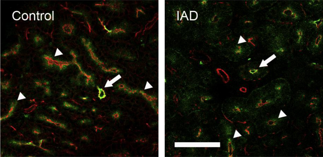
Fig. 6.
Immunofluorescence of CFTR-IR. Control: CFTR-IR (green) was present as punctuate aggregates within the apical cytoplasm of all acinar (arrowheads) and ductal cells (arrow), but the intensity in ductal cells was significantly higher. Rhodamine conjugated phalloidin, which stains F-actin, was used to outline the morphological profile (red). IAD: the distribution pattern of CFTR-IR in LGs from rabbits with IAD, both acinar (arrowheads) and ductal cells (arrow), was similar to those from control animals. Scale bar=50 µm.

