Abstract
Aims:
Evaluation of calcium ion and hydroxyl ion release and pH levels in various calcium hydroxide based intracanal medicaments.
Objective:
The purpose of this study was to evaluate calcium and hydroxyl ion release and pH levels of calcium hydroxide based products, namely, RC Cal, Metapex, calcium hydroxide with distilled water, along with the new gutta-percha points with calcium hydroxide.
Materials and Methods:
The materials were inserted in polyethylene tubes and immersed in deionized water. The pH variation, Ca++ and OH- release were monitored periodically for 1 week. Statistical Analysis Used: Statistical analysis was carried out using one-way analysis of variance and Tukey's post hoc tests with PASW Statistics version 18 software to compare the statistical difference.
Results:
After 1 week, calcium hydroxide with distilled water and RC Cal raised the pH to 12.7 and 11.8, respectively, while a small change was observed for Metapex, calcium hydroxide gutta-percha points. The calcium released after 1 week was 15.36 mg/dL from RC Cal, followed by 13.04, 1.296, 3.064 mg/dL from calcium hydroxide with sterile water, Metapex and calcium hydroxide gutta-percha points, respectively.
Conclusions:
Calcium hydroxide with sterile water and RC Cal pastes liberate significantly more calcium and hydroxyl ions and raise the pH higher than Metapex and calcium hydroxidegutta-percha points.
Keywords: Calcium hydroxide, calcium and hydroxyl ions, intracanal dressing, pH
Introduction
Endodontic treatment is directed toward the prevention and control of pulpal and periradicular infections. The outcome of the endodontic therapy depends on reduction or elimination of microorganisms. Complete chemo-mechanical preparation may be considered an essential step in root canal disinfection. However, total elimination of bacteria from the root canal is difficult to accomplish.[1,2] Hence, by remaining in the root canal in between appointments, intracanal medicaments help to eliminate surviving bacteria.
Since its introduction by Hermann in 1920, calcium hydroxide has been widely used in endodontics. It is a strong alkaline substance, which has a pH of approximately 12.5. In an aqueous solution, calcium hydroxide dissociates into calcium and hydroxyl ions. Various biological properties have been attributed to this substance, such as antimicrobial activity,[3] inhibition of tooth resorption[4] and induction of repair by hard tissue formation.[5] Because of such effects, calcium hydroxide has been recommended for use in several clinical situations.[6,7]
Most of the endodontic pathogens are unable to survive in the highly alkaline environment provided by calcium hydroxide.-[7] Antimicrobial activity of calcium hydroxide is related to the release of hydroxyl ions in an aqueous environment. Hydroxyl ions are highly oxidant free radicals that show extreme reactivity with several biomolecules.[8]
The aim of our study was first to compare the in vitro release of calcium and hydroxyl ionsfrom the various calcium hydroxide preparations used in endodontic treatment and secondly to determine whether their pH altered with time.
Materials and Methods
Materials used in the study were Metapex (META BIOMED, Chungbuk, Korea), RC Cal (Prime Dental products, Mumbai, India), calcium hydroxide points (Coltene Whaledent, Mahwah, NJ, USA), calcium hydroxide powder, distilled water, polyethylene tubes, deionized water, temporary sealer along with buffer solution, color reagent, pH meter, colorimeter and micropipette.
Methods
Calcium hydroxide based substances were divided into four groups:
Group A: Calcium hydroxide powder with distilled water
Group B: RC Cal
Group C: Metapex
Group D: Calcium hydroxide points
Ten-millimetre polyethylene cylindrical tubes were filled with the respective materials of each group. Before this, one end of all the tubes was sealed with a 1-mm layer of temporary sealer. An endodontic file was used to introduce carefully the calcium hydroxide pastes into the tubes through the opening, avoiding bubble formation. RC Cal and Metapex were placed according to manufacturer's instructions. Calcium hydroxide points (#40) were introduced until the tubes were completely filled. The tubes were then immersed in separate test tubes, each with 20 mL of deionized water which was used as the extraction solution. The extraction solutions were maintained at room temperature and without agitation. For each group, five samples were analyzed.
pH analysis
The pH value was measured with a calibrated pH meter. For each group, samples were analyzed after 0, 5, 10, 20 and 40 minutes; and after 1, 2, 8, 16, 24 and 48 hours till 1 week.
Calcium quantification
Calcium ion liberation was measured by a colorimetric method (o-cresolphthalein complexone calcium reaction) at the same time intervals used for pH readings. At each interval, 200 μL of solution was retrieved from the respective extraction solutions. For calcium quantification, 0.5 mL of buffered solution and 0.5 mL of color reagent were added. The cresolphthalein–calcium complex formation was followed by colorimetric method at 570 nm, which is directly proportional to calcium concentration. The linearity of the method was verified through calcium standards up to 25 mg/dL.
Analysis of liberated hydroxyl Ions
From the calculations of the calcium ions liberated, it was also possible to determine the amount of hydroxyl ions liberated. The molecular weight of 2 mol hydroxyl ion is 34, and the molecular weight of 1 mol calcium ion is 40.08, and therefore the molecular weight of the complete molecule is 74.08. The percent of the two in the total weight can be simply calculated to be 45.89% and 54.11%, respectively. Therefore, in 1 mol of calcium hydroxide, there are 45.89% hydroxyl ions and 54.11% calcium ions. After determining the quantity of liberated calcium ions, the quantity of hydroxyl ions can be calculated as they are in direct proportion.
Statistical analysis
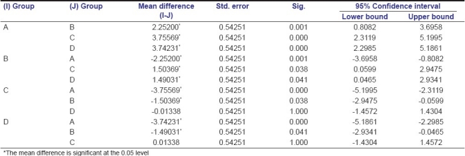
Statistical analysis was carried out using one-way analysis of variance (ANOVA) and Tukey's honestly significant difference (HSD) post hoc tests with PASW Statistics version 18 to compare the statistical difference [Tables 1, 2, 3].
Table 1.
Results of Tukey's HSD post hoc test for calcium ions released
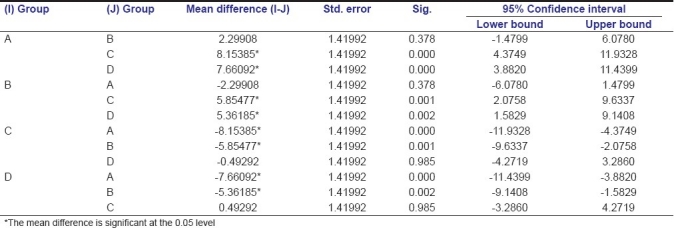
Table 2.
Results of Tukey HSD post- hoc test for hydroxylions released
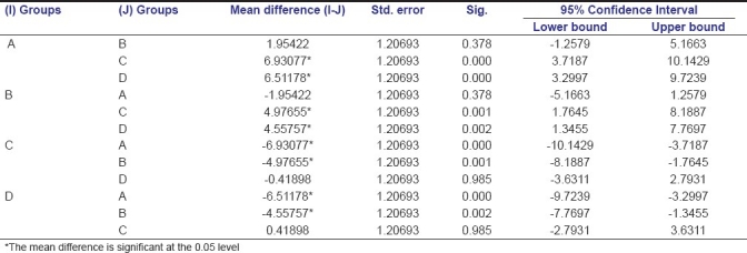
Table 3.
Results of Tukey's HSD post hoc test for pH levels
Results
Calcium ion release
The measurement of calcium release over 1 week was similar for calcium hydroxide powder with distilled water and RC Cal, with logarithmic release during the first 16 hours.
The delivery of RC Cal was constant for each interval, whereas calcium hydroxide powder with distilled water showed greater amounts of release in a short period, and a time-dependent increase of delivery was observed. Calcium hydroxide powder with distilled water released more calcium ions than other groups in the first hour (P > 0.01). Calcium hydroxide powder with distilled water and RC Cal showed a significant increase after 2 hours, reaching a maximum level after 16 hours and 1 week, respectively, followed by calcium hydroxide points [Figure 1].
Figure 1.
Colorimetric calcium determination of different intracanal dressings
Hydroxyl ion release
Hydroxyl ion release was calculated from the calcium ion release which was measured. The calculated percent of liberation of hydroxyl ions was thus 84.80% of the liberated calcium ions. The order of release of hydroxyl ions was RC Cal, where it was the highest, followed by calcium hydroxide powder with distilled water and calcium hydroxide points, and Metapex was the last [Figure 2].
Figure 2.
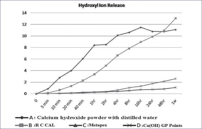
Hydroxyl ion determination of different intracanal dressings
pH measurement
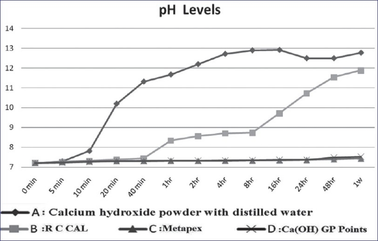
The pH of the samples increased gradually, reaching maximum values at 1 week, but calcium hydroxide powder with distilled water reached its maximum value after 8 hours. A positive correlation between pH and time was found for all materials. The highest was for calcium hydroxide powder with distilled water and RC Cal, and the lowest was for calcium hydroxide points followed by Metapex. The correlation between the pH values of almost all the test materials and time and the correlation between pH values in particular samples were statistically significant (P < 0.01) [Figure 3].
Figure 3.

Time course of pH variations using different intracanal dressings
Discussion
The therapeutic effect of calcium hydroxide is due to its ability to break down into calcium and hydroxyl ions. Hydroxyl ions show an affinity to various biologically active substances.[9] Their antimicrobial activity is important since microbes cause the majority of endodontic diseases. The antibacterial activity of hydroxyl ion is related to the formation of a potent alkaline medium leading to the destruction of lipids, the main component of bacterial cell membrane and causing structural damage to bacterial proteins and nucleic acids.[10]
Calcium hydroxide, apart from its bacterial enzymatic inhibition which represents an important antibacterial property, has the capability of activating tissue enzymes which favour tissue restoration through mineralization.[11]
The elevated pH of calcium hydroxide activates alkaline phosphatase. The best pH for the activation of this enzyme, varying with the type and concentration of substratum, the temperature, and the source of enzymes, ranges from 8.6 to 10.3.[12,13] Alkaline phosphatase is a hydrolytic enzyme that acts by liberation of inorganic phosphate from the esters of phosphate. It is believed to be intimately related to the process of mineralization.[14] This enzyme can separate the phosphoric esters, freeing phosphate ions which, once free, react with calcium ions from the blood stream to form a precipitate, calcium phosphate, in the organic matrix. This precipitate is the molecular unit of hydroxyapatite.[15]
Calcium hydroxide in direct contact with conjunctive tissue is the origin to a zone of necrosis, altering the physio-chemical state of intercellular substance which, through rupture of glycoprotein, seems to determine protein denaturation.[11] The formation of mineralized tissue after contact of calcium hydroxide with conjunctive tissue has been observed from about the 7th day to the 10th day.[11,12]
Time seems to be a vital factor in relation to the effective therapeutic action of calcium hydroxide preparations. In the present study, the pH of all the samples increased gradually with time, reaching the highest values on the 8th day. Zmener et al. tested the pH changes that occurred over a period of 30 days using a mixture of calcium hydroxide and distilled water and two commercial calcium hydroxide products in a simulated periapical environment. They found that there was a rapid increase in the pH at l hour and 24 hours, followed by continuous but more gradual increase from 15 to 30 days.[16]
Many substances have been added to the calcium hydroxide powder to improve its properties such as the antibacterial action, radiopacity, flow, and consistency. The ideal vehicle should allow a gradual and slow release of calcium and hydroxide ions. The vehicles suggested can be classified as aqueous and oily. Safavi et al. studied the effect of mixing vehicle on dissociation of calcium hydroxide in solution. They measured the conductivity values for saturated solutions of calcium hydroxide in water and in pure glycerine or propylene glycol. They found that the use of non-aqueous mixing vehicles may impede the effectiveness of calcium hydroxide as a root canal dressing.[17] In this study, we have used both aqueous and oily vehicles, along with a new type which was calcium hydroxide gutta-percha points.
Along with calcium hydroxide distilled water, we have also used RC Cal, which is water-based radiopaque calcium hydroxide paste with barium sulfate. Estrela et al.[18] described that liberation of calcium and hydroxide ions was faster and more significant when used as calcium hydroxide distilled water paste.[18] The diffusion of hydroxyl ions through dentin from different calcium hydroxide medicaments was determined by Sevimay et al.[19] They found that non-setting calcium hydroxide based materials have an effective release of hydroxyl ions compared with calcium hydroxide plus points. The results of present study are comparable to those observed in previous studies.
A gutta-percha point is a new device for calcium hydroxide delivery for intracanal dressing. Advantages include easy insertion and removal and fewer residues. Economides et al. evaluated the release of hydroxyl ions from calcium hydroxide gutta-percha points and found that calcium hydroxide containing gutta-percha points showed a significantly lower alkalinizing potential than the non-setting preparation and calcium hydroxide mixed with distilled water.[20] In Roeko points, gutta-percha matrix probably binds the hydroxyl ions and blocks their release at the site of application. In our study, the pH was not significantly increased by gutta-percha point. Economides et al. and Azabal-Arroyo et al. reported maximum pH values of 9.5 and 10.9, respectively.[21] Calt et al. showed that calcium hydroxide gutta-percha points did not induce any changes in pH and calcium ion levels.[22] Larsen and Horsted-Bindslev concluded that hydroxyl ion liberation from the gutta-percha points is limited compared to that from calcium hydroxide pastes. These results are comparable to those of our study. Lohbauer et al. evaluated calcium ion release and pH characteristics of calcium hydroxide plus points, conventional calcium hydroxide points and aqueous calcium hydroxide suspension. They found that calcium hydroxide plus points and conventional calcium hydroxide points increased the pH to more than 11 within 3 minutes. Calcium hydroxide plus points had a greater release of calcium ions compared to conventional calcium hydroxide points.[23]
Metapex contains calcium hydroxide with iodoform in silicon oil. It was observed that oil paste containing calcium hydroxide was largely lacking in both ion release and antimicrobial properties.Larsen and Bindslev observed that the aqueous suspension exhibited the highest pH and calcium ion liberation.[24] The low solubility and poor ability to diffuse make it difficult for oil paste containing calcium hydroxide compounds to reach maximum pH levels in a short period of time. The results of our in vitro studies show that they should be used for a minimum of 7 days to achieve maximum therapeutic effectiveness.
In accordance with the levels of pH, hydroxyl ion and calcium ion release observed in this study, the materials used can be ranked in the following order: 1) calcium hydroxide with sterile water 2) RC Cal pastes 3) calcium hydroxide gutta-percha points and 4) Metapex.
The present in vitro study suggests that to achieve maximum concentration of calcium and hydroxyl ions, two issues should be contemplated:
Aqueous based preparations of calcium hydroxide should be chosen rather than points or oil-based calcium hydroxide preparations.
The use of oil-based calcium hydroxide preparations and points should be considered in an in vivo study model as the current study model fails to simulate the clinical environment.
Conclusion
Calcium hydroxide with sterile water and RC Cal pastes showed a significant increase in pH, hydroxyl ion and calcium ion release and this increase was superior to that shown by Metapex and calcium hydroxide gutta-percha points.
Footnotes
Source of Support: Nil.
Conflict of Interest: None declared.
References
- 1.BystroÈm A, Sundqvist G. Bacteriologic evaluation of the efficacy of mechanical root canal instrumentation in endodontic therapy. Scand J Dent Res. 1981;89:321–8. doi: 10.1111/j.1600-0722.1981.tb01689.x. [DOI] [PubMed] [Google Scholar]
- 2.Siqueira JF, Jr, Goncalves RB. Antibacterial activities of root canal sealers against selected anaerobic bacteria. JOE. 1996;22:89–90. [Google Scholar]
- 3.Bystrom A, Sundqvist G. The antibacterial action of sodium hypochlorite and EDTA in 60 cases of endodontic therapy. Int Endod Jr. 1985;18:35–40. doi: 10.1111/j.1365-2591.1985.tb00416.x. [DOI] [PubMed] [Google Scholar]
- 4.Tronstad L. Root resorption etiology, terminology and clinical manifestations. Endod Dent Traumatol. 1988;4:241–52. doi: 10.1111/j.1600-9657.1988.tb00642.x. [DOI] [PubMed] [Google Scholar]
- 5.Foreman PC, Barnes F. A review of calcium hydroxide. Int Endod J. 1990;23:283–97. doi: 10.1111/j.1365-2591.1990.tb00108.x. [DOI] [PubMed] [Google Scholar]
- 6.Fava LR. Pastas de hidroxido de calcio.Consideraoes sobre seu emprego clõnico. Revista Paulista de Odontologia. 1991;13:36–43. [Google Scholar]
- 7.Heithersay GS. Calcium hydroxide in the treatment of pulpless teeth with associated pathology. J Br Endod Soc. 1975;8:74–92. doi: 10.1111/j.1365-2591.1975.tb01000.x. [DOI] [PubMed] [Google Scholar]
- 8.Freeman BA, Crapo JD. Biology of disease: Free radicals and tissue injury. Lab Investig. 1982;47:412–24. [PubMed] [Google Scholar]
- 9.Siqueira JF, Lopes HP. Mechanisms of antimicrobial activity of calcium hydroxide: A critical review. Int Endod J. 1999;32:361–9. doi: 10.1046/j.1365-2591.1999.00275.x. [DOI] [PubMed] [Google Scholar]
- 10.Tamburic SD, Vuleta GM, Ognjanovic JM. In vitro release of calcium and hydroxyl ions from two types of calcium hydroxide preparations. Int Endod J. 1993;26:125–30. doi: 10.1111/j.1365-2591.1993.tb00554.x. [DOI] [PubMed] [Google Scholar]
- 11.Carlos, Blitzkow G, Luescke L, Oswaldo Mechanism of action of calcium and hydroxyl ions of calcium hydroxide on tissue and bacteria. Braz Dent J. 1995;6:85–90. [PubMed] [Google Scholar]
- 12.Binnie WH, Mitchell DF. Induced calcification in the subdermal tissues of the rat. J Dent Res. 1973;52:1087–91. doi: 10.1177/00220345730520051701. [DOI] [PubMed] [Google Scholar]
- 13.Tronstad L, Andreassen JO, Haselgreen G, Kristerson L, Riis I. pH changes in dental tissues after root canal filling with calcium hydroxide. J Endod. 1981;7:17–21. doi: 10.1016/S0099-2399(81)80262-2. [DOI] [PubMed] [Google Scholar]
- 14.Granstrom G, Linde A. A biochemical study of alkaline phosphatase in isolated rat incisor odontoblast. Arch Oral Biol. 1972;17:1213–24. doi: 10.1016/0003-9969(72)90092-1. [DOI] [PubMed] [Google Scholar]
- 15.Seltzer S, Bender IB. Philadelphia: Ishiyaku Euro America Inc; 1979. The dental pulp. [Google Scholar]
- 16.Zmener O, Pameijer CH, Banegas G. An in vitro study of the pH of three calcium hydroxide dressing materials. Dent Traumatol. 2007;23:21–5. doi: 10.1111/j.1600-9657.2005.00447.x. [DOI] [PubMed] [Google Scholar]
- 17.Safavi K, Nakayama TA. Influence of mixing vehicle on dissociation of calcium hydroxide in solution. J Endod. 2000;26:649–52. doi: 10.1097/00004770-200011000-00004. [DOI] [PubMed] [Google Scholar]
- 18.Estrela C. Chemical analysis of the liberation of calcium and hydroxyl ions from calcium hydroxide pastes in connective tissue in the dog. Braz Dent J. 1996;7:41–6. [PubMed] [Google Scholar]
- 19.Sevimay S, Kalayci A, Yelmaz S. In vitro diffusion of hydroxyl ions through root dentin from various calcium hydroxide medicaments. J Oral Rehabil. 2003;30:1047–51. doi: 10.1046/j.1365-2842.2003.01081.x. [DOI] [PubMed] [Google Scholar]
- 20.Economides N, Koulaouzidou EA, Beltes P, Kortsaris AH. In vitro release of hydroxyl ions from calcium hydroxide gutta-percha points. J Endod. 1999;23:481–2. doi: 10.1016/S0099-2399(99)80285-4. [DOI] [PubMed] [Google Scholar]
- 21.Azabal-Arroyo M, Menasalvas-Ruiz G, Martín-Alonso J, Arroquia JJ, Vega-del Barrio JM. Loss of hydroxyl ions fromgutta-percha points with calcium hydroxide in their composition: An in vivo study. J Endod. 2002;28:697–8. doi: 10.1097/00004770-200210000-00005. [DOI] [PubMed] [Google Scholar]
- 22.Calt S, Serper A. Time dependant effects of EDTA on dentin structures. J Endod. 2002;28:17–9. doi: 10.1097/00004770-200201000-00004. [DOI] [PubMed] [Google Scholar]
- 23.Lohbauer U, Gambarini G, Ebert J, Dasch W, Petschelt A. Calcium release and pH characteristics of calcium hydroxide plus points. Int Endod J. 2005;38:683–9. doi: 10.1111/j.1365-2591.2005.00972.x. [DOI] [PubMed] [Google Scholar]
- 24.Larsen MJ, Horsted-Bindslev P. A laboratory study evaluating the release of hydroxyl ions from various calcium hydroxide products in narrow canal-like tubes. Int Endod J. 2000;33:238–42. doi: 10.1046/j.1365-2591.2000.00279.x. [DOI] [PubMed] [Google Scholar]


