Abstract
Papillon–Lefevre syndrome (PLS) is a rare disease characterized by skin lesions, which includes palmar-plantar hyperkeratosis and hyperhidrosis with severe periodontal destruction involving both the primary and the permanent dentitions. It is transmitted as an autosomal-recessive condition, and consanguinity of parents is evident in about one-third of the cases. This paper describes a 13-year-old male patient who presented to the department of pedodontics, with rapidly progressing periodontitis. A general physical examination revealed scaling on the hands and feet, which had been medically diagnosed as PLS. The incidence of this rare entity is increasing in the recent times, which is associated with irreparable periodontal destruction at an early age, with not so prominent skin lesions in some cases. In such instances, the dentist has a more important role in diagnosing, treatment planning and preservation of the periodontal tissues and, at the same time, referring for the treatment of the skin lesions. This paper emphasizes the combined effort of the two specialities in order to maintain skin as well as dental conditions in health by early intervention and a synergistic treatment approach.
Keywords: Hyperkeratosis, periodontosis, Papillon–Lefevre
Introduction
Papillon–Lefevre syndrome (PLS), also termed as juvenile (precocious) periodontosis with palmar plantar hyperkeratosis, was first described in the literature by two French physicians Papillon and Lefevre in 1924.[1,2] Till date, more than 200 cases have subsequently been reported.[1] The syndrome is a rare autosomal-recessive trait, with an incidence between one and four per million.[1] Parental consanguinity is demonstrated in between 20% and 40% of the cases.[1,3] Papillon–Lefevre is characterized by hyperkeratosis of the palms and soles combined with precocious periodontal destruction and shedding of the deciduous and permanent dentitions.[2] The etiology of the syndrome is not clearly understood, and was found to be related to generalized epithelial dysplasia or to be an endocrinopathy, suggesting the possibility of vitamin A deficiency.[4] Recently, two new aspects of Papillon–Lefevre have been discovered: the deep sub-gingival flora is composed of a great number of motile gram negative anaerobic rods, including Bacteroides gingivalis, Capnocytophaga and a number of spirochetes. Secondly, some PLS patients have exhibited a cellular immune defect with a deficient chemotactic and phagocytic function of the neutrophilic granulocytes.[4] The syndrome is easily misdiagnosed at initial presentation because the skin lesions can be mistaken for eczema.[1] Skin lesions are usually present from 6 months to 3 years of age, approximating the time of tooth eruption. There can be occasional involvement of the eyelids, cheeks, labial commissures, knees, elbows, thighs, external malleoli, toes and dorsal fingers.
Periodontal effects appear almost immediately after tooth eruption, when the gingiva becomes erythematous and edematous. Plaque accumulates in the deep crevices and halitosis can ensue. The primary incisors are usually affected first, and can display marked mobility by the age of 3 years. By the age of 4 or 5 years, all the primary teeth may have exfoliated. Following such tooth loss, the gingival appearance resolves and may well return to health only for the process to be repeated as the permanent dentition starts to erupt. The majority of the teeth are lost by the age of 14–15 years. There is dramatic alveolar bone destruction, often leaving atrophied jaws.[1] Resorption of the alveolar bone gives the teeth a “floating-in-air” appearance on dental radiographs.[5]
Case Report
A 13-year-old male patient has visited the Department of Pedodontics, with a chief complaint of loosening teeth and discomfort on eating. General physical examination revealed symmetrical, well-demarcated hyperkeratotic plaques of the palms of the hands [Figure 1] and soles, which extended onto the dorsal surface of the feet and hands [Figure 2]. An intraoral examination revealed edentulous ridges in the maxillary and mandibular anterior regions due to loss of permanent incisors [Figure 3]. Oral hygiene was poor, with significant plaque accumulation and a gingival swelling associated with the maxillary left first permanent molar. All teeth showed mobility, with periodontal pockets, bleeding on probing and gingival recession in the region of the mandibular canine region. Past dental history revealed that since childhood the teeth exfoliated one by one by the age of 7 years. In the present case, the family history was not significant.
Figure 1.
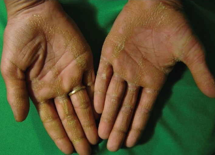
Keratotic plaques on the palms extending over the dorsal surface of the finger joints of the hands
Figure 2.
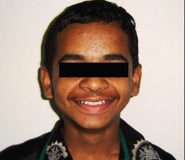
Post-treatment photograph of the patient with a removable partial prosthesis
Figure 3.
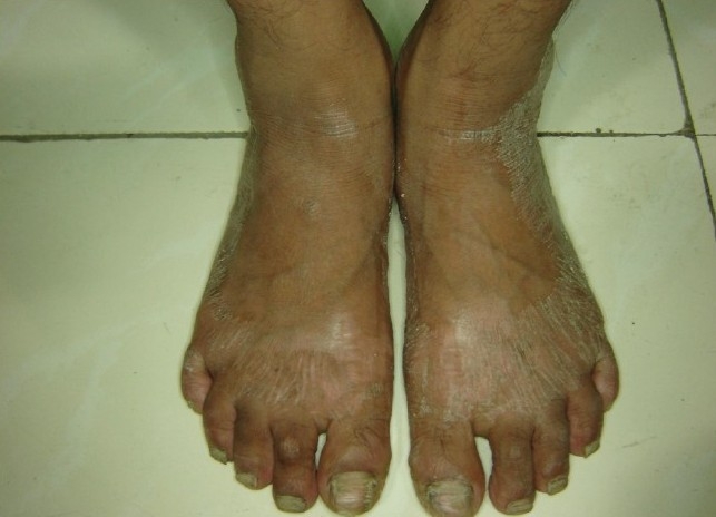
Keratotic plaques involving the entire plantar surface of the feet, with extension of the keratotic plaques over the skin overlying the dorsal aspect of the feet. The skin appears dry, scaly and rough
Radiographic examination (orthopantomogram) confirmed the presence of generalized destruction of the alveolar bone around the permanent dentition, giving a floating in air appearance of the teeth [Figure 4].
Figure 4.
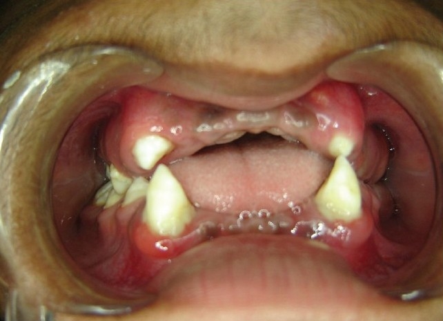
Intraoral photograph showing missing permanent teeth anteriorly in the upper and lower arch
In view of the above findings, the case was diagnosed as PLS. Dental treatment done for this patient included oral prophylaxis, extraction of teeth with poor prognosis followed by replacement with removable prosthesis [Figure 5]. Consultation of a dermatologist was taken for evaluation of hyperkeratosis of the palmar and plantar surfaces.
Figure 5.
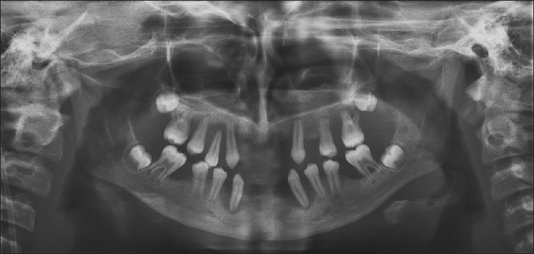
Orthopantogram showing a “floating-in-air” appearance of the teeth
Discussion
PLS is a disorder of keratinization,[6] noticed by hyperkeratosis of palms and soles with severe destructive periodontitis affecting both the deciduous and the permanent dentitions. Some other findings of PLS cases have shown inconsistent manifestations, like calcification of the falx cerebri and choroid plexus, calcification of the dura, attachment of the tentorium, pyogenic infections, nail dystrophy and generalized hyperhidrosis.[6] In our case, the symptoms, clinical findings and past dental history bore resemblance to the classical syndrome. Clinically, the patient showed typical characteristic hyperkeratotic skin lesions of varied thickness on the palmar and plantar surfaces, giving a rough, dry appearance. Secondly, intraoral findings showed missing teeth, mobility of present teeth and characteristic features of periodontosis described for the PLS. Moreover, our patient was also suffering from hyperhidrosis and nail dystrophy.
The underlying cause for PLS is not well understood, but is thought to be a genetically transmitted trait, suggesting a multigenic etiology for PLS.[3] Recent studies have suggested a greater incidence of mutations of the cathepsin-C gene located on the 11q14-q21 region of the chromosome encoding a lysosomal protease in the interval between D11S4082 and D11S931.[1,7] It is expressed at high levels in various immune cells, including polymorphonuclear leukocytes, macrophages and their precursors.[8] The cathepsin C gene is expressed in the epithelial region (palms, soles, knees) and keratinized oral gingiva which is important in the structural growth and development of skin. Subsequently, inactivating mutations were identified in this gene and an almost total loss of cathepsin C activity was shown in patients with PLS.[8–10] Impaired function of cathepsin C gene in epithelial regions can lead to skin abnormalities like thickening on the palms and soles of the feet. Loss of function of the cathepsin C gene results in altered immune response to infection, which may effect the integrity of the junctional epithelium surrounding the tooth. Moreover, this leads to failure of clearance of bacteria from the gingiva, leading to destructive periodontosis and, ultimately, tooth loss. Despite these advances in characterization of the genetic basis of the syndrome, the pathogenetic mechanisms leading to periodontal involvement remain elusive.[11] Microbiological studies have demonstrated a greater prevalence of Actinobacillus actinomycetemcomitans in patients with PLS, and these findings have suggested that it has a significant role in the pathogenesis and progression of rapid periodontal breakdown.[1]
Hiam-Munk syndrome is similar to PLS with respect to prepubertal periodontitis and palmo-plantar hyperkeratosis, but sufferers also exhibit arachnodactly, acroosteolysis and deformity of the phalanges of the hand.[1] When there is premature loss of the deciduous and/or permanent teeth, one should also consider Acrodynia, which is also known as Feer's syndrome. This condition is usually caused by mercury intoxication. Here, one may observe a red desquamative process involving both the extremities. But, in addition, there is erythocyanosis, muscle pain, insomnia, sweating, tachycardia and psychic disturbances. The teeth erupt prematurely, have dystrophic enamel and are shed prematurely. Acrodynia is seen in children between the ages of 6 months and 4 years.[12,13]
Another condition of interest in differential diagnosis is hypophosphatasia. In addition to the clinical features of knock-knee, bowing of femur and tibia and enlarged wrists, the teeth are prematurely shed and are hypoplastic. Diagnosis can be made on the basis of increased amounts of phosphoethanolamine in the urine.[10–11] Another condition characterized by progressive gangrenous lesions involving the gingiva and alveolar bone, resulting in exfoliation of the teeth, is Acatalasemia or Takahara's syndrome. This is also transmitted as an autosomal-recessive trait, but has rarely been observed outside Japan.[12,13] In cyclic neutropenia, severe periodontal disease may also be present, but palmoplantar hyperkeratosis is absent.[12–14] Other conditions to be considered in the differential diagnosis of PLS are palmoplantar hyperkeratosis of Unna Thost, mal de Meleda, Howel-Evans syndrome, keratosis punctata, keratoderma hereditarium mutilans (Vohwinkel's syndrome) and Greither's syndrome. However, while these entities are associated with palmoplantar hyperkeratosis, there is no periodontopathy.[12,14] A multidisciplinary approach is important for the care of patients with PLS. The skin manifestations of PLS are usually treated with emollients. Oral retinoids such as acitretin, etretinate, and isotretinoin were reported to be beneficial for both dental and cutaneous lesions of PLS. However, isotretinoin has to be used carefully as the teratogenic effects have been evident.[15] About 40% of the infants exposed to isotretinoin in the first trimester will have major malformations.[16] In February 2004, the Food and Drug Administration (FDA) advisory committee recommended that in view of continuing safety issues, a mandatory registration for isotretinoin users and prescribers should be implemented.[17] Retinoid treatment may end up with normal dental development if started during the eruption of permanent teeth.[1] To control periodontal destruction, several treatment modalities have been suggested: conventional periodontal therapy, oral hygiene instructions and systemic antibiotics.[18] In conclusion, the Pedodontist and Dermatologist are the first members of the health team to see this interesting and challenging diagnostic problem, and must differentiate it from other entities on the basis of the unique clinical findings. Dermatologists have a key role in diagnosing this syndrome as well as in educating the patient about the importance of preservation of the dental tissues and referring the patient to a dentist. A synergistic treatment plan from the two specialities at an early age can help the patient to maintain skin as well as dental conditions in health.
Footnotes
Source of support: Nil
Conflict of Interest: Nil.
References
- 1.Patel S, Davidson LE. Papillon–Lefevre syndrome: A report of two cases. Int J Paediatr Dent. 2004;14:288–94. doi: 10.1111/j.1365-263X.2004.00559.x. [DOI] [PubMed] [Google Scholar]
- 2.Mahajan VK, Thakur NS, Sharma NL. Papillon–Lefevre syndrome. Indian Pediatr. 2003;40:1197–200. [PubMed] [Google Scholar]
- 3.Gorlin RJ, Cohen MM, Levin LS. Syndromes of the Head and Neck. 3rd ed. Oxford: Oxford University Press; 1990. pp. 853–5. [Google Scholar]
- 4.Deshpande N, Nayak AU, Sudha P. Periodontal diseases. In: Tandon S, editor. Textbook of Pedodontics. 2nd ed. Hyderabad: Paras Medical Publishers; 2008. pp. 77–80. [Google Scholar]
- 5.Nagaveni, Suma, Shashikiran, reddy Subba. Papillon lefevre syndrome-report of two cases in the same family. J Indian soc pedod prev dent. 2008;26:78–81. doi: 10.4103/0970-4388.41622. [DOI] [PubMed] [Google Scholar]
- 6.Dhadka SV, Kulkarni PM, Dhadke VN, Deshpande NS, Wattamwar PR. Papillon Lefevre Syndrome. Case report. J Assoc Physicians India. 2006;54:246–7. [PubMed] [Google Scholar]
- 7.Thakker N. Genetic analysis of Papillon –Lefevre syndrome. Oral Dis. 2000;6:263. doi: 10.1111/j.1601-0825.2000.tb00135.x. [DOI] [PubMed] [Google Scholar]
- 8.Angel TA, Hsu S, Kornbleuth SI, Kornbleuth J, Kramer EM. Papillon–Lefevre syndrome: A case report of four affected siblings. J Am Acad Dermatol. 2002;46:S8–10. doi: 10.1067/mjd.2002.104968. [DOI] [PubMed] [Google Scholar]
- 9.Allende LM, Moreno A, de Unamuno P. A genetic study of cathepsin C gene in two families with Papillon-Lefèvre syndrome. Mol Genet Metab. 2003;79:146–8. doi: 10.1016/s1096-7192(03)00070-2. [DOI] [PubMed] [Google Scholar]
- 10.Pilger U, Hennies HC, Truschnegg A, Aberer E. Late-onset Papillon-Lefèvre syndrome without alteration of the cathepsin C gene. J Am Acad Dermatol. 2003;49:S240–3. doi: 10.1016/s0190-9622(03)01558-5. [DOI] [PubMed] [Google Scholar]
- 11.Lundgren T, Parhar RS, Renvert S, Tatakis DN. Impaired Cytotoxicity in Papillon-Lefèvre Syndrome. J Dent Res. 2005;84:414–7. doi: 10.1177/154405910508400503. [DOI] [PubMed] [Google Scholar]
- 12.Singla A, Sheikh S, Jindal SK, Brar R. Papillon Lefevre syndrome: Bridge between Dermatologist and Dentist. J Clin Exp Dent. 2010;2:e43–6. [Google Scholar]
- 13.Ashri NY. Early diagnosis and treatment options for the periodontal problems in Papillon-Lefèvre syndrome: A literature review. J Int Acad Periodontol. 2008;10:81–6. [PubMed] [Google Scholar]
- 14.Hattab FN, Amin WM. Papillon-Lefèvre syndrome with albinism: A review of the literature and report of 2 brothers. Oral Surg Oral Med Oral Pathol Oral Radiol Endod. 2005;100:709–16. doi: 10.1016/j.tripleo.2004.08.030. [DOI] [PubMed] [Google Scholar]
- 15.Adverse effect of isotretenoin. FDA Drug Bull. 1983;13:21–2. [PubMed] [Google Scholar]
- 16.Korean G, Avner M, Shear N. Generic isotretinoin: A new risk for unborn children. CMAJ. 2004;170:1567–8. doi: 10.1503/cmaj.1040317. [DOI] [PMC free article] [PubMed] [Google Scholar]
- 17.Elliott VS. FDA panel back patient registry for acne drug. AM News. 2004. Mar 22/29, [Last accessed on 2004 Apr 17]. Available from: http://www.ama assn.org/amednews/2004/03/22/hlsc0322.htm#w1 .
- 18.Shah J, Goel S. Papillon–Lefevre syndrome - two case reports. Indian J Dent Res. 2007;18:210–3. doi: 10.4103/0970-9290.35834. [DOI] [PubMed] [Google Scholar]


