Abstract
A primary advantage of functional magnetic resonance imaging (fMRI) over other techniques in neuroscience is its flexibility. Researchers have used fMRI to study a remarkable diversity of topics, from basic processes of perception and memory, to the complex mechanisms of economic decision making and moral cognition. The chief contributor to this experimental flexibility – indeed, to the growth of fMRI itself – has been the development of event-related experimental designs and associated analyses. The core idea of an event-related design, as first articulated in the late 1990s, is the separation of cognitive processes into discrete points in time (i.e., “events”) allowing differentiation of their associated fMRI signals. By modeling brain function as a series of transient changes, rather than as an ongoing state, event-related fMRI allowed researchers to create much more complex paradigms and more dynamic analysis methods. Yet, this flexibility came with a cost. As the complexity of experimental designs increased, fMRI analyses became increasingly abstracted from the original data, which in turn has had consequences both positive (e.g., greater use of model-based fMRI) and negative (e.g., fewer articles plot the timing of activation). And, as event-related methods have become ubiquitous, they no longer represent a distinct category of fMRI research. In a real sense, event-related fMRI has now become, simply, fMRI.
Keywords: fMRI, event-related, design, analysis, neuroimaging, hemodynamic
Introduction
No other advance – not stronger magnetic fields, not improved pulse sequences, nor even sophisticated new analyses – has contributed more to the popularization of fMRI than event-related approaches to experimental design. Broadly considered, “event-related fMRI” involves separating the elements of an experiment into discrete points in time, so that the cognitive processes (and associated brain responses) associated with each element can be analyzed independently (Huettel et al., 2009). A typical event-related approach to studying decision making, for example, presents a different pair of options on each trial (e.g., between safer and riskier options); whereupon the participant indicates their choice via a button press. By isolating the fMRI activation associated with these different events (e.g., stimuli and responses), researchers have characterized how brain regions contribute to individual processes within decision making (Huettel et al., 2006; O’Doherty et al., 2003). A remarkable variety of event-related experiments has arisen: the infrequently presented targets of the oddball task (McCarthy et al., 1997), the sorting of stimuli into remembered and forgotten categories in the subsequent-memory paradigm (Brewer et al., 1998; Wagner et al., 1998), the cue-target pairs of selective attention studies (Hopfinger et al., 2000), among many others. Using these and other approaches, researchers gained information about brain function not accessible using older “blocked” designs.
Why was event-related fMRI so influential? In retrospect, it seems an obvious step – and even relatively minor compared to the many technical developments over the first two decades of fMRI. Its effects, however, have been anything but obvious and minor. In a very deep sense, event-related designs have become inextricably intertwined with fMRI itself. To understand why, consider what makes fMRI such a powerful technique within modern neuroscience. The core advantage of fMRI does not come from the nature of its data – many other techniques provide more direct measures of brain function – but from the flexibility of its experiments. A wider array of experimental designs has been used with fMRI than with all other neuroscience techniques combined. Conversely, the core challenge of fMRI no longer involves data collection itself, given the straightforwardness of modern scanners. Instead, fMRI remains a challenging technique because of the complexity of the analyses it allows – largely because of event-related designs.
Where do we come from? Antecedents of Event-Related fMRI
When one first encounters fMRI, the complexity of its technology can be overwhelming. The very idea of peering into the working human brain seems magical… save perhaps to the jaded neuroscientist! To be sure, modern MR scanners incorporate a host of advances in hardware design, engineering, and signal processing – all now cloaked within their plastic casings and buried under their polished interfaces. That MR data collection works so transparently reflects one of the triumphs of recent biomedical science. It also has made MR data trivially simple to collect. At our center, like many others, undergraduate students in neuroscience run fMRI experiments as a part of their class exercises. As the students stare at the scanner console much like engineering students in front of their oscilloscopes, they watch a real-time measure of primary motor cortex activation rise and fall with the rhythmic finger tapping of the experimental participant. Yet, the same technique that can be appreciated by naïve students on their first day of class also can bedevil neuroscientists with a decade of experience – especially when they struggle with the many ways to analyze their latest study.
Early blocked-design experiments
Experiments conducted during the first few years of fMRI tended to use simple blocked designs. A prototypical design would alternate extended blocks of the experimental task (e.g., tapping the index finger for 30s) with similarly long blocks of a control task (e.g., resting for 30s). As discussed elsewhere in this special issue, the several groups simultaneously implementing fMRI for functional studies (Bandettini et al., 1992; Kwong et al., 1992) converged on this design because of a confluence of several factors.
First, blocked designs were already prevalent within the main neuroscience techniques of that time. Positron Emission Tomography (PET) measures the radioactive decay of an injected isotope whose density in time provides an estimate of ongoing metabolism throughout the brain. These decay events arise from a stochastic and slow process following each injection, and thus blocked designs were (and remain) necessary. Even electroencephalography (EEG), which tracks ongoing and high-frequency oscillatory activity, was historically used to provide a rough measure of the overall state of the brain during some time period. Second, at the outset of fMRI experimentation, very little was known about its signal-to-noise properties. Researchers anticipated that the endogenous blood-oxygenation-level-dependent (BOLD) signal would have relatively low amplitude, compared to background noise. Aggregating events into blocks promised to maximize the associated hemodynamic changes, thus providing early researchers with the largest possible signals for measurement (up to 2–5% in early studies of sensory or motor cortices). Third, very little was known about how to analyze fMRI data. No other neuroscience technique provided such a complex four-dimensional dataset. Nor was there a clear model for estimating the underlying neuronal activity from the hemodynamic response it evoked. Comparison of the BOLD signal between two blocks provided a coarse estimate of the overall effect of an experimental manipulation – just as similar subtraction analyses shaped early PET research, and even early psychological research a century before.
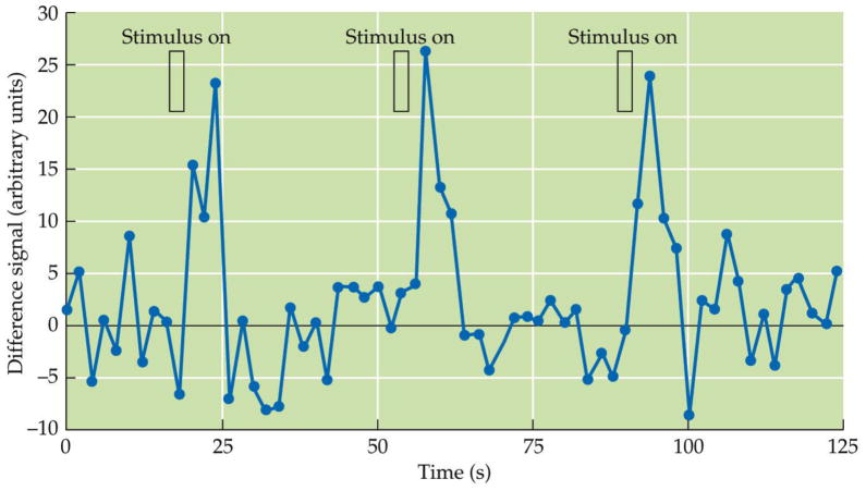
Even from the earliest days of fMRI, however, it was clear that its promise could not be fulfilled solely through traditional blocked designs. In the first, striking example of an event-related fMRI study, Blamire and colleagues presented flashing checkerboard stimuli for varying durations, from 1s to 45s (Blamire et al., 1992). When presented at long durations, the stimuli evoked box-car shaped changes in BOLD signal similar to those found in other blocked-design experiments. The short-duration stimuli, in contrast, evoked a punctate BOLD response that was slightly delayed from stimulus presentation, rose to a peak a few seconds thereafter, and then returned to baseline (Figure 1). This provided an initial example of what would later be known as the event-related “BOLD hemodynamic response” – the fundamental element of modern fMRI analyses. Analogously, the inflow of researchers with backgrounds in electrophysiology brought new methods for event-related analyses. These individuals already analyzed EEG data using the method of event-related potentials (ERPs), which had long been recognized as important markers of neural events associated with attention (Woldorff and Hillyard, 1991) and cognition (Sutton et al., 1965): extracting individual events, sorting them according to category, and averaging similar events to improve signal to noise. The time series of BOLD signal changes collected during an experiment had much lower temporal resolution – by about three orders of magnitude! – than that of the ongoing EEG signal. Nevertheless, the same principles of design and analysis could be applied.
Figure 1. Hemodynamic responses measured within the first study using an event-related fMRI design.
In this very early study, participants viewed simple visual stimuli that flashed for one second and then turned off for a long duration. These short stimuli evoked a detectible increase in the measured fMRI signal, now known as the BOLD hemodynamic response. Data from Blamire and colleagues (1992); figure from Huettel and colleagues (2009).
Modeling events: Not a solution but a new sort of problem
Scientific advances occur, the stereotype holds, when someone sees an unexpected solution to an ongoing problem. Event-related fMRI, however, did not solve problems – it introduced them. Consider the simplest sort of slow event-related design, as used in the canonical oddball task (Clark et al., 2000; McCarthy et al., 1996). Participants monitor a stream of frequently presented stimuli (e.g., squares of varying size and color) for the occurrence of a rare target (e.g., a circle); when that target appears, the participant presses a button as quickly as possible. Early versions of this design separated target events by intervals of 15 seconds or more, which minimized the overlap between the BOLD hemodynamic responses evoked by successive targets and allowed identification of target-related activity in prefrontal cortex (Kirino et al., 2000). Yet, the same regions of prefrontal cortex contribute to much more than detection of infrequent targets. As we later showed, activation within prefrontal cortex changes from moment to moment based on the preceding pattern of events (Huettel et al., 2002). To characterize those effects, and many others within the brain, requires a different approach to event-related fMRI.
In a fast event-related design, events occur so rapidly that their hemodynamic responses overlap (i.e., the events are presented within a few seconds of each other). To current researchers, placing events in close temporal proximity may seem obvious – how else could one design fMRI experiments? But, in the early days of fMRI, it was not clear that fast designs were even possible, much less practical. Important foundations were laid by early studies of the linearity of the fMRI hemodynamic response. A seminal study by Boynton and colleagues found that the hemodynamic response followed two key properties of a linear system (Boynton et al., 1996). It showed scaling, in that its amplitude increased proportionally to the (presumed) amplitude of the underlying neuronal activity. And, it showed superposition, in that the response to longer duration visual stimuli (e.g., a 24s stimulus) could be estimated by adding a series of responses from shorter stimuli (e.g., two 12s stimuli). Work from other groups (Buckner, 1998; Dale and Buckner, 1997) corroborated this basic result: The fMRI hemodynamic responses to multiple stimuli combined in a roughly linear manner. This meant that even complex designs could be analyzed through a simple sort of deconvolution. As shown by Burock and colleagues, the fMRI activation associated with two types of randomly interleaved stimuli (e.g., flashes to either the left or right visual field) could be separated at inter-stimulus intervals of as little as 500ms – and the detection power even increased the faster the stimuli were presented (Burock et al., 1998)! For the first time, fMRI designs could be optimized by adjusting the timing of events to maximize experimental power (see article by Liu in this collection).
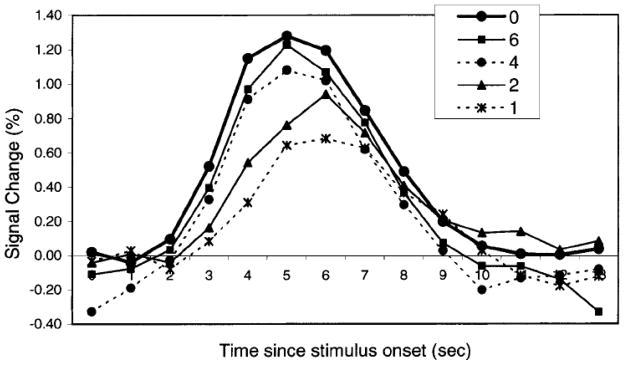
This simple story – the fMRI hemodynamic response responds to stimuli in a linear manner – itself turned out to be only roughly true. Some deviations from linearity were evident in even the earliest reports (Boynton et al., 1996; Dale and Buckner, 1997), particularly when the interval between successive stimuli was only 2 or 3 seconds. In a series of studies with Gregory McCarthy and other colleagues (Huettel and McCarthy, 2000; Huettel and McCarthy, 2001; Huettel et al., 2004a; Huettel et al., 2001), we showed that when stimuli are separated by less than about 6s, the hemodynamic response has attenuated amplitude and a slightly delayed peak, compared to a single stimulus in isolation (Figure 2). The magnitude of these refractory effects differs across brain regions (Birn et al., 2001), perhaps in a way that is selective to local computations (Huettel et al., 2004a). Research on the nature and consequence of these nonlinearities remains ongoing (Liu et al., 2010). Nevertheless, it is now recognized that the nonlinearities in the fMRI response are small enough to not preclude fast event-related designs, but large enough to influence analysis methods.
Figure 2. Effects of the interval between successive events on the amplitude of the fMRI hemodynamic response.
Participants viewed visual checkerboard stimuli presented either in temporal isolation (0s) or with a short interval between their onset times (1, 2, 4, or 6 s). For the paired event conditions, shown here is the estimated hemodynamic response associated with the second event in the pair (i.e., the combined response minus the mean single-event response, temporally aligned to the presentation of the second event). If the hemodynamic response to multiple events were equivalent to a linear combination of their individual responses, then all of the curves should be similar in form and amplitude. Instead, the fMRI hemodynamic response exhibits a refractory effect: its amplitude decreases and its latency increases when events are separated by short intervals. Figure from Huettel and McCarthy (2000).
Who are we? Impact of “neural events”
Importantly, the aspects of event-related fMRI that were considered problems a decade ago (e.g., overlap between events, modeling processes across trials) now have become key features of fMRI research. Most generally, the explosion of new fast event-related paradigms has shifted the primary goal of fMRI research away from simple localization of function. (Note that the misconception that fMRI merely identifies “where the brain lights up” remains common and destructive, particularly in accounts of fMRI research by the popular press.) Researchers now study how complex brain functions arise from a sequence of activated brain regions. The first event-related studies – which merely separated trials into their components – set the stage for the enormously influential later methods for understanding functional connectivity among brain regions (see the article on connectivity by Smith in this collection).
In addition to this general contribution, advances in event-related designs have become central to many of the recent developments in the field. As introduced above, trial sorting has become so central to fMRI research that it often goes unnoticed. That is, events are no longer defined by the experimenter a priori, based on the pattern of stimulus presentation. Instead, events are sorted into categories (or assigned values on some continuum) based on the participants’ behavior. Close links between fMRI activation and behavior have been extraordinarily important, not least because they provide strong evidence that fMRI does more than show epiphenomenal blobs of activation. Yet, linking fMRI data to behavior is often not enough. For many topics in neuroscience (e.g., decision making), the key events of interest are not experimental stimuli, nor even observable behavior, but inferred internal states. Analyzing the neural representation of those states has been called “model-based fMRI”, in that the researchers create a model for a process that translates stimuli into behavior (e.g., the subjective value currently assigned to two choice options) and then enter the moment-to-moment states of that process into their fMRI analyses (O’Doherty et al., 2007). And, the influence of event-related fMRI can be felt even in non-event-related paradigms. FMRI-adaptation experiments use repeated presentations of similar stimuli to uncover the sensitivity of a brain region to one or more stimulus features (Grill-Spector and Malach, 2001). Their underlying concepts (e.g., refractory effects to repeated presentations, separating distinct neural events) build upon the foundation laid by early event-related studies. And, the analytic tools developed for event-related designs can themselves be applied to improve the precision of blocked analyses (Mechelli et al., 2003).
Not all of the contributions of event-related fMRI have been so salutary. Those who have long histories with fMRI might lament the complexity of current designs (and analysis models). When one runs a simple alternating-block design – now, typically as part of a class project – it is easy to see the desired effect in the raw data. Consider the workhorse paradigms used by early fMRI researchers: finger-tapping vs. rest, flashing checkerboard vs. darkness. A simple blocked design might evoke 2–5% signal change within voxels in the primary motor or visual cortex; looking at the timecourses of such effects, one only needs the “interocular trauma test” for significance. Given the complexity of many paradigms, it is now common to display estimated model functions instead of raw data. In the best cases, this provides a principled method for extracting and displaying the brain changes caused by one part of a complex experiment; in the worst cases, it can hide meaningful effects behind an imperfect model.
In summary, event-related approaches have had paradoxical effects on fMRI. They have empowered fMRI researchers to investigate new topics, both within cognitive psychology and throughout other disciplines. Yet, they have also embedded fMRI analyses within layers of abstraction – pulling those same researchers ever farther away from their data. They allow consideration of subtle and transient effects of experimental manipulations, even if those manipulations only evoke changes in the BOLD signal of less than a tenth of a percent. (Consider that such effect sizes are almost two orders of magnitude smaller than those from the very first fMRI studies!) And, they have catalyzed fMRI research on evocative interactions among brain regions – functional connectivity, causality, and adaptation – all of which are difficult to visualize except as simple spots on the brain.
Where are we going? Future Directions
In the near future, “event-related fMRI” will largely disappear. What was once the hallmark of cutting edge fMRI research will be relegated to a historical curiosity. To be clear, I am not predicting that researchers will abandon rapid, multi-stage designs and return to the simple blocked paradigms of the 1990s. On the contrary, the complexity of fMRI experiments will only continue to increase. I am instead predicting that the very concept of “event-related” will become so central to fMRI that it will no longer carry meaning.
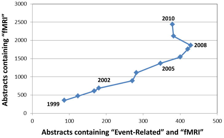
Evidence for this assertion can be gleaned from the ways fMRI researchers describe their experiments (Figure 3). From 1999 until 2008 there was continual year-to-year growth both in the number of studies conducted using fMRI and in the number of studies using event-related designs. In 2008, approximately 1860 articles used the term “fMRI” (or a variant) in their abstracts – and approximately 430 of those articles reported using “event related” methods. Since that time, however, there have been dramatic year to year increases in the overall number of fMRI studies, but an actual decline in the number of abstracts including the phrase “event related”. This change is becoming even starker with each passing year. At the time of this literature search (in July 2011), only about 14% of the current year’s fMRI abstracts use the term “event related” – compared to 25–30% throughout most of the 2000s. As event-related designs became ubiquitous, they also became unremarkable, at least in abstracts of fMRI research.
Figure 3. Changes over time in the use of “event-related” in fMRI abstracts.
For each year since 1999, two literature searches were conducted using PubMed: the total number of abstracts containing the terms “fMRI”, “functional MRI”, or “functional magnetic resonance” (y-axis), and the number of abstracts within that set that also included the term “event related” (x-axis). The number of research articles that describe fMRI research has increased every year since 1999, with a mean year-over-year increase of about 20%. In contrast, the number of those abstracts that also label their research as “event-related” has actually declined over the past five years. This reflects the ubiquity of event-related designs, which are now sufficiently well accepted that they less likely to be noted within research abstracts.
Consider also how fMRI methods are taught to those new to the field. In the first edition of our fMRI textbook (Huettel et al., 2004b), we devoted a number of pages to comparing blocked and event-related designs – as in the section titled “Advantages and Disadvantages of Event-Related Designs”. By the second edition (Huettel et al., 2009), the title of that section was two words shorter: “Advantages of Event-Related Designs”. A particular point of emphasis was that manipulating the timing of events provides more flexible experimental designs and carries minimal costs. In fact, intercalated within that very section was a new box on efficient design of fMRI experiments, which described several approaches to optimizing event-related designs to maximize their power to detect and estimate changes in the BOLD signal. Blocked designs are still important for instruction, but primarily for engaging students with basic principles of design and analyses, before they turn to the more difficult concepts that guide current practice. With each new edition of this textbook, fMRI experimental design will become increasingly isomorphic with efficient implementation of event-related concepts.
Will the disappearance of “event-related fMRI” matter? What if the concepts it introduced – efficient design, trial sorting, and many others – become so intrinsic to fMRI practice that no formal distinction can be made between “fMRI” and “event-related” fMRI? My sense is that the field has progressed sufficiently that this term could vanish from the literature without anyone noticing. (By contrast, and with much irony, the term “blocked design” will likely be viable for much longer. It now describes a special case of fMRI experimentation that will remain important for experiments where maximal detection power is needed.) By the 40th year of fMRI, researchers will still be manipulating the timing of events, analyzing individual elements of complex trials, and evaluating the relative order of brain functions. But to them, those practices will just be “fMRI”.
Acknowledgments
The author thanks Gregory McCarthy and Allen Song for many discussions about fMRI methods over the past 15 years, and David Smith for comments on this manuscript. Support is provided by the National Institutes of Health (NINDS P01-41328, NIMH RC1-88680) and by an Incubator Award from the Duke Institute for Brain Sciences.
Footnotes
Publisher's Disclaimer: This is a PDF file of an unedited manuscript that has been accepted for publication. As a service to our customers we are providing this early version of the manuscript. The manuscript will undergo copyediting, typesetting, and review of the resulting proof before it is published in its final citable form. Please note that during the production process errors may be discovered which could affect the content, and all legal disclaimers that apply to the journal pertain.
References
- Bandettini PA, Wong EC, Hinks RS, Tikofsky RS, Hyde JS. Time Course Epi of Human Brain-Function during Task Activation. Magnetic Resonance in Medicine. 1992;25:390–397. doi: 10.1002/mrm.1910250220. [DOI] [PubMed] [Google Scholar]
- Birn RM, Saad ZS, Bandettini PA. Spatial heterogeneity of the nonlinear dynamics in the FMRI BOLD response. Neuroimage. 2001;14:817–826. doi: 10.1006/nimg.2001.0873. [DOI] [PubMed] [Google Scholar]
- Blamire AM, Ogawa S, Ugurbil K, Rothman D, McCarthy G, Ellerman JM, Hyder F, Rattner Z, Shulman RG. Dynamic mapping of the human visual cortex by high-speed magnetic resonance imaging. Proceedings of the National Academy of Sciences. 1992;89:11069–11073. doi: 10.1073/pnas.89.22.11069. [DOI] [PMC free article] [PubMed] [Google Scholar]
- Boynton GM, Engel SA, Glover GH, Heeger DJ. Linear systems analysis of functional magnetic resonance imaging in human V1. Journal of Neuroscience. 1996;16:4207–4221. doi: 10.1523/JNEUROSCI.16-13-04207.1996. [DOI] [PMC free article] [PubMed] [Google Scholar]
- Brewer JB, Zhao Z, Desmond JE, Glover GH, Gabrieli JD. Making memories: brain activity that predicts how well visual experience will be remembered. Science. 1998;281:1185–1187. doi: 10.1126/science.281.5380.1185. [DOI] [PubMed] [Google Scholar]
- Buckner RL. Event-related fMRI and the hemodynamic response. Human Brain Mapping. 1998;6:373–377. doi: 10.1002/(SICI)1097-0193(1998)6:5/6<373::AID-HBM8>3.0.CO;2-P. [DOI] [PMC free article] [PubMed] [Google Scholar]
- Burock MA, Buckner RL, Woldorff MG, Rosen BR, Dale AM. Randomized event-related experimental designs allow for extremely rapid presentation rates using functional MRI. Neuroreport. 1998;9:3735–3739. doi: 10.1097/00001756-199811160-00030. [DOI] [PubMed] [Google Scholar]
- Clark VP, Fannon S, Lai S, Benson R, Bauer L. Responses to rare visual target and distractor stimuli using event-related fMRI. Journal of Neurophysiology. 2000;83:3133–3139. doi: 10.1152/jn.2000.83.5.3133. [DOI] [PubMed] [Google Scholar]
- Dale AM, Buckner RL. Selective averaging of rapidly presented individual trials using fMRI. Human Brain Mapping. 1997;5:329–340. doi: 10.1002/(SICI)1097-0193(1997)5:5<329::AID-HBM1>3.0.CO;2-5. [DOI] [PubMed] [Google Scholar]
- Grill-Spector K, Malach R. fMR-adaptation: a tool for studying the functional properties of human cortical neurons. Acta Psychologica. 2001;107:293–321. doi: 10.1016/s0001-6918(01)00019-1. [DOI] [PubMed] [Google Scholar]
- Hopfinger JB, Buonocore MH, Mangun GR. The neural mechanisms of top-down attentional control. Nature Neuroscience. 2000;3:284–291. doi: 10.1038/72999. [DOI] [PubMed] [Google Scholar]
- Huettel SA, Mack PB, McCarthy G. Perceiving patterns in random series: Dynamic processing of sequence in prefrontal cortex. Nature Neuroscience. 2002;5:485–490. doi: 10.1038/nn841. [DOI] [PubMed] [Google Scholar]
- Huettel SA, McCarthy G. Evidence for a refractory period in the hemodynamic response to visual stimuli as measured by MRI. Neuroimage. 2000;11:547–553. doi: 10.1006/nimg.2000.0553. [DOI] [PubMed] [Google Scholar]
- Huettel SA, McCarthy G. Regional differences in the refractory period of the hemodynamic response: an event-related fMRI study. Neuroimage. 2001;14:967–976. doi: 10.1006/nimg.2001.0900. [DOI] [PubMed] [Google Scholar]
- Huettel SA, Obembe J, Song A, Woldorff M. The BOLD fMRI refractory effect is specific to stimulus attributes: Evidence from a visual motion paradigm. Neuroimage. 2004a;21:402–408. doi: 10.1016/j.neuroimage.2004.04.031. [DOI] [PubMed] [Google Scholar]
- Huettel SA, Singerman J, McCarthy G. The effects of aging upon the hemodynamic response measured by functional MRI. Neuroimage. 2001;13:161–175. doi: 10.1006/nimg.2000.0675. [DOI] [PubMed] [Google Scholar]
- Huettel SA, Song AW, McCarthy G. Functional Magnetic Resonance Imaging. Sinauer; Sunderland, Massachusetts: 2004b. [Google Scholar]
- Huettel SA, Song AW, McCarthy G. Functional Magnetic Resonance Imaging. 2. Sinauer; Sunderland, Massachusetts: 2009. [Google Scholar]
- Huettel SA, Stowe CJ, Gordon EM, Warner BT, Platt ML. Neural signatures of economic preferences for risk and ambiguity. Neuron. 2006;49:765–775. doi: 10.1016/j.neuron.2006.01.024. [DOI] [PubMed] [Google Scholar]
- Kirino E, Belger A, Goldman-Rakic P, McCarthy G. Prefrontal activation evoked by infrequent target and novel stimuli in a visual target detection task: an event-related functional magnetic resonance imaging study. Journal of Neuroscience. 2000;20:6612–6618. doi: 10.1523/JNEUROSCI.20-17-06612.2000. [DOI] [PMC free article] [PubMed] [Google Scholar]
- Kwong KK, Belliveau JW, Chesler DA, Goldberg IE, Weisskoff RM, Poncelet BP, Kennedy DN, Hoppel BE, Cohen MS, Turner R. Dynamic magnetic resonance imaging of human brain activity during primary sensory stimulation. Proceedings of the National Academy of Sciences. 1992;89:5675–5679. doi: 10.1073/pnas.89.12.5675. [DOI] [PMC free article] [PubMed] [Google Scholar]
- Liu Z, Rios C, Zhang N, Yang L, Chen W, He B. Linear and nonlinear relationships between visual stimuli, EEG and BOLD fMRI signals. Neuroimage. 2010;50:1054–1066. doi: 10.1016/j.neuroimage.2010.01.017. [DOI] [PMC free article] [PubMed] [Google Scholar]
- McCarthy G, Luby M, Gore J, Goldman-Rakic P. Infrequent events transiently activate human prefrontal and parietal cortex as measured by functional MRI. Journal of Neurophysiology. 1997;77:1630–1634. doi: 10.1152/jn.1997.77.3.1630. [DOI] [PubMed] [Google Scholar]
- McCarthy G, Luby M, Gore JC, Goldman-Rakic P. Functional magnetic resonance imaging in a visual oddball task. Neuroimage. 1996;3:S548. [Google Scholar]
- Mechelli A, Henson RN, Price CJ, Friston KJ. Comparing event-related and epoch analysis in blocked design fMRI. Neuroimage. 2003;18:806–810. doi: 10.1016/s1053-8119(02)00027-7. [DOI] [PubMed] [Google Scholar]
- O’Doherty JP, Critchley H, Deichmann R, Dolan RJ. Dissociating valence of outcome from behavioral control in human orbital and ventral prefrontal cortices. Journal of Neuroscience. 2003;23:7931–7939. doi: 10.1523/JNEUROSCI.23-21-07931.2003. [DOI] [PMC free article] [PubMed] [Google Scholar]
- O’Doherty JP, Hampton A, Kim H. Model-based fMRI and its application to reward learning and decision making. Ann N Y Acad Sci. 2007;1104:35–53. doi: 10.1196/annals.1390.022. [DOI] [PubMed] [Google Scholar]
- Sutton S, Braren M, Zubin J, John ER. Evoked-potential correlates of stimulus uncertainty. Science. 1965;150:1187–1188. doi: 10.1126/science.150.3700.1187. [DOI] [PubMed] [Google Scholar]
- Wagner AD, Schacter DL, Rotte M, Koutstaal W, Maril A, Dale AM, Rosen BR, Buckner RL. Building memories: remembering and forgetting of verbal experiences as predicted by brain activity. Science. 1998;281:1188–1191. doi: 10.1126/science.281.5380.1188. [DOI] [PubMed] [Google Scholar]
- Woldorff MG, Hillyard SA. Modulation of early auditory processing during selective listening to rapidly presented tones. Electroencephalography & Clinical Neurophysiology. 1991;79:170–191. doi: 10.1016/0013-4694(91)90136-r. [DOI] [PubMed] [Google Scholar]


