Abstract
Parasitic flatworms of the genus Schistosoma are the causative agents of schistosomiasis, a highly prevalent, neglected tropical disease that causes significant morbidity in hundreds of millions of people worldwide. The current treatment of choice against schistosomiasis is praziquantel (PZQ), which is known to affect Ca2+ homeostasis in schistosomes, but which has an undefined molecular target and mode of action. PZQ is the only available antischistosomal drug in most parts of the world, making reports of PZQ resistance particularly troubling. Voltage-gated Ca2+ (Cav) channels have been proposed as possible targets for PZQ, and, given their central role in the neuromuscular system, may also serve as targets for new anthelmintic therapeutics. Indeed, ion channels constitute the majority of targets for current anthelmintics. Cav channel subunits from schistosomes and other platyhelminths have several unique properties that make them attractive as potential drug targets, and that could also provide insights into structure-function relationships in, and evolution of, Cav channels.
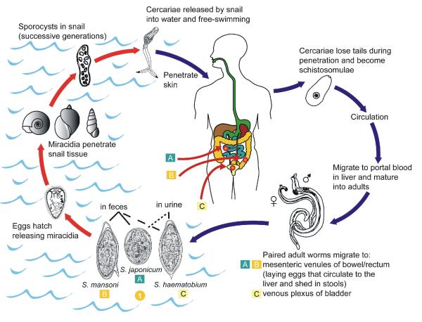
Schistosomes are trematode flatworms that parasitize humans and other mammals (as well as birds), and cause schistosomiasis, a prevalent tropical disease. Schistosomes have a complex life cycle that requires freshwater snails as intermediate hosts, and they infect the mammalian host via water-borne contact with the free-swimming larvae shed from those snails (Figure 1)1-3. According to the World Health Organization in its publication “Preventive chemotherapy in human helminthiasis” (http://whqlibdoc.who.int/publications/2006/9241547103_eng.pdf), there are an estimated 200 million people whose quality of life is severely impaired by schistosomiasis. More recent estimates suggest the number may be closer to 450 million, with a burden on human health similar to that of tuberculosis or malaria (http://iom.edu/~/media/Files/Activity%20Files/PublicHealth/MicrobialThreats/2010-SEP-21/King%20CH.pdf). These facts alone provide an important impetus for research into the basic neuromuscular physiology of these parasites. Indeed, the majority of anthelmintic drugs in current use target ion channels of the worm’s neuromuscular system, where they typically act as agonists or as positive allosteric modulators4 (eg, ivermectin, levamisole). Our lab focuses on the structure, function, and modulation of schistosome voltage-gated calcium (Cav) channels, as Cav channels are widely recognized as targets in pharmacotherapy5-7. However, our interests go beyond identification of possible therapeutic targets, and focus also on using a comparative approach to understand how these channels fit into the biology and life cycle of parasitic platyhelminths, and perhaps add insights into mammalian channel function.
Figure 1. Life cycle of schistosomes.
Shown are the life cycles of S. japonicum (A), S. mansoni (B), and S. haematobium (C), the three major schistosome species that parasitize humans. The overall life cycles are quite similar, requiring both a mammalian host and an intermediate fresh-water snail host. Differences between the three species are in the species of intermediate snail host and in the predilection sites in the definitive mammalian host. Other differences include egg morphology, range of definitive mammalian hosts, and levels of egg production. Unlike most Digeneans, schistosomes have two separate sexes. Male and female adult worm pairs reside in the vasculature of the mammalian host in preferential locations depending on the species. There, they undergo sexual reproduction and deposit hundreds (S. mansoni) to thousands (S. japonicum, S. haematobium) of eggs per female, per day. Note that no increase in worm numbers occurs within the mammalian host. Eggs move to the lumen of the intestine or bladder, and are excreted in feces or urine. Eggs which remain within the host are the cause of the majority of pathology of chronic schistosomiasis. Excreted eggs that reach fresh water will hatch into a miracidium, a free-swimming larva that parasitizes an intermediate host snail. Within the snail host, the worms undergo developmental changes and asexual reproduction, emerging in a few weeks as free-living cercariae, the larval form that parasitizes the definitive human host. The cercariae attach to the host skin, and then penetrate it and shed their forked tail to become schistosomules. The schistosomules migrate through several tissues and mature into adults, which take up residence in their predilection sites. Adult S. mansoni can reside within the mammalian host for many years. Figure adapted from an image provided by the Parasitology Diagnostic Web Site (DPDx) at the Centers for Disease Control and Prevention (http://www.dpd.cdc.gov/dpdx/Default.htm).
Cav channels initiate the contraction of the schistosome musculature8. Interestingly, neuropeptide-based signalling, which is well established as of major importance in the neuromuscular system of flatworms 9, 10, appears to be functionally coupled with Cav channel activity in schistosome muscle11, emphasizing their physiological significance in this system. In addition, schistosome Cav channels are also almost certainly key players in other important Ca2+-dependent events, such as synaptic transmission, enzyme activity, and gene expression.
Currently, the treatment of choice against schistosomiasis is praziquantel, which is highly effective against all schistosome species, has minimal side effects, and has been demonstrated repeatedly to control schistosomiasis in large-scale treatment efforts12-17. Due to these advantages, as well as steadily reduced costs, PZQ has become the only commercially available antischistosomal treatment in most parts of the world15, 18. However, this success comes at a potential cost. Reliance on a single drug to treat such a hugely prevalent disease represents an ultimately untenable situation8, as there is no readily available alternative should drug resistance develop. In that light, reports of PZQ resistance in the field9-11 and in the laboratory after drug selection12, 13 are particularly troubling. Furthermore, treatment failures can arise because juvenile schistosomes are refractory to PZQ, and do not become sensitive until egg deposition begins at approximately 6 weeks following infection23-26.
In this review we will focus on what we know about Cav channel expression and function in native schistosome cells and on the function of schistosome Cav channel subunits, including their apparent role in PZQ action, as surmised by expression in heterologous systems.
Ca2+ CURRENTS IN NATIVE SCHISTOSOME CELLS
Cav channel activity in schistosomes was first indicated by experiments showing that rapid contraction of worm muscle was dependent upon the presence of Ca2+ in the bathing medium8. In that study, the reported speed at which schistosomes contracted following introduction of external Ca2+ was consistent with a Ca2+ gate in the plasma membrane that is opened by a characteristically rapid change in membrane voltage. However, the first electrophysiological study geared towards recording voltage-gated ion currents from schistosome muscle cells, which used the whole-cell patch-clamp technique, detected the prominent outward K+ currents in these cells but no Ca2+ or other inward currents14. In contrast, Ca2+ currents of dissociated cell preparations from ectoparasitic and free-living platyhelminths were more readily obtainable. Thus, neurons from the polyclad flatworm Notoplana acticola express a typical complement of ionic currents, including cadmium-sensitive Ca2+ currents15. Similarly, both nerve and muscle cells of the triclad ectoparasitic flatworm Bdelloura candida express Ca2+ currents that activate at −30 mV, reach peak amplitude in approximately 5 ms, and inactivate slowly16, 17. The neuronal Ca2+ current is relatively insensitive to Cav channel blockers such as nifedipine and verapamil, is not blocked by ω-conotoxin GVIA, and exhibits no sensitivity to 10 μM praziquantel18. The muscle Ca2+ current was too unstable for determination of pharmacological sensitivities. Muscle cells from the free-living freshwater flatworm Girardia tigrina also express Ca2+ currents that tend to run-down under whole-cell patch clamp19. Interestingly, although 10 μM PZQ had no apparent effect on intact B. candida, it did produce a rapid Ca2+-dependent contracture in approximately 20% of the isolated B. candida muscle fibers when applied within one hour of dispersion of the cells16. Our preliminary data indicate that Dugesia spp. respond to PZQ, though with less sensitivity and rapidity than schistosomes (unpublished observations), suggesting free-living planarians may have potential for use as models to study PZQ targets.
More than a decade after the first patch-clamp studies on schistosomes, the schistosome muscle preparation was revisited in two studies11, 20. In the first33, voltage-gated Ca2+ currents were detected in muscle fibers, but only after extensive block of prominent outward K+ currents in these cells. These Ca2+ currents peaked at approximately +20 mV and were relatively small (less than 100 pA), activated within 30 ms after depolarization, and did not inactivate for at least 250 ms if the depolarization was maintained. This time-course of the Ca2+ current in schistosome muscle was not unlike that of the L-type Ca2+ currents of mammalian muscle. Similar to the Ca2+ currents recorded from other flatworms16, 19, the Ca2+ currents from schistosome muscle fibers ran down rapidly, within a few minutes of establishing the whole-cell configuration, thus complicating pharmacological analysis. However, depolarization-initiated contractions of the muscle fibers, which are presumably dependent on Cav channel activity, were blocked by nicardipine with an IC50 of 4.1 μM. Surprisingly, other dihydropyridines such as nifedipine and nitrendipine were largely ineffective at blocking these depolarization-induced contractions. Diltiazem was also relatively ineffective, as were conotoxins that inhibit non-L type mammalian Cav channels. The more recent study11 investigated neuropeptide enhancement of Ca2+ currents. As with depolarization-induced muscle fiber contraction, peptide (YIRFamide)-induced contractions were sensitive to nicardipine, and also to high concentrations of the phenylalkylamines verapamil and methoxyverapamil. Recording of robust Cav currents was aided by using a combination of Ca2+ and Ba2+ as charge carrier, and rundown was managed by recording within a brief window of time during which currents were relatively stable. The currents recorded under these conditions were enhanced by the peptide YIRFamide. Due to technical issues, the effect of dihydropyridines such as nicardipine and nifedipine were not tested, but these currents were partially inhibited (~50%) by 10 μM verapamil11. Thus, what we know so far about the pharmacological profile of schistosome and other platyhelminth Cav channels sets them apart from their mammalian homologues.
Clearly, studying native Ca2+ currents in schistosome cells is technically challenging. Furthermore, as in any native system, but particularly in invertebrates where channel pharmacology is not as well established, Ca2+ currents are often contaminated by other ionic currents that are not always possible to eliminate. An alternative and complementary approach is to use heterologous expression of cloned channel genes in Xenopus oocytes or mammalian cells. Prior to the availability of a genome database, cDNAs encoding three Cav channel α1 (SmCav1, SmCav2A SmCav2B) and two β (SmCavβ, SmCavβvar) subunits were cloned by Greenberg and collaborators21, 225. Publication of the S. mansoni genome23 confirmed these results and also uncovered genes for additional subunits. A summary of Cav channel subunits found in the S. mansoni genome is shown in Sidebar 1.
Sidebar 1.
Predicted Cav channel subunits in the S. mansoni genome
| Accession number | Putative subtype | Predicted size (kDa) |
|---|---|---|
| 1. Smp_020170 | Non L-type α1 (SmCav2A) | 236 |
| 2. Smp_004730 | Non L-type α1 (SmCav2B) | 156* |
| 3. Smp_020270 | L-type α1 (SmCav1A) | 181 |
| 4. Smp_159990 | L-type α1 (SmCav1B) | 230 |
| 5. Smp_134050 | α 2 δ | 129 |
| 6. Smp_124530 | α 2 δ | 48* |
| 7. Smp_135140 | β (SmCavβvar) | 88 |
| 8. Smp_141660 | β (SmCavβ) | 56** |
partial sequence
the gene prediction algorithm excludes the NPAM-containing N-terminus
STRUCTURE OF SCHISTOSOME Cav CHANNEL α1 SUBUNITS
Original cloning of cDNAs using RT-PCR with degenerate primers revealed three high voltage-activated (HVA) Cav channel α1 subunits in S. mansoni35. One of these cDNAs (SmCav1) has highest similarity to L-type (Cav1) channels, and the other two (SmCav2A, SmCav2B) appear to be non L-type (Cav2) channels. Analysis of the genome of S. mansoni confirmed those three sequences, as well as a second L-type-like α1 subunit (Smp_159990), making four Cavα1 genes in total. Other invertebrates that have been examined typically contain three α1 subunit genes24. Despite the larger number of α1 subunit genes in S. mansoni, there may in fact be less functional diversity, as all four of the S. mansoni α1 subunits appear to be HVA channels; in other invertebrates, one of their three α1 subunits is typically a low voltage-activated (LVA; T-type; Cav3) channel sequence. Surprisingly, neither the S. mansoni nor the S. japonicum genomes appear to contain LVA channel-like sequences. Thus, schistosome excitable cells may uniquely lack a requirement for the functions normally carried out by LVA channels. Alternatively, schistosomes may recruit HVA subunits to perform the roles typically fulfilled by LVA subunits, and such a change in channel properties could likely be dependent upon specific interaction with auxiliary subunits. In either case, schistosomes, and perhaps other platyhelminths, clearly differ from other metazoans in their repertoire of Cav channel α1 subunits. The Schmidtea mediterranea genome contains a sequence fragment that appears to have highest similarity to LVA subunits, as well as other α1 (and β) subunit representatives that are found in schistosomes. Whether these sequence fragments are within genes that in fact code for LVA channels remains to be determined.
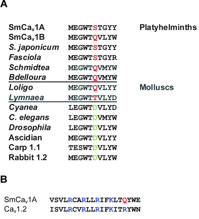
The predicted structures for the schistosome α1 subunits are overall very similar to their mammalian counterparts, though there are some interesting differences35. For example, flatworm L-type channels, including both L-type SmCav channels, substitute a non-charged amino acid for an aspartic acid residue that is absolutely conserved in the Domain I pore region of other L-type α1 subunits. Interestingly, L-type-like α1 subunits from molluscs (Loligo bleekeri), also members of the Lophotrochozoa, show a similar substitution (Fig 2). Whether this change affects ion selectivity or other channel properties, and whether it might be a potential target for highly specific antiparasitics remains to be determined. SmCav1A also contains one less positively charged residue in the fourth transmembrane segment of Domain II than homologous L-type mammalian Cav1 subunits, which may suggest a slight weakening of the voltage dependence of this channel. Furthermore, the C-terminal tail of SmCav1A contains two, instead of the normal one, IQ-like calmodulin binding domains, perhaps indicating important variation or redundancy in Ca2+-dependent regulation. The non L-type SmCav2A subunit does not have sites for interactions with syntaxin 1A and SNAP-25 in the II-III loop. These interaction sites are, however, present in SmCav2B, suggesting its involvement in synaptic transmission in neurons, whereas SmCav2A plays other roles. Consistent with the data from schistosome muscle fiber contraction studies that indicate at most mild sensitivity to various dihydropyridines20, the SmCav1A subunit contains only 6 residues of the 13 thought to be involved in determining dihydropyridine sensitivity in L-type Cav channels25. In contrast, the SmCav1B sequence is identical at 10 of these 13 residues, suggesting greater sensitivity.
Figure 2. Examples of structural differences between schistosome and other L-type Cav channel α1 subunits.
A. Charge change in the Domain I pore region. The Domain I pore region of L-type α1 subunits is shown. Note the shaded residues four spots C-terminal from the absolutely conserved glutamic acid (E) that forms part of the selectivity gate. In L-type channels from most phyla, this residue is a negatively-charged aspartic acid (D, green). However, in platyhelminths and molluscs, both of which are classed as lophotrochozoans, this residue is uncharged (red). This is interesting, because a non-charged residue at this position is characteristic of some non L-type channels (eg, Cav2.3). Accession numbers for sequences are: SmCav1A, AF361884; SmCav1B, Smp_159990; S. japonicum, Sjp_0099010; Fasciola (F. hepatica), Schmidtea (S. meditteranea), and Bdelloura (B. candida) are from DNA fragments we sequenced; Loligo (L. bleekeri), D86600; Lymnaea (L. stagnalis), AF484081; Cyanea (C. capillata), AAC63050; C. elegans, AAC47755; Drosophila, AAA81883; Ascidian (Halocynthia roretzi), BAA34927; Carp (Cyprinus carpio) Cav1.1, P22316; Rabbit (Oryctolagus cuniculus) Cav1.2, NM_001136522. B. SmCav1A contains one fewer positively charged residue in II-S4. Shown are residues in the fourth transmembrane region of domain II from SmCav1A and rat Cav1.2 (M67515). SmCav1A contains one fewer positively charged residue than Cav1.2. Positively charged arginines (R) and lysines (K) are highlighted in blue, and the substituted glutamine (Q) in SmCav1A is highlighted in red.
To date, none of the schistosome Cavα1 subunits have been functionally expressed, making characterization problematical. In both Xenopus oocytes (Kohn and Greenberg, unpublished data) and in mammalian cells (Salvador-Recatalà and Greenberg, unpublished data), attempts at expression using a variety of approaches, including co-expression with schistosome or mammalian auxiliary subunits, were unsuccessful. Possible causes for this lack of expression include: unusual and/or extensive endoplasmic reticulum retention signals; the need for a specific, non-channel, schistosome chaperoning factor; the high A/T content of the schistosome coding regions; requirement of specific lipids in the plasma membrane. The development of immortalized platyhelminth cell lines could help in this regard. These would be expected to contain helminth-specific chaperones and cell membranes with a composition more similar to that of schistosome cells, with transcription/translation machinery likely more suitable for expression of the schistosome Cavα1 subunits than the vertebrate systems. Planarian stem cells may offer a means to achieve this end26, and recent results using cells from Echinococcus multilocularis have also been encouraging27. Current efforts to develop schistosome cell lines, including immortalization by transfection with oncogenes, have recently been reviewed28.
SCHISTOSOME CaVβ SUBUNITS
Schsitosomes and other platyhelminths, express at least two Cavβ subunit genes39 instead of the single β subunit characteristic of other invertebrates. One of these subunits is structurally similar to Cavβ subunits from other vertebrate and invertebrate species. whereas the other, though clearly a β subunit based on sequence alignment, is different, and we have dubbed it a “variant” β subunit (SmCavβvar). For example, at ~85 kDa, SmCavβvar is larger than other β subunits, with most of the extra sequence in the C-terminal region. Most notably, all platyhelminth βvar subunits lack two otherwise absolutely conserved protein kinase C (PKC) sites in the highly conserved, ~30 amino acid region of the subunit known as the BID. When co-expressed in Xenopus oocytes with a mammalian (Cav2.3) or jellyfish (CyCav1) α1 subunit Cavβvar reduces the current amplitude instead of increasing it, one of the “hallmark” effects of Cavβ subunits21. Furthermore, SmCavβvar confers PZQ sensitivity to an otherwise PZQ-insensitive mammalian α1 subunit. When coexpressed with SmCavβvar, voltage-gated peak currents through Cav2.3 are increased 1.5-2-fold in the presence of 100 nM PZQ compared to those in the absence of PZQ21. Cav2.3 expressed alone, or co-expressed with a conventional β subunit (eg, SmCavβ), does not show this responsiveness to PZQ. Addition by mutagenesis of either one or both of the missing PKC sites in the SmCavβvar BID region induces a more conventional modulatory phenotype, i.e. one that increases the Ca2+ current and that does not confer PZQ sensitivity29. The complementary experiment of eliminating those PKC sites in a mammalian β subunit results in a subunit that now has the ability to confer PZQ sensitivity to Cav2.341. How these consensus PKC sites in the BID, a part of β subunit structure thought to be non-accessible30-32, influence behaviour of the β subunit remains an open question, as does the mechanism by which SmCavβvar may be mediating these effects. To help resolve these issues, we have also tried expressing SmCavβvar with Cav2.3 in a mammalian cell line (HEK), but it shows no effect on the α1 subunit, and indeed appears not to express at the protein level based on imaging experiments using a Cavβvar–GFP chimera (Salvador-Recatalà and Greenberg, unpublished data). Interestingly, however, in the free-living flatworm Dugesia japonica, PZQ disrupts normal polarity during regeneration of the worm, but this PZQ effect can be eliminated by knockdown of either Cavβ subunit33. How this fascinating, long-term effect on developmental polarity that is apparently mediated by Cavβ subunits relates to the shorter-term antischistosomal activity of PZQ is not clear.
Does SmCavβvar play a role in development of PZQ resistance? To date, the only published examination of this question has found no changes in the sequence or expression levels in S. mansoni isolates with reduced PZQ susceptibility34. Furthermore, there appears to be no significant difference in β subunit expression between PZQ-sensitive adults and PZQ-refractory juveniles (our unpublished data and ref. 31). These findings, though perhaps disappointing in terms of finding a useful marker for emergence of PZQ resistance, do not eliminate the prospect that Cavβvar and schistosome Cav channels are targeted by PZQ. Indeed, although juvenile worms are far less sensitive to the drug, they still respond to PZQ exposure with an influx of and an initial muscular contraction35. However, they recover from exposure to concentrations of PZQ that are lethal to adult parasites. Thus, although PZQ may initially be targeting Cav channels (or other schistosome receptors), some downstream component of the cascade that is initiated by this interaction must differ between juvenile and adult worms, and between isolates with differential PZQ susceptibility. Screens for stage- and gender-specific differences in gene expression have revealed possible candidates36, and we have found that higher expression of multidrug resistance transporters is one factor which correlates with reduced PZQ sensitivity37.
The more conventional schistosome β subunit (SmCavβ) has a predicted molecular weight of ~67.5 kDa and a predicted isoelectric point of 5.8, similar to mammalian β3 subunits. It was initially expressed in Xenopus oocytes with Cav2.3 as the reporter α1 subunit, where it predictably increased Cav2.3 amplitude and shifted steady-state inactivation to more hyperpolarized potentials29. In contrast to SmCavβvar, SmCavβ does not confer PZQ sensitivity to Cav2.3. Subsequently, we showed that SmCavβ induces run-down of Cav2.3 channels expressed in HEK cells38. A systematic characterization of this phenomenon revealed that (1) it occurred even if Ca2+ was substituted by Ba2+ as the charge carrier, suggesting that it is calmodulin-independent; (2) it was dependent on chelated forms of ATP that were added to the patch pipette solution that perfuses the cell during whole-cell patch-clamp recordings, ironically with the goal of preventing run-down (more typically, rundown occurs in the absence of ATP); and (3) the structure responsible for the rapid rundown effect resides in the first 44 amino acids of SmCavβ, a region that contains a long polyacidic motif of 15 aspartate and glutamate residues. It is tempting to suggest that the run-down that occurs in this heterologous system corresponds to the run-down of the Ca2+ currents recorded from platyhelminth muscle fibres11, 16, 19, 20. Interestingly, we have found this N-terminal polyacidic motif (NPAM) in β subunits from other parasitic platyhelminths, but not in free-living platyhelminths, nor, indeed, in any β subunits from other phyla.
In a follow-up study39, we discovered that NPAM has the additional role of accelerating calmodulin-independent inactivation of a Cav2 subunit (Cav2.3). By constructing chimeric β subunits, we have shown that this function of the acidic motif is portable to mammalian β subunits. Perhaps by reducing Ca2+ entry to a necessary minimum, these atypical β subunits act as part of the unique fine-tuning of Ca2+ influx essential for the parasite’s success. The physiological constraints that necessitate this unusually tight regulation of Ca2+ homeostasis remain an open question, but could provide clues to vulnerable points of attack by new antischistosomal agents.
SUMMARY AND FUTURE PERSPECTIVES
Cav channels of schistosomes have piqued the curiosity of researchers at least since the early 1980s. This relatively small area of ion channel research comprises similarly few investigators, but the number of questions and technical caveats is relatively large. After several decades of research, we have been able to recognize the importance of these channels in the worm musculature and nervous system, though full characterization in situ remains elusive. We have been able to express and characterize the function of some subunits (Cavβ) but not others (Cavα1). Some of the findings highlight conserved features of Cav channels, and some highlight intriguing, non-conserved features such as in the structure of the L-type SmCav1A, which has features in the pore and C-terminus that differ from other L-type channels. Moreover, whereas the hallmark of mammalian Cavβ subunits is to increase the currents that pass through α1 subunits, the β subunits of schistosomes appear to have the opposite function of reducing or limiting Ca2+ currents. Thus, schistosomes show us that the structure/function of Cavβ subunits can be profoundly modified to remodel the physiology of the neuromuscular system. Future investigations into the function of schistosome α1/β subunit combinations are likely to reveal more exciting features of Cav channels from these phylogenetically distant and clinically important organisms.
Conclusion
The majority of current anthelmintic drugs act as agonists on ion channels of the neuromuscular system of parasitic worms4. Additionally, Cav channels are validated targets of several drugs that are used to treat a variety of clinical conditions. By gaining an understanding or the structure, function, and modulation of schistosome Cav channels, we hope to provide information that will be useful for rational drug design against schistosomiasis. Furthermore, these types of comparative studies on the ion channels of a phylogenetically distant set of organisms are likely to provide important information about the evolution of ion channels, as well as additional insights into the structure-function relationships of mammalian Cav channels. The implementation of new technologies and research strategies may surprise us with further unexpected answers to lingering and unresolved questions about the Cav channels of these fascinating organisms. []
Acknowledgements
The work from our laboratory described in this review was supported in part, and at different times, by NIH grants R01 AI73660, R01 AI40522, and R21 AI82390. Schistosome-infected mice and snails used for these studies were obtained from the Schistosomiasis Resource Center (Rockville, MD), through NIH-NIAID Contract N01 AI30026.
APPENDIX I. ARTICLE LIST BY SUBJECT CATEGORY
VOLTAGE-GATED ION CHANNELS
CALCIUM ION CHANNELS
Voltage-gated calcium ion channels
Molecular Structure of Voltage-gated Ion Channels and comparison among different species
Calcium Channels as therapeutic targets
Calcium channels in Excitation secretion coupling
Calcium channels trafficking and auxiliary subunits
Calcium channels signaling to the nucleus
Calcium channels: Modulation
Calcium channels: Interfacing with other proteins
Calcium channels and disease
Calcium channels in endocrine tissue
CaV3 T-type calcium channels
Calcium channels: Novel roles
Calcium channels: Stress channels
L-type calcium channels -Role in cardiac and hearing
Calcium channels - presynaptic
LIGAND-GATED ION CHANNELS AND RECEPTORS
PURINERGIC SIGNALING
Purinergic Signaling
Molecular structure of P2X receptors
Biophysics of P2X receptors
Pharmacology of P2X receptors
P2X-mediated synaptic transmission
P2X-mediated signaling in neuroglia
P2X receptors and inflammation
Evolution of P2X receptors
P2X receptors and pain
P2X4/7 receptors and neuropathic pain
P2X receptors in the cardiovascular system
P2X receptors in endocrine organs
P2X receptors in the bone
The pore-forming 2X7 receptor
P2X receptors in cancerous growth
P2X receptors in muscle
P2X receptors in reproductive system: the role in fertility
P2X receptors in kidneys
P2X receptors in the gut
P2X receptors in liver
METABOTROPIC GLUTAMATE (mGlu) RECEPTORS
Metabotropic glutamate receptor signaling
Molecular structure of mGlu receptors
Pharmacology of mGlu receptors
mGlu receptor-mediated synaptic plasticity
mGlu receptor-mediated signaling in neuroglia
mGlu receptors in the retina
Evolution of mGlu receptors
mGlu receptors and pain
mGlu receptors in endocrine organs
mGlu receptors in the bone
mGlu receptors in cancerous growth
mGlu receptors in neurodegeneration
mGlu receptors in drug addiction behaviors
mGlu receptors in schizophrenia
SEROTONERGIC SIGNALING
Serotonergic signaling
SAR of 5-HT receptors
Biophysics of 5-HT receptors
Pharmacology of 5-HT receptors
5-HT signaling in eating disorders
5-HT signaling in affective disorders
5-HT7 receptors in sleep and diurnal rhythm
5-HT receptors in the gut
In vivo imaging of the 5-HT system
Internalization of 5-HT4 receptors
5-HT signaling in decision making
Localization, trafficking, signaling and ligand bias of 5-HT receptors
Differential regulation of serotonin transporter cell surface expression
Internalization of 5-HT1A receptors
AMPA/KAINATE RECEPTORS
Molecular biology of kainate receptor subunits
Pharmacology and functions of kainite receptors
Presynaptic kainate receptors in hippocampal interneurons
Metabotropic functions of kainate receptors
Functions of kainate receptors at mossy fiber-CA3 pyramidal neuron synapses
Kainate receptors in developing presynaptic terminals
Pharmacology of kainite receptors
Kainate receptors in epilepsy
Regulation of kainite receptors by the small ubiquitin-like modifier protein (SUMO)
Modulation of GABAergic transmission by Kainate receptors
Regulation of kainate receptors by fatty acids
Gating of kainate receptors
INTRACELLULAR CHANNELS AND RECEPTORS
INOSITOL-1,4,5 TRIPHOSPHATE RECEPTORS (IP3)
Molecular biology of the InsP3Rs
Structure of InsP3R
Physiology and biophysics of InsP3R
Pathophysiology of InsP3R
Modulation of InsP3R
Mammalian InsP3R isoforms
InsP3R in Drosophila
InsP3R in C. elegans
InsP3R binding partners
InsP3R function in the brain
InsP3R function in immune cells
Footnotes
Further Reading/Resources Excellent summaries of the basics of schistosomiasis can be found at the CDC website, http://www.cdc.gov/parasites/schistosomiasis/, and at the World Health Organization website, http://www.who.int/topics/schistosomiasis/en/. Additional review articles on the parasites and the disease are cited in the references.
| Subtopic | Article title |
|---|---|
| CALCIUM ION CHANNELS | Calcium channels as therapeutic targets |
| CALCIUM ION CHANNELS | Calcium channels: Novel roles |
| CALCIUM ION CHANNELS | Voltage-gated calcium ion channels |
Contributor Information
Vicenta Salvador-Recatalà, Department of Pathobiology, University of Pennsylvania, Philadelphia, PA, 19104, USA.
Robert M. Greenberg, Department of Pathobiology, University of Pennsylvania, Philadelphia, PA, 19104, USA.
References
- 1.Gryseels B, Polman K, Clerinx J, Kestens L. Human schistosomiasis. Lancet. 2006;368:1106–1118. doi: 10.1016/S0140-6736(06)69440-3. [DOI] [PubMed] [Google Scholar]
- 2.Olds GR, Dasarathy S. Schistosomiasis. Current Treatment Options in Infectious Diseases. 2000;2:88–99. [Google Scholar]
- 3.Pearce EJ, Macdonald CA. The immunobiology of schistosomiasis. Nature Reviews Immunology. 2002;2:499–511. doi: 10.1038/nri843. [DOI] [PubMed] [Google Scholar]
- 4.Robertson AP, Martin RJ. Ion-channels on parasite muscle: pharmacology and physiology. Invertebrate Neuroscience. 2007;7:209–217. doi: 10.1007/s10158-007-0059-x. [DOI] [PubMed] [Google Scholar]
- 5.Triggle DJ. Calcium channel antagonists: clinical uses--past, present and future. Biochemical Pharmacology. 2007;74:1–9. doi: 10.1016/j.bcp.2007.01.016. [DOI] [PubMed] [Google Scholar]
- 6.Twede VD, Miljanich G, Olivera BM, Bulaj G. Neuroprotective and cardioprotective conopeptides: an emerging class of drug leads. Current Opinion in Drug Discovery and Development. 2009;12:231–239. [PMC free article] [PubMed] [Google Scholar]
- 7.Lewis RJ. Conotoxins: molecular and therapeutic targets. Progress in Molecular and Subcellular Biology. 2009;46:45–65. doi: 10.1007/978-3-540-87895-7_2. [DOI] [PubMed] [Google Scholar]
- 8.Caffrey CR. Chemotherapy of schistosomiasis: present and future. Current Opinion in Chemical Biology. 2007;11:433–439. doi: 10.1016/j.cbpa.2007.05.031. [DOI] [PubMed] [Google Scholar]
- 9.Melman SD, Steinauer ML, Cunningham C, Kubatko LS, Mwangi IN, Wynn NB, Mutuku MW, Karanja DM, Colley DG, Black CL, et al. Reduced susceptibility to praziquantel among naturally occurring Kenyan isolates of Schistosoma mansoni. PLoS Neglected Tropical Diseases. 2009;3:e504. doi: 10.1371/journal.pntd.0000504. [DOI] [PMC free article] [PubMed] [Google Scholar]
- 10.Day TA, Botros S. Drug resistance in schistosomes. In: Maule A, Marks NJ, editors. Parasitic Flatworms: Molecular Biology, Biochemistry, Immunology and Physiology. CAB International; Oxfordshire, UK: 2006. pp. 256–268. [Google Scholar]
- 11.Doenhoff MJ, Pica-Mattoccia L. Praziquantel for the treatment of schistosomiasis: its use for control in areas with endemic disease and prospects for drug resistance. Expert Review of Anti-infective Therapy. 2006;4:199–210. doi: 10.1586/14787210.4.2.199. [DOI] [PubMed] [Google Scholar]
- 12.Couto FF, Coelho PM, Araujo N, Kusel JR, Katz N, Mattos AC. Use of fluorescent probes as a useful tool to identify resistant Schistosoma mansoni isolates to praziquantel. Parasitology. 2010;137:1791–1797. doi: 10.1017/S003118201000065X. [DOI] [PubMed] [Google Scholar]
- 13.Fallon PG, Doenhoff MJ. Drug-resistant schistosomiasis: resistance to praziquantel and oxamniquine induced in Schistosoma mansoni in mice is drug specific. American Journal of Tropical Medicine and Hygiene. 1994;51:83–88. doi: 10.4269/ajtmh.1994.51.83. [DOI] [PubMed] [Google Scholar]
- 14.Day TA, Orr N, Bennett JL, Pax RA. Voltage-gated currents in muscle cells of Schistosoma mansoni. Parasitology. 1993;106(Pt 5):471–477. doi: 10.1017/s0031182000076769. [DOI] [PubMed] [Google Scholar]
- 15.Keenan L, Koopowitz H. Ionic bases of action-potentials in identified flatworm neurons. Journal of Comparative Physiology. 1984;155:197–208. [Google Scholar]
- 16.Blair KL, Anderson PAV. Physiological and pharmacological properties of muscle cells isolated from the flatworm Bdelloura candida (Tricladia) Parasitology. 1994;109:325–335. [Google Scholar]
- 17.Blair KL, Anderson PAV. Properties of voltage-activated ionic currents in cells from the brains of the triclad flatworm Bdelloura candida. Journal of Experimental Biology. 1993;185:267–286. [Google Scholar]
- 18.Blair KL, Anderson PAV. Physiology and pharmacology of turbellarian neuromuscular systems. Parasitology. 1996;113:S73–S82. [Google Scholar]
- 19.Cobbett P, Day TA. Functional voltage-gated Ca2+ channels in muscle fibers of the platyhelminth Dugesia tigrina. Comp Biochem Physiol A Mol Integr Physiol. 2003;134:593–605. doi: 10.1016/s1095-6433(02)00350-1. [DOI] [PubMed] [Google Scholar]
- 20.Mendonca-Silva DL, Novozhilova E, Cobbett PJ, Silva CL, Noel F, Totten MI, Maule AG, Day TA. Role of calcium influx through voltage-operated calcium channels and of calcium mobilization in the physiology of Schistosoma mansoni muscle contractions. Parasitology. 2006;133:67–74. doi: 10.1017/S0031182006000023. [DOI] [PubMed] [Google Scholar]
- 21.Kohn AB, Anderson PA, Roberts-Misterly JM, Greenberg RM. Schistosome calcium channel β subunits. unusual modulatory effects and potential role in the action of the antischistosomal drug praziquantel. Journal of Biological Chemistry. 2001;276:36873–36876. doi: 10.1074/jbc.C100273200. [DOI] [PubMed] [Google Scholar]
- 22.Kohn AB, Lea J, Roberts-Misterly JM, Anderson PA, Greenberg RM. Structure of three high voltage-activated calcium channel α1 subunits from Schistosoma mansoni. Parasitology. 2001;123:489–497. doi: 10.1017/s0031182001008691. [DOI] [PubMed] [Google Scholar]
- 23.Berriman M, Haas BJ, LoVerde PT, Wilson RA, Dillon GP, Cerqueira GC, Mashiyama ST, Al-Lazikani B, Andrade LF, Ashton PD, et al. The genome of the blood fluke Schistosoma mansoni. Nature. 2009;460:352–358. doi: 10.1038/nature08160. [DOI] [PMC free article] [PubMed] [Google Scholar]
- 24.Littleton JT, Ganetzky B. Ion channels and synaptic organization: analysis of the Drosophila genome. Neuron. 2000;26:35–43. doi: 10.1016/s0896-6273(00)81135-6. [DOI] [PubMed] [Google Scholar]
- 25.Striessnig J, Grabner M, Mitterdorfer J, Hering S, Sinnegger MJ, Glossmann H. Structural basis of drug binding to L Ca2+ channels. Trends in Pharmacological Sciences. 1998;19:108–115. doi: 10.1016/s0165-6147(98)01171-7. [DOI] [PubMed] [Google Scholar]
- 26.Alvarado A Sanchez. Stem cells and the planarian Schmidtea mediterranea. Comptes Rendus Biologies. 2007;330:498–503. doi: 10.1016/j.crvi.2007.05.005. [DOI] [PMC free article] [PubMed] [Google Scholar]
- 27.Brehm K. Echinococcus multilocularis as an experimental model in stem cell research and molecular host-parasite interaction. Parasitology. 2010;137:537–555. doi: 10.1017/S0031182009991727. [DOI] [PubMed] [Google Scholar]
- 28.Quack T, Wippersteg V, Grevelding CG. Cell cultures for schistosomes - Chances of success or wishful thinking? International Journal for Parasitology. 2010;40:991–1002. doi: 10.1016/j.ijpara.2010.04.013. [DOI] [PubMed] [Google Scholar]
- 29.Kohn AB, Roberts-Misterly JM, Anderson PA, Khan N, Greenberg RM. Specific sites in the Beta Interaction Domain of a schistosome Ca2+ channel β subunit are key to its role in sensitivity to the anti-schistosomal drug praziquantel. Parasitology. 2003;127:349–356. doi: 10.1017/s003118200300386x. [DOI] [PubMed] [Google Scholar]
- 30.Opatowsky Y, Chen CC, Campbell KP, Hirsch JA. Structural analysis of the voltage-dependent calcium channel β subunit functional core and its complex with the α1 interaction domain. Neuron. 2004;42:387–399. doi: 10.1016/s0896-6273(04)00250-8. [DOI] [PubMed] [Google Scholar]
- 31.Chen YH, Li MH, Zhang Y, He LL, Yamada Y, Fitzmaurice A, Shen Y, Zhang H, Tong L, Yang J. Structural basis of the α1-β subunit interaction of voltage-gated Ca2+ channels. Nature. 2004;429:675–680. doi: 10.1038/nature02641. [DOI] [PubMed] [Google Scholar]
- 32.Van Petegem F, Clark KA, Chatelain FC, Minor DL., Jr. Structure of a complex between a voltage-gated calcium channel β-subunit and an α-subunit domain. Nature. 2004;429:671–675. doi: 10.1038/nature02588. [DOI] [PMC free article] [PubMed] [Google Scholar]
- 33.Nogi T, Zhang D, Chan JD, Marchant JS. A novel biological activity of praziquantel requiring voltage-operated Ca2+ channel β subunits: subversion of flatworm regenerative polarity. PLoS Neglected Tropical Diseases. 2009;3:e464. doi: 10.1371/journal.pntd.0000464. [DOI] [PMC free article] [PubMed] [Google Scholar]
- 34.Valle C, Troiani AR, Festucci A, Pica-Mattoccia L, Liberti P, Wolstenholme A, Francklow K, Doenhoff MJ, Cioli D. Sequence and level of endogenous expression of calcium channel beta subunits in Schistosoma mansoni displaying different susceptibilities to praziquantel. Molecular and Biochemical Parasitology. 2003;130:111–115. doi: 10.1016/s0166-6851(03)00171-3. [DOI] [PubMed] [Google Scholar]
- 35.Pica-Mattoccia L, Orsini T, Basso A, Festucci A, Liberti P, Guidi A, Marcatto-Maggi AL, Nobre-Santana S, Troiani AR, Cioli D, et al. Schistosoma mansoni: lack of correlation between praziquantel-induced intra-worm calcium influx and parasite death. Experimental Parasitology. 2008;119:332–335. doi: 10.1016/j.exppara.2008.03.012. [DOI] [PubMed] [Google Scholar]
- 36.Aragon AD, Imani RA, Blackburn VR, Cupit PM, Melman SD, Goronga T, Webb T, Loker ES, Cunningham C. Towards an understanding of the mechanism of action of praziquantel. Molecular and Biochemical Parasitology. 2009;164:57–65. doi: 10.1016/j.molbiopara.2008.11.007. [DOI] [PMC free article] [PubMed] [Google Scholar]
- 37.Kasinathan RS, Greenberg RM. Pharmacology and potential physiological significance of schistosome multidrug resistance transporters. Experimental Parasitology. 2011 doi: 10.1016/j.exppara.2011.03.004. in press. [DOI] [PMC free article] [PubMed] [Google Scholar]
- 38.Salvador-Recatalà V, Schneider T, Greenberg RM. Atypical properties of a conventional calcium channel β subunit from the platyhelminth Schistosoma mansoni. BMC Physiology. 2008;8:6. doi: 10.1186/1472-6793-8-6. [DOI] [PMC free article] [PubMed] [Google Scholar]
- 39.Salvador-Recatalà V, Greenberg RM. The N terminus of a schistosome β subunit regulates inactivation and current density of a Cav2 channel. Journal of Biological Chemistry. 2010;285:35878–35888. doi: 10.1074/jbc.M110.144725. [DOI] [PMC free article] [PubMed] [Google Scholar]


