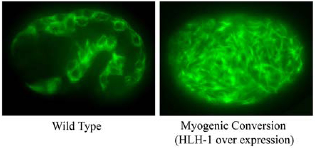
Figure 1.
Comparison of myosin staining in a wild-type and a manipulated embryo that has undergone myogenic conversion. The embryo at left is a left lateral view of a wild-type embryo at the 1.5-fold stage of embryogenesis showing the typical myosin heavy chain antibody staining pattern in the body-wall muscles. At right is a transgenic embryo treated with a pulse of heat shock to induce the expression of the master myogenic regulator HLH-1 resulting in efficient myogenic conversion of most blastomeres. The body-wall muscle-like cells are visible throughout the manipulated embryo with robust myosin levels in disorganized filament-like structures.

