Abstract
Background
In order to verify the infectivity of rodents with endoparasites in Germi (Dashte-Mogan, Ardabil Province) the current study was undertaken.
Methods
Using live traps, 177 rodents were trapped during 2005–2007. In field laboratory, all rodents were bled prior to autopsy, frozen at −20°C, and shipped to the School of Public Health, Tehran University of Medical Sciences, Iran. In parasitological laboratory, every rodent was dissected and its different organs were examined for the presence of any parasite. Blood thick and thin smears as well as impression smears of liver and spleen were stained with Geimsa and examined microscopically.
Results
Two species of rodents were trapped; Meriones persicus (90.4%) and Microtus socialis (9.6%). The species of parasites found in M. persicus and their prevalences were as follows: Hymenolepis diminuta (38.8%), Hymenolepis nana (2.5%), Trichuris sp.(40.6), Mesocestoides larva (=tetrathyridium) (3.1%), Capillaria hepatica (6.9%), Moniliformis moniliformis (11.3%), Syphacia obvelata (2.5%), Taenia endothoracicus larva (0.6%), Physaloptera sp. (0.6%), Dentostomella translucida (0.6%), Heligmosomum mixtum (0.6%), Strobilocercus fasciolaris (0.6%),and Aspiculuris tetraptera (0.6%). The species of parasites found in M. socialis and their prevalences were as follows: H. diminuta (17.6%), Trichuris sp. (5.9%), Mesocestoides larva (5.9%), S. obvelata (11.8%), S. syphacia (11.8%), H. mixtum (17.6%), and Aspiculuris tetraptera (11.8%). There were no statistical differences between male and female for infectivity with parasites in either M. persicus or M. socialis. No blood or tissue protozoan parasite was found in any of the rodents examined.
Conclusion
Among different species identified, some had zoonotic importance. Therefore, the potential health hazard of these species needs to be considered to prevent infectivity of humans.
Keywords: Rodents, Meriones persicus, Microtus socialis, Endoparasites, Iran
Introduction
Rodents have important role as host of many parasitic agents. Study on their parasites in every geographical area has medical and veterinary importance to prevent transmission of diseases to human and domestic animals. In Iran, there are some reports on the infectivity of rodents with parasites in some areas (1–5). In addition, some rodent species have been reported as reservoir of cutaneous leishmaniasis (6–8) and visceral leishmaniasis (9). However, compare to the vast extent of the country and variations of zoogeographical condition there are still much work to undertake in order to verify the species of parasites of these small mammals in different geographical areas.
This study was performed in rodents of Germi, Ardabil Province to identify the species of endoparasites with emphasize on zoonotic species.
Materials & Methods
Study area
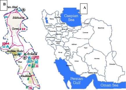
Ardabil Province is located in the North-West Iran. Its coordinate is 38°15′05″N and 48°17′50″E. This province with total area of 18634 km2 is divided in two geographical parts, mainly mountainous and 1/3 as plateau (Fig. 1). This study was carried out in Germi, a city in mountainous zone of northern part of this province. The annual precipitation of Germi is 300 mm and a temperature of −10 °C to 36 °C.
Fig. 1.
Map of the study area; A: Map of Iran, B: Map of Ardabil Province
Rodents' collection and identification
According to the map of the study area, several rodent live traps were set at outdoor places in agriculture and horticulture farms, dry riverbeds, and by the walls from mid of spring to mid of autumn, during three consecutive years from 2005 to 2007.
The fresh cucumber and walnuts were used as baits in the traps. The traps were set each afternoon during trapping occasions and were collected next early mornings. In field laboratory of Meshkinshahr Health Research Center, morphological characteristics of every rodent and their sex were registered and using valid identification key (10) species identification was performed.
Rodents' examinations and parasites identification
Rodents were anesthetized and after taking some blood and mounting on microscopic slides, they were killed. After dissection, from liver and spleen impression smears were prepared. In case of presence of any papule on the rodents' ears, in addition to preparation of two smears, some samples was also collected from the lesion and cultured in aseptic condition on special media for cutaneous leishmaniasis. Then, rodents’ carcasses were frozen at −20 °C and transferred to the School of Public Health, Tehran University of Medical Sciences for further examinations and parasites identification. In the laboratory, different organs of each rodent, including esophagus, stomach, small intestine, large intestine and cecum, peritoneum, muscles, bladder, liver, lung, brain and skin were examined under stereomicroscope and parasites were removed. After applying specific clearing and staining techniques, the parasites were identified using appropriate systematic keys (11, 12). Thick and thin blood smears and impression smears of liver and spleen were stained with Geimsa and examined microscopically with high power.
After recording the data in a file, statistical analysis was performed using Epi Info software.
Results
During the study period, 177 rodents were captured, among which 160 (90.4%) were identified as Meriones persicus (89 female and 71 male), and 17 (9.6%) as Microtus socialis (4 females and 13 males). The infection rates of these species with endoparasites are shown in Table 1. Accordingly, 74% of the rodents were infected at least with one species of parasite. Table 2 and 3 are correspondent with the infectivity of these rodents with different parasites according to the organ involvement in M. persicus and M. socialis, respectively. As these tables indicate, in M. persicus, 13 species of helminth parasites and in M. socialis, 7 species of helminth parasites were detected. No blood or tissue protozoan parasite was found in any of the rodents examined.
Table 1.
Infectivity of captured rodents with endoparasites in Germi, Ardabil Province, Iran
| Rodent species | Number infected | Percentage of infection |
|---|---|---|
| Meriones persicus (n=160) | 120 | 75 |
| Microtus socialis (n=17) | 11 | 64.7 |
| Total=177 | 131 | 74 |
Table 2.
Infectivity of Meriones persicus with different helminthes according to the sex of the rodent and internal organs
| Organ | Helminth species | Male (71) | Female (89) | Total n(%) |
|---|---|---|---|---|
| Liver | Capillaria hepatica | 3 | 8 | 11 (6.9) |
| *Mesocestoides larva (=tetrathyridium) | - | 4 | 4 (2.5) | |
| Taenia endothoracicus larva | 1 | - | 1 (0.6) | |
| Stomach | Physaloptera sp. | - | 1 | 1 (0.6) |
| Dentostomellatranslucida | - | 1 | 1 (0.6) | |
| Small intestine | Hymenolepis diminuta | 24 | 38 | 62(38.8) |
| Hymenolepis nana | 3 | 1 | 4 (2.5) | |
| Heligmosomum mixtum | - | 1 | 1 (0.6) | |
| Moniliformis moniliformis | 6 | 12 | 18(11.3) | |
| Large intestine and cecum | Syphacia obvelata | 2 | 2 | 4 (2.5) |
| Aspiculuris tetraptera | 1 | - | 1 (0.6) | |
| Trichuris sp. | 28 | 37 | 65(40.6) | |
| Peritoneum | *Mesocestoides larva | - | 4 | 4 (2.5) |
| Taenia taeniaformis larva (=Cysticercus fasciolaris) | 1 | - | 1 (0.6) |
In 3 cases the parasite occurred both in liver and peritoneum
Table 3.
Infectivity of Microtus socialis with different helminthes according to the sex of the rodent and internal organs
| Organ | Helminth species | Male (13) | Female (4) | Total n (%) |
|---|---|---|---|---|
| Stomach | Heligmosomum mixtum | 1 | - | 1 (5.9) |
| Small intestine | Hymenolepis diminuta | 3 | - | 3 (17.6) |
| Heligmosomum mixtum | 1 | 1 | 2 (11.8) | |
| Large intestine and cecum | Syphacia syphacia | 2 | - | 2 (11.8) |
| Syphacia obvelata | 2 | - | 2 (11.8) | |
| Aspiculuris tetraptera | 2 | - | 2 (11.8) | |
| Trichuris sp. | 1 | - | 1 (5.9) | |
| Peritoneum | Mesocestoides larva | - | 1 | 1 (5.9) |
Discussion
There are seven genera of Merioens in Iran. M. persicus has a wide distribution in the country. It has been known since long time ago as probable reservoir of zoonotic cutaneous leishmaniasis in North West Iran (6). In a study carried out by Mohebali et al. in the adjacent city of Germi, Meshkinshahr, M. persicus comprised 89% of the rodents trapped (3).
In the present study, overall 74 % of the rodents were infected with at least one helminth species. The rate of infection in male and female of M. persicus and M. socialis were 69% and 79.8%, and 69.2% and 50%, respectively. There were no statistical differences between male and female for infectivity with parasites either in M. persicus (P=0.16) or M. socialis (P=0.58).
Considering the variation of parasite, 13 species of helminth parasites were found in M. persicus and 7 species in M. socialis. The higher variations in M. persicus are mainly due to higher number of M. persicus examined. In M. persicus the two most prevalent species were Trichuris sp. (40.6%) and Hymenolepis diminuta (38.8%). Statistical analysis showed no significant differences between male and female of M. persicus for infectivity with Trichuris sp. (P=0.91) and H. diminuta (P=0.32).
Comparison of helminth variations in the present study with the results of similar studies on rodents’ helminth parasites in other parts of the country (1–5) indicates that H. diminuta is the most common parasite in different species of rodents. However, the prevalence of infection is variable in different studies. The rate of infection with this helminth in M. persicus in this study (38.8%) is more similar to the result of the study carried out in Meshkinshahr reporting 32.7% infection with H. diminuta in M. persicus (3).
Among 14 species of helminth parasites recovered from M. persicus and M. socialis in the study area, following species are considered as zoonotic helminthes (13); H. diminuta, H. nana, Mesocestoides sp. Larva (=tetrathyridium), Capillaria hepatica, Moniliformis moniliformis, Syphacia obvelata, Physaloptera sp., Taenia taeniaformis larva (Strobilocercus fasciolaris). In the present study, among these zoonotic species, the most prevalent one was H. diminuta. In general, 36.7% of all rodents were infected with this helminth. This high prevalence is a health threat for human in the study area. Regarding to the human infectivity with above-mentioned zoonotic species in Iran, H. nana is commonly reported throughout the country (14); and H. diminuta (15) as well as M. moniliformis (14) have already been reported as case reports in human. Additionally, recently a case of H. diminuta in a 16-month old female infant (16) and M. moniliformis in a 2-year old girl have also been reported (17). Most human cases of all these species have been occurred in children. C. hepatica has also been reported in different species of rodents in the country (2–3), so it should be considered a life-threatening parasite for human.
In conclusion, the role of rodents in spread of infectious agents in environment and needs for implementation of control measures to prevent disease transmission to human is emphasized.
Acknowledgments
This study was financially supported by the National Institute of Health Research and Tehran University of Medical Sciences, Project No: 240/5144. Thanks to Dr. S. Shojaee, Mrs B Akhoundi, Mrs. A. Mohammadiha and Mr. A. Rahimi from the Dept. of Medical Parasitology and Mycology, Tehran University of Medical Sciences for their kind assistance. The authors declare that they have no conflicts of interest.
References
- 1.Sadjjadi SM, Massoud J. Helminth parasites of wild rodents in Khuzestan Province, south west of Iran. J Vet Parasitol. 1999;13(1):55–56. [Google Scholar]
- 2.Mowlavi Gh. Study on the parasitic infections of rats in Tehran; Tehran, Iran: School of Public Health and Institute of Public Health Research, Tehran University of Medical Sciences; 1991. MSPH Thesis. [Google Scholar]
- 3.Mohebali M, Rezaei H, Farahnak A, Kanani Nootash A. A survey on parasitic fauna (helminths and ectoparasites) of the rodents in Meshkin Shahr district, North West Iran. J Fac Vet Med Univ Tehran. 1997;52(3):23–25. [Google Scholar]
- 4.Kia EB, Homayouni MM, Farahnak A, Mohebali M, Shojai S. Study on endoparasites of rodents and their zoonotic importance in Ahvaz, south west Iran. Iran J Public Health. 2001;30(1–2):49–52. [Google Scholar]
- 5.Fasihi-Harandi M. Study on the fauna of parasites of wild rodents in northern Isfahan; Tehran, Iran: School of Public Health and Institute of Public Health Research, Tehran University of Medical Sciences; 1992. MSPH Thesis. [Google Scholar]
- 6.Edrissian GhH, Ghorbani M, Tahvildar- Bidruni GH. Meriones persicus, another probable reservoir zoonotic cutaneous leishmaniasis in Iran. Trans R Soc Trop Med Hyg. 1975;69(5 & 6):517–9. doi: 10.1016/0035-9203(75)90113-3. [DOI] [PubMed] [Google Scholar]
- 7.Yaghoobi- Ershadi MR, Akhavan AA, Mohebali M. Rhombomys opimus and Meriones libycus (Rodentia: Gerbillidae) are the main reservoir hosts in a new focus of zoonotic cutaneous leishmaniasis in Iran. Trans R Soc Trop Med Hyg. 1996;90:503–504. doi: 10.1016/s0035-9203(96)90295-3. [DOI] [PubMed] [Google Scholar]
- 8.Javadian E, Dehestani M, Nadim A, et al. Confirmation of Tatera indica (Rodentia: Gerbillidae) as the main reservoir host of zoonotic cutaneous leishmaniasis in the west of Iran. Iranian J Public Health. 1998;27(1–2):55–60. [Google Scholar]
- 9.Mohebali M, Poormohammadi B, Kanani P, et al. Rodents- Gerbillida- Cricetidae. Another animal host of visceral leishmaniasis in Meshkinshahr district, I.R. of Iran. East Mediterr Health J. 1998;4(2):376–8. [Google Scholar]
- 10.Etemad E. Mammals of Iran. Vol I: Rodents and key to their identification; National Society of Natural Source and Human Environment Protection Publication; 1978. p. 288. Tehran. [Google Scholar]
- 11.Eslami A. Tehran University Publications; 1997. Veterinary Helminthology (2nd ed.). Vol.II Cestoda, Vol III Nematoda and Acanthocephala. [Google Scholar]
- 12.Yamaguti S. INC New York: Interscience Publishers; 1958–1963. Systema Helminthum (1st ed.). Vol I-IV. [Google Scholar]
- 13.Ashford RW, Crewe W. Taylor & Francis; 2003. The parasites of Homo sapiens (2nd ed.) p. 142. [Google Scholar]
- 14.Rokni MB. The present status of human helminthic diseases in Iran. Ann Trop Med Parasitol. 2008;102(4):283–295. doi: 10.1179/136485908X300805. [DOI] [PubMed] [Google Scholar]
- 15.Chadirian E, Arfaa F. Human infection with Hymenolepis diminuta in villages of Minab, Southern Iran. Int J Parasitol. 1972;2:481–482. doi: 10.1016/0020-7519(72)90093-8. [DOI] [PubMed] [Google Scholar]
- 16.Mowlavi GH, Mobedi I, Mamishi S, et al. Hymenolepis diminuta (Rudolphi, 1819) infection in a child from Iran. Iranian J Public Health. 2008;37(2):120–121. [Google Scholar]
- 17.Salehabadi A, Mowlavi GH, Sadjjadi SM. Human infection with Moniliformis moniliformis (Bremser, 1811) (Travassos, 1915) in Iran: Another case report after three decades. Vector Borne Zoonotic Dis. 2008;8(1):101–103. doi: 10.1089/vbz.2007.0150. [DOI] [PubMed] [Google Scholar]


