Abstract
Background
The aim of this paper was to study the prevalence and intensity of Anisakids larvae in the long tail tuna fish captured from Iranian shores of Persian Gulf.
Methods
Different organs including skin, abdominal cavity, stomach and intestinal contents, stomach sub serous tissues, liver, spleen, gonads and 20 grams of muscles of 100 long tail tuna fish (Thannus tonggol) caught from waters of the north parts of Persian Gulf were searched for anisakid nematodes larvae. Twenty grams of around the body cavity muscles were digested in artificial gastric juice. Different organs and digested muscles were examined with naked eyes for the presence of anisakids larvae. The collected larvae were preserved in 70% alcohol containing 5% glycerin, and cleared in lactophenol for identification.
Results
Our findings revealed that 89% of fish harbored 3rd stage larvae of Anisakis sp. of which 2% were infected with both Anisakis and Raphidascaris. All inspected organs except that of skin were found to be infected, while stomach sub serous tissues were the most infected organ (80%) followed by abdominal cavity (10%), liver (4%), testicle (3%), stomach contents and spleen (2%) and intestinal contents (1%). Intestine and abdominal cavity were the organs harbored Raphidascaris sp. Digested muscles were free of parasite. Mean intensity was low for both species and ranged between 1.5 for Raphidascaris sp. and 3.67 for Anisaki sp.
Conclusion
Anisakids larvae especially Anisakis are very prevalent in some fish including tunas of Persian Gulf, and consumption of infected fish if it is not properly cooked may lead to human anisakiasis.
Keywords: Anisakiasis, Long tail tuna fish, Persian Gulf, Iran
Anisakis larvae were reported from several species of marine fish throughout the world. The adult worms live in the intestine of marine mammals and fish. The small 3rd stage larvae are found encapsulated or free in abdominal cavity and other organs. The larvae will migrate to abdominal muscle if the fish are not quickly eviscerated. Consumption of raw, salted, smoked, fermented or undercooked fish could produces human anisakiasis.
Although 3rd stage larvae have been reported from several species of fish from south Caspian Sea(1–3), north Persian Gulf (4–6) and from abdominal cavity muscles of tuna fish (Euthynus sp.) from Persian Gulf and pikeperch (Lucioperca lucioperca) from Caspian Sea (5), no human anisakiasis is yet reported from Iran. Since the first case report in the Netherland in 19607, several cases have been reported throughout 27 countries including Japan until 1998 (8). In Japan, the annual incidence has been estimated as 2000-3000 (9). The induced clinical symptoms are due to physical effects of larval invasion into the mucosa of stomach or because of hypersensitivity to the larval secretions (10).
The aim of this paper was to study the prevalence and intensity of anisakids larvae in the long tail tuna fish captured from Iranian shores of Persian Gulf.
Materials and Methods
One hundred long tail tuna fish (Thannus tonggol) caught from waters of north parts of Persian Gulf were purchased from local fish market in Bandar -Abbas in Hormozgan Province, south of Iran, and examined for the presence of anisakid nematodes. The fish were examined within 24 hours of being caught. Each fish was eviscerated and abdominal cavity and different viscera were washed under running water into a 100 mesh sieve to remove adhering larvae. Then skin, abdominal cavity, stomach sub serous tissues, the contents of stomach and intestine and sliced livers, spleens and gonads were searched for anisakids larvae through naked eyes and under dissecting microscope. Meanwhile, 20 grams of muscles taken from around the body cavity of each fish were digested in artificial gastric juice and examined under dissecting microscope. All collected larvae were preserved in 70% alcohol containing 5% glycerin and cleared in lactophenol for identification using Chai et al. key (11).
Results
Eighty nine percent of examined fish harbored anisakids larvae in different organs, the results of which are summarized in Table 1.
Table 1.
Prevalence and intensity of Anisakis sp. in 100 long tail tuna fish from north Persian Gulf, Iran
| Organ | Infected (No.) | Infected (%) | Range | Mean No. larvae |
|---|---|---|---|---|
| Abdominal cavity | 10 | 10 | 1-42 | 8 |
| Stomach sub serous tissues | 80 | 80 | 1-5 | 3 |
| Intestinal contents | 1 | 1 | 1 | 1 |
| Stomach contents | 2 | 2 | 2-6 | 4 |
| Liver | 4 | 4 | 2-6 | 4 |
| Spleen | 2 | 2 | 1-5 | 3 |
| Testicles | 3 | 3 | 1-10 | 5 |
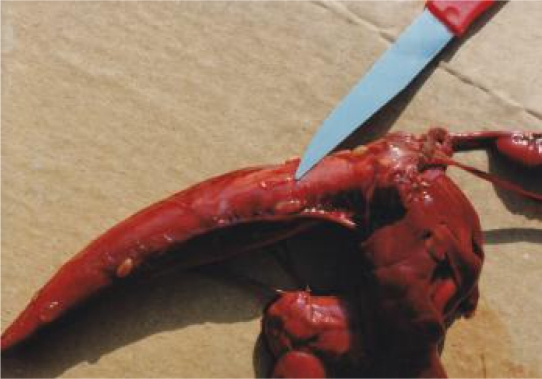
A total 367 larvae were collected from infected fish. The data in Table 1 would indicate that stomach sub serous tissues (Fig. 1) were the most and intestine the least infected organs. The high rate of prevalence and low intensity may indicate that the anisakids larvae are dispersed within their hosts. Meanwhile two fish (2%) harbored a low number of Raphidascaris sp. (ranged 1-2) in the intestine and Anisakis sp. in body cavity. No larva was found in the digested muscles.
Fig. 1.
3rd stage larvae of Anisakis spp. on the viscera of tuna fish (Authors preparation)
Discussion
Since anisakids are not host specific at the larval stage, they may be found in a wide range of different available host species, and this may result in a high probability of transmission (12, 13). In Iran, several species of fish from Caspian Sea (1–3, 5) and Persian Gulf (5, 6, 14) as well as lagoons of Khuzestan (4) have been reported to be infected with Anisakis sp. It seems likely that Anisakis is prevalent among tuna fish in the world (15).The prevalence rate of anisakiasis in long tail tuna fish in the present study was very high (89%), and the highest rate reported from fish in Iran. Meanwhile it is in harmony with other species of tuna fish, e.g. Euthynus sp. (79%) (5), other species of fish of Persian Gulf e.g. Epinephlus tauvina (14), other workers on tropical fish, (67-100%) (16), and Norwegian herring (17) (98-100%). Low intensity of anisakiasis in this study and Chai et al. study (11), (3.67 and 35.6 respectively) may indicate that Anisakis is dispersed within the hosts.
Anisakis larvae can infect several tissues of fish, a phenomenon which was observed in the present study. In contrast to our findings the migration of the larvae to the skin without encysting in fish flesh has been reported by Abollo et al. (18). Apparently anisakiasis does not harm the health of the fish, because no histhopathological lesions were found associated with the presence of encapsulated nematodes in the stomach wall and infected tissues (19). However, the pathological effects depend on the number of larvae present and the size (age) of the fish (20). Juvenile fish nematode infections may lead to some mortality(21).Apart from mortalities, the presence of encapsulated larvae in fish tissues, may lead to devaluation of the quality of the fish and significantly lower the aesthetical quality of products or pose a consumer health risk (16, 19). No larvae were found in the flesh of fish in the present study, whereas Anisakis larvae were previously found in the muscles around the body cavity of 20% of other species of Tuna fish (Euthynnus sp.) from north Persian Gulf and 15% of pikeperch (Lucioperca lucioperca) from Caspian Sea (5).
At the end of 1999, “the Food Sanitation Law Enforcement Regulation "was amended and anisakid larvae were newly added to the causative agents of food poisoning (22). The absence of anisakiasis in human in Iran may be mainly due to cooking habitat of fish in studied areas as well as other parts of the country. Smoking has little detritus effects on the larvae, although smoking of salted fish at high temperature (50 °C) may prove lethal to some larvae (23). Meanwhile it was shown that insufficient fried fish, if infected, could cause visceral larva migrans in man (10). Van thiel et al. (7) showed that heating at 50 °C will kill easily the larvae, but this temperature could not destroy encysted larvae and those protected by fish musculature. Raphidascaris sp. another anisakids larvae collected from the intestines of 2 fish (2%) with mean intensity of 1.5 has been reported from Pike (Esox lucius) of Caspian Sea (1). There are some reports on its zoonotic importance (16), but according to Cheng (24) since the adults of Raphidascaris group are intestinal parasite of fish, it is doubtful whether they can cause human infection.
Acknowledgements
This work has not been supported by any foundation. The authors declare that they have no conflict of interest.
References
- 1.Eslami A, Anwar M, Khatibi SH. Incidence and intensity of helminthose in pike (Esox lucius) of Caspian Sea (northern Iran) Riv It Piscic Ittiop. 1972;7(1):1–14. [Google Scholar]
- 2.Eslam A, Kohnehshahri M. Study on the helminthiasis of Rutilus frisii katum from the south Caspian Sea. Acta Zool Patho Antverpienia. 1978;70:153–155. [PubMed] [Google Scholar]
- 3.Mokhayer B. A list of parasites of eusturgeon (Acipenseridae) in Iran. J Vet Fac Univ Tehran. 1973;29(1):11–12. [Google Scholar]
- 4.Farahnak A, Mobedi I, Tabibi R. Fish anisakid helminthes in Khuzestan province, south west of Iran. Iranian J Publ Health. 2002;31(3,4):129–132. [Google Scholar]
- 5.Eslami A, Mokhayer B. Nematode larvae of medical importance found in market fish in Iran. Pahlavi Med J. 1977:345–348. [PubMed] [Google Scholar]
- 6.Mokhayer B. Pseudotangue in snapper fish of Persian Gulf. Proc. 3rd Int Cong Parasitol.; Monchen; pp. 345–8. [Google Scholar]
- 7.Van Thiel PH, Kuipers FC, Roskam RT. A nematode parasitic to herring causing acute abdominal syndrome in man. Trop Georg Med. 1960:97–113. [PubMed] [Google Scholar]
- 8.Ishikura PH, Takahanshi S, Yagi K, Nakamura K, Kon S, Matsuura A. Epidemiology: global aspect of anisakidosis, Chiba (Japan) Int Cong Parasitol. Report No. ICOPA IX; 1998. pp. 379–382. [Google Scholar]
- 9.Umehara A, Kawakami Y, Araki J, Uchida A. Molecular identification of the etiological agent of the human anisakiasis in Japan. Parasitol Int. 2007;58(3):211–5. doi: 10.1016/j.parint.2007.02.005. [DOI] [PubMed] [Google Scholar]
- 10.Kim SG, Jo YJ, Park YS, Kim SH, Song MH, Lee HH, Kim JS, Ryou JW, Joo JE, Kim DH. Four cases of gastric submucosal mass suspected as anisakiasis. Korean J Parasitol. 2006;44(1):81–86. doi: 10.3347/kjp.2006.44.1.81. [DOI] [PMC free article] [PubMed] [Google Scholar]
- 11.Chai JY, Chu YM, Sohn WM, Lee SH. Larval anisakids collected from the yellow corvine in Korea. Korean J Parasitol. 1986;24(1):1–11. doi: 10.3347/kjp.1986.24.1.1. [DOI] [PubMed] [Google Scholar]
- 12.Smith JW, Wootten R. Experimental study on the migration of Anisakis larvae (Nematoda, Ascaridida) into the flesh of herring, Clupea harengus L. Int J Parasitol. 1978;5:133–136. doi: 10.1016/0020-7519(75)90019-3. [DOI] [PubMed] [Google Scholar]
- 13.Mattiucci S, Nscetti G, Cianchi R, Paggi L, Arduino P, Margolis L., Brattey J, Webb SD, 'Amelio S, Orecchia P, Bullini L. Genetic and ecological data on Anisakis simplex complex with evidence for a new species (Nematoda, Anisakidae) J Parasitol. 1997;83:401–416. [PubMed] [Google Scholar]
- 14.Radfar. MH, Eslami A. Study on the helminth infections of Epinephlus tauvina of Persian Gulf, Iranian shores. 1st. Cong Dis Aqua Org Iran; 2000. pp. 11–14. [Google Scholar]
- 15.Munday BL, Sawada Y, Cribb T, Hyward CJ. Diseases of Tunas, Thunnus spp. J Fish Dis. 2003:187–206. doi: 10.1046/j.1365-2761.2003.00454.x. [DOI] [PubMed] [Google Scholar]
- 16.Doupe RG, Lymbery AJ, Wong S, Hobbs RP. Larval anisakid infections of some tropical fish species fro north-west Australia. J Helminthol. 2003;77:363–365. doi: 10.1079/joh2003193. [DOI] [PubMed] [Google Scholar]
- 17.Levsen A, Tore B. Anisakis simplex third stage larvae in Norwegian spring spawning herring (Clupea herringus L.) with emphasis on larval distribution in the flesh. Vet Parasitol. 2010;171(3,4):247–253. doi: 10.1016/j.vetpar.2010.03.039. [DOI] [PubMed] [Google Scholar]
- 18.Abollo E, Pascul S. SEM study of Anisakis brevispiculata Dollfus, 1966 and Pseudoterranova ceticola (Deardoff Overstreet,1981) (Nematoda, Anisakidae), parasites of pigmy sperm wale, Kogia breviceps . Sci Mar. 2002;66:249–255. [Google Scholar]
- 19.Costa G, Madeira A, Pontes T, Amelio D. Anisakid nematodes of blockspot seabream, Pagellus bogaraveo, from Madeiran waters, Portugal. Acta Parasitol. 2004;49(2):156–161. [Google Scholar]
- 20.Lester RJG, Adams JR. Gyrodactylus alexandri reproduction, mortality, and effect on its host Gasterosteus aculeatus . Canadian J Zool. 1974;52:827–833. doi: 10.1139/z74-112. [DOI] [PubMed] [Google Scholar]
- 21.Rohde K. Ecology of marine parasites. Walingford: CAB International; 1993. [Google Scholar]
- 22.National Institute of infectious Diseases and Tuberculosis and Infectious Diseases Control Division, Ministry of Health, Labour and Welfare. Foodborn helminthiases as emerging diseases in Japan. Infect Agents Surveillance Rep. 2004;25:114–115. [Google Scholar]
- 23.Euzeby J. Sur quelque nematodes ascaridida, agent d'une zoonose helminthique d'actualite la “maladie du ver harrenge, Bull. Soc Vet Med Comp. 1972;74:359–371. [Google Scholar]
- 24.Cheng TC. Anisakiosis. In: Palmer SR, Lord Soulsby EJl, Simpson DIH, editors. Zoonosis. Oxford. Oxford University Press; 1998. pp. 827–831. [Google Scholar]


