Abstract
Purpose
To evaluate the safety and efficacy of combined transepithelial topography-guided photorefractive keratectomy (PRK) therapeutic remodeling, combined with same-day, collagen cross-linking (CXL). This protocol was used for the management of cornea blindness due to severe corneal scarring.
Methods
A 57-year-old man had severe corneal blindness in both eyes. Both corneas had significant central scars attributed to a firework explosion 45 years ago, when the patient was 12 years old. Corrected distance visual acuity (CDVA) was 20/100 both eyes (OU) with refraction: +4.00, −4.50 at 135° in the right eye and +3.50, −1.00 at 55° in the left. Respective keratometries were: 42.3, 60.4 at 17° and 35.8, 39.1 at 151.3°. Cornea transplantation was the recommendation by multiple cornea specialists as the treatment of choice. We decided prior to considering a transplant to employ the Athens Protocol (combined topography-guided partial PRK and CXL) in the right eye in February 2010 and in the left eye in September 2010. The treatment plan for both eyes was designed on the topography-guided wavelight excimer laser platform.
Results
Fifteen months after the right eye treatment, the right cornea had improved translucency and was topographically stable with uncorrected distance visual acuity (UDVA) 20/50 and CDVA 20/40 with refraction +0.50, −2.00 at 5°. We noted a similar outcome after similar treatment applied in the left eye with UDVA 20/50 and CDVA 20/40 with −0.50, −2.00 at 170° at the 8-month follow-up.
Conclusion
In this case, the introduction of successful management of severe cornea abnormalities and scarring with the Athens Protocol may provide an effective alternative to other existing surgical or medical options.
Keywords: Athens Protocol, collagen cross-linking, cornea blindness, cornea scarring, photorefractive keratectomy, vision
Video abstract
Video
Case report
A 57-year-old man had severe corneal blindness from cornea scarring attributed to a firework explosion accident that occurred 45 years ago, when the patient was 12 years old. When we first evaluated the patient in 2009, the uncorrected distance visual acuity (UDVA) was 20/400 in both eyes, with pinhole improvement to 20/100 in the right eye and 20/70 in the left eye. There was no improvement to his visual function with spectacle refraction or soft contact lenses, and there was intolerance to gas-permeable contact lenses in both eyes.
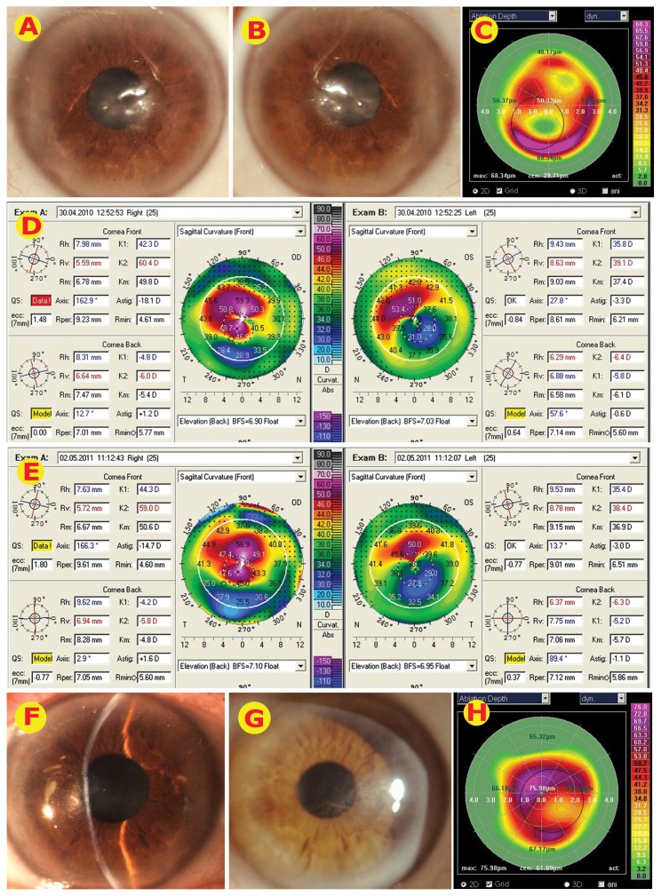
Due to the dense scarring in the right eye, an endothelial cell count (ECC) was not possible. The ECC was 2000 cells/mm2 in the left eye. Slit lamp biomicroscopy revealed severe horizontal central corneal scars in both eyes (see Figure 1A is the right eye and B the left eye). Dilated fundus examination revealed no cataracts, normal disks, macula, and retina vessels.
Figure 1.
(A) Slit-lamp image of the OD at presentation, showing a significant horizontal cornea scar. (B) Slit-lamp picture of the OS; there was central corneal scarring similar to the OD. (C) The treatment plan on the wavelight excimer platform for topography-guided partial PRK employed for the OD treatment. The treatment plan, pivotal to the application of the Athens Protocol, combines a myopic ablation over the elevated cornea and a partial hyperopic application peripheral to the flattened (by scarring) inferior cornea. This combination treatment enhances the normalization of the severe irregularity with small ablation (35 μm) over the thinnest cornea. (D) Tomography maps (Oculyzer, Wavelight, Erlangen, Germany) of the OD and OS preoperative to the Athens Protocol. (E) Tomography maps of the OD and OS postoperative to the Athens Protocol, 15 months following the OD and 8 months following the OS. (F) Slit-lamp picture of the OD, 15 months following treatment; cornea regularity and improvement in translucency is evident (when compared to A). (G) Slit-lamp picture of the OS, 8 months following treatment; cornea regularity and improvement in translucency is evident (when compared to B). (H) The treatment plan on the wavelight excimer platform employed for topography-guided partial PRK of the OS.
Abbreviations: OD, right eye; OS, left eye; PRK, photorefractive keratectomy.
CDVA was 20/100 both eyes (OU) with refraction: +4.00, −4.50 at 135 in the right eye (OD), +3.50, −1.00 at 55 in the left eye (OS). Respective keratometries were: 42.3, 60.4 at 17 and 35.8, 39.1 at 151.3.
Tomographic evaluation (Oculyzer II, Wavelight, Erlangen, Germany) of both eyes is noted in Figure 1D and shows the thinnest pachymetry of 467 μm in the right eye and 448 μm in the left eye, respectively. Considering the options of lamellar and penetrating keratoplasty, we discussed with the patient the possibility of normalizing the cornea surface, removing some of the scar and additionally utilizing collagen cross-linking (CXL) to biomechanically reinforce the thinned corneas and potentially reduce scarring by suppressing stromal keratocytes.
Potential complications were extensively discussed. The patient decided to proceed with our recommendation and we employed the Athens Protocol (combined topography-guided partial photorefractive keratectomy [PRK] and CXL) in the right eye in February 2010 and in the left eye 7 months later, in September 2010. We have previously reported on this technique1–5 for the management of cornea ectasia. The excimer laser treatment plan for both eyes was designed on the wavelight excimer laser platform and is demonstrated in Figure 1C and H. The treatment plan, pivotal to the application of the Athens Protocol, combines a myopic ablation over the elevated cornea and a partial hyperopic application peripheral to the flattened (by scarring), inferior cornea. This combination treatment enhances the normalization of the severe irregularity with small ablation (35 μm) over the thinnest cornea. Fifteen months after the treatment of the right eye, the cornea had cleared and was topographically stable with UDVA at 20/50 and corrected distance visual acuity (CDVA) of 20/40 with refraction +0.50, −2.00 at 5°. We noted a similar outcome for the left eye 8 months after the treatment with UDVA of 20/50 and CDVA of 20/40 with −0.50, −2.00 at 170°.
The postoperative slit-lamp photos of the anterior segment are seen in Figure 1 (right eye: F and left eye: G). His tomographic keratometry in the right eye has improved to 44.3, 59.0 at 13.7°and in the left eye to 35.4, 38.4 at 166.3°(Figure 1E shows the right eye on the left and the left eye on the right). Endothelial cell counts (ECC) were possible postoperatively in the right eye, most likely due to improved cornea clarity, and were 1600 cells/mm2. The postoperative ECC in the left eye was measured at 2010 cells/mm2.
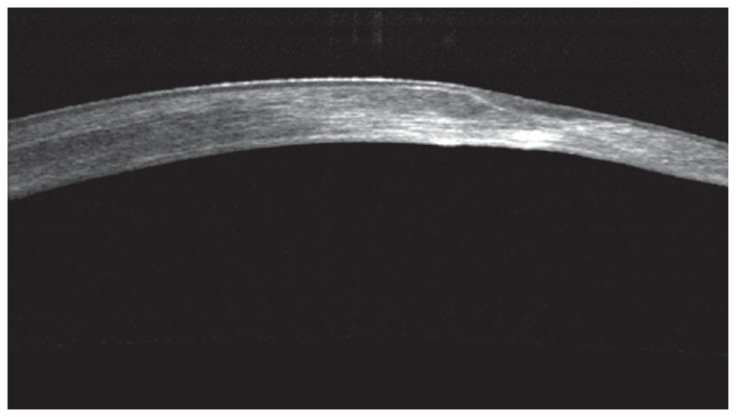
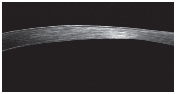
The preoperative cornea optical coherence tomography (OCT) is shown in Figure 2 and the postoperative in Figure 3. Due to improved visual rehabilitation, the patient has recently obtained a driver’s license and has assumed a more independent lifestyle.
Figure 2.
Preoperative cornea OCT showing the dense, deep stromal scarring (hyper-reflective spots) along with the very irregular stromal surface, partly masked by epithelium thinning over the peaks and thickening over the deep valleys.
Abbreviation: OCT, optical coherence tomography.
Figure 3.
Picture showing the same portion of cornea as in Figure 2, in a cornea OCT 12 months following the treatment. The scar has been significantly reduced, the stromal surface smoothed, the epithelium has become more uniform in thickness, and the overall cornea thickness has been reduced.
Abbreviation: OCT, optical coherence tomography.
In this particular patient, the therapeutic aim of the topography- guided PRK was to attempt to normalize the highly irregular corneal surface, and the employment of the CXL had a two-fold objective: to reduce corneal scarring by eliminating keratocytes, and to stabilize the thinner cornea produced by the removal of corneal tissue with the therapeutic topography-guided ablation.
We feel that in this case the introduction of successful management of severe cornea abnormalities and scarring with the Athens Protocol may provide an effective alternative to other surgical options such as lamellar or penetrating keratoplasty. Further studies in a large cohort of patients with a longer follow-up are needed to further establish the effectiveness and safety of this technique.
Footnotes
Disclosure
The author reports no conflicts of interest in this work.
References
- 1.Kanellopoulos AJ, Skouteris VS. Secondary ectasia due to forceps injury at childbirth. management with combined topography-guided partial PRK and collagen cross-linking (the Athens protocol), followed by the implantation of a phakic intraocular lens (IOL) J Refract Surg. 2011 doi: 10.3928/1081597X-20110901-05. [DOI] [PubMed] [Google Scholar]
- 2.Kanellopoulos AJ, Binder PS. Management of corneal ectasia after LASIK with combined, same-day, topography-guided partial transepithelial PRK and collagen cross-linking: the Athens protocol. J Refract Surg. 2011;27(5):323–331. doi: 10.3928/1081597X-20101105-01. [DOI] [PubMed] [Google Scholar]
- 3.Krueger RR, Kanellopoulos AJ. Stability of simultaneous topography- guided photorefractive keratectomy and riboflavin/UVA cross-linking for progressive keratoconus: case reports. J Refract Surg. 2010;26(10):S827–S832. doi: 10.3928/1081597X-20100921-11. [DOI] [PubMed] [Google Scholar]
- 4.Kanellopoulos AJ. Comparison of sequential vs same-day simultaneous collagen cross-linking and topography-guided PRK for treatment of keratoconus. J Refract Surg. 2009;25(9):S812–S818. doi: 10.3928/1081597X-20090813-10. [DOI] [PubMed] [Google Scholar]
- 5.Kanellopoulos AJ, Binder PS. Collagen cross-linking (CCL) with sequential topography-guided PRK: a temporizing alternative for keratoconus to penetrating keratoplasty. Cornea. 2007;26(7):891–895. doi: 10.1097/ICO.0b013e318074e424. [DOI] [PubMed] [Google Scholar]


