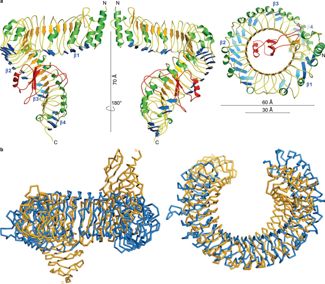
Figure 1. The BRI1 ectodomain forms a superhelical assembly.
a, Ribbon diagram of the BRI1 LRR domain (front, back and top). The canonical LRR β-sheet is shown in orange, and the additional plant-specific β-sheets in blue. Helices are shown in green, and the island domain is depicted in red. b, Structural comparison of the BRI1 (shown as yellow Cα trace) and TLR3 (in blue, pdb-id: lziw)27 ectodomains. The structures superimpose with an r.m.s.d. of 4.2 Å between 341 corresponding Cα atoms. Side and top views are shown. The island domain has been omitted for clarity.

