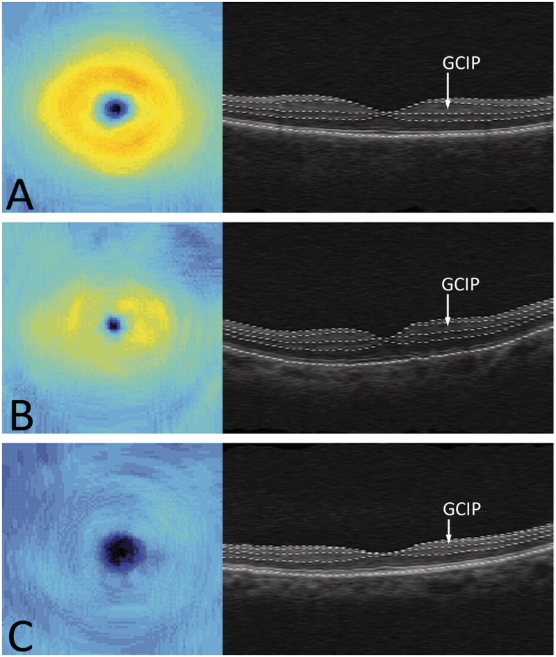
Figure 2.
Ganglion cell layer plus inner plexiform layer (GCIP) thickness maps and retinal B-scans in the horizontal direction showing layer segmentations for a healthy control (A), a multiple sclerosis eye with a history of optic neuritis (B), and a neuromyelitis optica eye with a history of optic neuritis (C).

