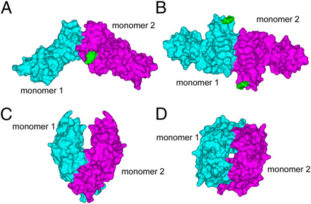
FIGURE 1.
Surface representations of the two distinct crystallographic dimers of FcγRIIa. A, Side view of the FcγRIIa-LR dimer with monomer 1 in cyan, monomer 2 in magenta, and the GlcNAc residue attached to N145 shown in green. B, End-on view of the FcγRIIa-LR dimer, looking down onto the surfaces involved in IgG binding. C, Side view of the FcγRIIa-HR dimer. D, End-on view of the FcγRIIa-HR dimer.

