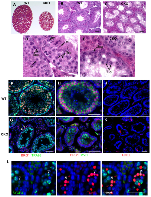
Fig. 2.
Loss of spermatocytes and increased apoptosis in Brg1CKO testes. (A-E) Hematoxylin and Eosin staining of wild-type (WT) and mutant (CKO) testes from 10-week-old mice. (F,G) Double immunostaining of BRG1 (red) and TRA98, a pan-germ cell marker (green), in 4-week-old wild-type (F) and Brg1CKO (G) testes (Sertoli cells marked by arrows). (H,I) Mouse vasa homolog (MVH, green) activation in differentiating germ cells. (J,K) Fluorescence TUNEL assay of wild-type (J) and mutant (K) testes at 3 weeks. (L) Co-staining of TUNEL (red) with SYCP3 (green) in apoptotic Brg1CKO spermatocytes. Scale bars: 50 μm. el, elongated spermatid; ro, round spermatid; Spc, spermatocyte; Spg, spermatogonia.

