Abstract
CD40L is essential for the development of adaptive immune responses. It is generally thought that CD40L expression in CD4+ T cells is regulated transcriptionally and made from new mRNA following antigen recognition. However, imaging studies show that the majority of cognate interactions between effector CD4+ T cells and APCs in vivo are too short to allow de novo CD40L synthesis. We previously showed that Th1 effector and memory cells store preformed CD40L (pCD40L) in lysosomal compartments and mobilize it onto the plasma membrane immediately after antigenic stimulation, suggesting that primed CD4+ T cells may use pCD40L to activate APCs during brief encounters. Indeed, our recent study showed that pCD40L is sufficient to mediate selective activation of cognate B cells and trigger DC activation in vitro. In this study, we show that pCD40L is present in Th1 and follicular helper T cells developed during infection with lymphocytic choriomeningitis virus, Th2 cells in the airway of asthmatic mice, and Th17 cells from the CNS of animals with experimental autoimmune encephalitis (EAE). pCD40L is nearly absent in both natural and induced Treg cells, even in the presence of intense inflammation such as occurs in EAE. We also found pCD40L expression in CD4 single positive thymocytes and invariant NKT cells. Together, these results suggest that pCD40L may function in T cell development as well as an unexpectedly broad spectrum of innate and adaptive immune responses, while its expression in Treg cells is repressed to avoid compromising their suppressive activity.
Introduction
T cell help for APCs is essential for adaptive immune responses [1], [2]. Effector CD4+ T cells deliver help to antigen-specific B cells in an MHC class II-restricted manner [3]. CD40L, a membrane-bound cytokine in the TNF superfamily, plays a crucial role in this process. CD40L is also required for generating optimal CD4+ T and CD8+ T cell responses through activation of dendritic cells (DCs) [4]. Thus, lack of CD40L expression causes defective humoral and cellular immunity [5]. In contrast, dysregulated CD40L expression causes autoimmunity, lymphoma, and premature termination of humoral immunity [6], [7], [8], [9]. A recent clinical trial of recombinant CD40L failed to restore B cell responses whereas it successfully elicited Th1 responses in patients who harbor mutations in the genes encoding CD40L [10]. A precise understanding of CD40L regulation, including its expression and manner of delivery, could assist in the development of effective vaccines, immunological interventions for inflammatory diseases, and successful treatment of CD40L deficient patients.
It is generally thought that CD40L is synthesized from new mRNA (de novo CD40L) and delivered while effector CD4+ T cells are engaged in intimate interactions with cognate APCs in the time frame of a few hours [11]. This notion has been challenged by studies utilizing two-photon microscopy. While the initial, stable interactions of naïve CD4+ T cells and DCs can last for several hours, the majority of interactions between effector CD4+ T cells and cognate APCs in vivo are surprisingly short, ranging from several minutes up to 30 minutes [12], [13], [14], [15]. Although these short interactions are antigen-specific and presumed to be productive, 30 minutes is not enough time for effector CD4+ T cells to make de novo CD40L.
We propose that effector CD4+ T cells activate cognate APCs during brief interactions using preformed CD40L (pCD40L). We and others have demonstrated that human and mouse effector and resting memory CD4+ T cells retain pCD40L intracellularly, and that pCD40L can come to the cell surface within a few minutes of antigenic stimulation [16], [17]. Th1 cells store pCD40L in lysosome-related organelles known as secretory lysosomes [17], a category of secretory vesicles which includes the lytic granules containing perforin and granzyme B in cytotoxic T-lymphocytes (CTLs) and natural killer (NK) cells [18]. The existence of cytotoxic Th1 cells in humans and mice which resemble CD8+ CTLs in function also supports the idea of antigen-specific execution of CD4+ T cell effector functions by controlled, directional secretion of preformed effector molecules through delivery of secretory compartment to the immunological synapse [19], [20], [21]. In fact, our recent study demonstrates that pCD40L is sufficient to mediate selective activation of cognate B cells and trigger DC activation in vitro [22].
Many subsets of effector CD4+ T cells have been described: Th1 cells control intracellular pathogens, Th2 cells contain extracellular parasites, Th17 cells counteract extracellular bacteria and fungi, T follicular helper (TFH) cells promote antibody production, and regulatory T (Treg) cells prevent uncontrolled tissue damage by dampening APC activation [23]. Although other groups have reported selective expression of pCD40L in certain subsets of effector CD4+ T cells in disease states and healthy animals [16], [17], [24], [25], [26], [27], [28], this report is the first to systematically examine surface mobilization of pCD40L in each subset of effector CD4+ T cells and Treg cells, using physiologically relevant antigen-pulsed APCs to trigger surface mobilization in an effort to shed light on the role of pCD40L in vivo.
In the present study, we investigated TCR-regulated surface expression of pCD40L in Th1, Th2, Th17, and TFH cells, thymocytes, invariant natural killer T (iNKT) cells and Treg cells. Our results show that pCD40L is stored in all tested subsets of effector CD4+ T cells from lymphoid organs and non-lymphoid effector sites as well as CD4 single positive (SP) thymocytes and iNKT cells, but is undetectable or nearly undetectable in natural or induced Treg cells, NK cells, or CD8+ T cells. These results provide the first comprehensive description of cells that store and mobilize pCD40L in vivo, and suggest that pCD40L may function in CD4+ T cell development and a broad range of immune responses in vivo.
Materials and Methods
Mice
Mice were housed under specific pathogen–free conditions. These studies were approved by the Institutional Animal Care and Use Committee at Oregon Health & Science University. BALB/c, C57BL/6, DO11.10, Cd40lg−/−, β2m−/−, and IL-4/enhanced GFP reporter (4get) mice were from the Jackson Laboratory (Bar Harbor, ME). DO11.10 Rag2−/− and BALB/c nu/nu mice were obtained from Taconic Farms (Germantown, NY). 4get/DO11.10 mice were bred in-house. SMARTA mice were obtained from Dr. J. Lindsay Whitton (The Scripps Research Institute).
Antibodies and reagents
Biotin–anti-CXCR5 was purchased from BD Biosciences (San Jose, CA). FITC-anti-T1/ST2 was purchased from MD Biosciences (Saint Paul, MN). Allophycocyanin-labeled tetramers consisting of CD1d folded with PBS57 were provided by the NIH Tetramer Core Facility (Atlanta, GA). All other antibodies for flow cytometry were purchased from eBioscience (San Diego, CA). The Foxp3 staining kit was purchased from Biolegend (San Diego, CA). Recombinant cytokines were purchased from Peprotech (Rocky Hill, NJ). Anti-IFN-γ and anti-IL-4 were from Bio X Cell (West Lebanon, NH). Papain was from Calbiochem (San Diego, CA). Endotoxin-free ovalbumin (OVA) protein was from Profos AG (Regensburg, Germany). OVA peptide (323–339) was from AnaSpec, Inc. (Fremont, CA). All-trans retinoic acid, BSA, OVA protein, PMA and ionomycin were from Sigma-Aldrich (St Louis, MO).
T cell differentiation in vitro
Th1, Th2, and Th17 cells were prepared by culturing spleen cells from DO11.10 mice in the presence of 1 µM of antigenic peptide (OVA 323–339) for 4 days with combinations of cytokines and antibodies as follows: Th1: 1 ng/ml IL-12 and 10 µg/ml anti-IL-4; Th2: 10 µg/ml anti-IFN-γ and 100 ng/ml IL-4; Th17: 20 µg/ml anti-IFN-γ, 20 µg/ml anti-IL-4, 20 µg/ml anti-IL-2, 100 ng/ml IL-6, 5 ng/ml human TGF-β1, 20 ng/ml IL-1β, and 20 ng/ml TNF-α. Differentiation of Th1, Th2, and Th17 cells was confirmed by intracellular cytokine staining of IFN-γ, IL-4, and IL-17A upon 5 hour stimulation with PMA plus ionomycin in the presence of brefeldin A using the Cytofix/Cytoperm kit from BD Biosciences (data not shown). In some experiments, Th2 cells were restimulated with antigen-pulsed purified B cells in the absence or presence of recombinant IL-4 or anti-IL-4 for 4 days. Stability of Th2 cells after the second round of proliferation was assessed by FACS analysis of IL-4/eGFP levels and by detection of IL-4 in the culture media with ELISA kits (BD Biosciences). To obtain induced Treg (iTreg) cells, DO11.10 cells and B cells were purified with EasySep mouse CD4+ T cell enrichment and B cell enrichment kits (Stemcell Technologies: Vancouver, Canada), respectively, and were co-cultured in the presence of 1 µM antigenic peptide, 100 U/ml IL-2, 20 ng/ml TGF-β, and 10 nM all-trans retinoic acid for 4 days [29].
Lymphocytic choriomeningitis virus (LCMV) infection
To obtain in vivo-generated Th1 cells, spleen cells were prepared from recipient C57BL/6 mice that had been given 2×10⁴ spleen cells from SMARTA mice followed by i.p. infection with 2×10⁵ PFU of LCMV (Armstrong 53b strain) [17]. To assess endogenous, polyclonal Th1 and TFH cells, spleen cells were harvested 12 days after LCMV infection.
Papain plus OVA immunization
To obtain in vivo-generated Th2 and TFH cells, one million 4get/DO11.10 cells (CD25-depleted, >95% purity) were transferred into BALB/c mice. The next day, recipients were immunized subcutaneously with papain (50 µg) plus endotoxin-free OVA protein (50 µg) [30]. On day 7, Th2 and TFH cells from draining lymph nodes (dLNs) were prepared for flow cytometric analysis.
Asthma model
To obtain polyclonal Th2 cells, C57BL/6 mice were sensitized twice by i.p. injection of OVA protein emulsified in aluminum hydroxide (Pierce, Rockford, IL) and challenged intratracheally with OVA protein/PBS three times. Mice were sacrificed, tracheas were cannulated, and lungs were lavaged three times with 0.5 ml PBS per wash to obtain bronchoalveolar lavage fluid (BALF) cells [31].
Experimental autoimmune encephalomyelitis (EAE) model
For analysis of in vivo-generated Th17 cells and Treg cells from an inflammatory site, active EAE was induced and CNS infiltrating leukocytes were obtained as described [32]. Briefly, C57BL/6 mice were immunized by subcutaneous injection in the lower back with 200 µg myelin oligodendrocyte glycoprotein (MOG)35–55 (MEVGWYRSPFSRVVHLYRNGK) peptide emulsified at a 1∶1 ratio with complete Freund's adjuvant containing 150 µg Mycobacterium tuberculosis H37RA (Difco, Detroit, MI). Pertussis toxin (List Biological Laboratories, Campbell CA) was administered on day 0 (200 ng) and day +2 (200 ng) with respect to the immunization day. Only symptomatic mice were used at 14 days after immunization.
Oral tolerance model
In vivo-generated iTreg cells were prepared from mesenteric LNs of BALB/c nu/nu recipients which had received i.v. injection of 5×10⁵ naive CD4+ T cells purified from DO11.10 Rag2−/− mice followed by feeding with BSA- or OVA-containing water for 6 days. [33].
Flow cytometry for detection of pCD40L
The surface mobilization assay and intracellular staining were described previously [17] and are explained at the beginning of the Results section. To detect pCD40L in Th17 cells generated in vivo, CNS infiltrating leukocytes from EAE animals were analyzed by the CD40L mobilization assay followed by intracellular cytokine staining. Cells were incubated in the presence or absence of MOG peptide-pulsed APCs with either isotype-PE or PE-labeled anti-CD40L at 37°C for 30 minutes. After washes, cells were incubated for 4.5 hours in the presence of brefeldin A and MOG peptide-pulsed APCs. After fixation, intracellular IL-17A and IFN-γ were stained. Data were obtained with an LSR II (BD Biosciences) and analyzed with FlowJo software (Tree Star, Inc., Ashland, OR).
Results
Detecting pCD40L using the mobilization assay and intracellular staining
We previously reported successful use of the mobilization assay and intracellular staining to assess existence of pCD40L in Th1 effector and memory cells as well as memory-phenotype CD4+ T cells [17], [22]. It has been reported that CD40 engagement induces CD40L internalization [34] and that inhibition of CD40-CD40L engagement with blocking anti-CD40L increases CD40L detection [35]. In the mobilization assay, fluorochrome-labeled anti-CD40L mAb is included in the culture at 37°C in the presence or absence of stimulation. Compared to the “snap shot” nature of conventional staining at 4°C after completion of stimulation, the mobilization assay provides the “long exposure” view of CD40L surface expression by capturing and stabilizing CD40L that has been delivered to the cell surface during incubation (Fig. S1) [36], [37]. We found negligible amounts of pre-existing surface CD40L on resting effector CD4+ T cells [17]. By limiting the stimulation period to 30 minutes, we were able to exclude surface expression of de-novo CD40L made following stimulation. Although a contribution of new CD40L protein expression from stable pre-existing CD40L mRNA cannot be fully excluded by limiting the assay to 30 minutes [38], we showed in a previous report with T effectors that complete inhibition of protein synthesis did not diminish surface expression of CD40L following 30 minutes of stimulation [17]. Intracellular staining can be seen as an “x-ray”, and could distinguish defective mobilization of pCD40L from absence of stored pCD40L in cases where no mobilization of pCD40L is observed.
In vitro-generated Th1 and Th17, but not Th2 or iTreg cells, store and mobilize pCD40L upon stimulation
We previously showed that effector and resting memory Th1 cells, differentiated either in vitro or in vivo, store intracellular pCD40L and mobilize it to the cell surface within 30 minutes of stimulation [17]. A finding that pCD40L is stored in certain Th subsets would suggest that pCD40L serves in defined immune responses, just as signature cytokines do. Therefore, we examined the distribution of pCD40L among subsets of effector CD4+ T cells and Treg cells.
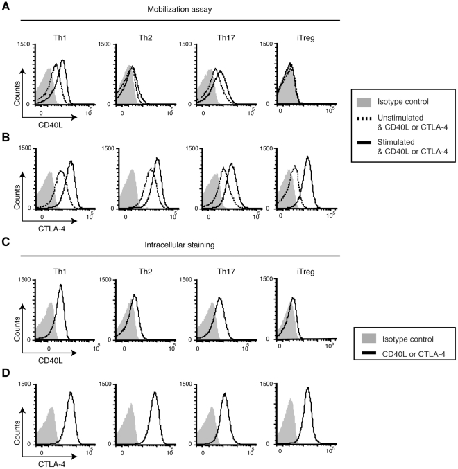
We generated Th1, Th2, Th17, and iTreg cells in vitro and measured pCD40L. While Th1 and Th17 cells clearly mobilize pCD40L, Th2 cells mobilize much less pCD40L (Fig. 1A ) and possess significantly less intracellular CD40L (Fig. 1C ). pCD40L is at the limit of detection in iTreg cells (Figs. 1A, 1C ). The decreased surface mobilization in Th2 cells, and the near absence of pCD40L in Treg cells, are not due to suboptimal stimulation or permeabilization of these cells because Th2 and iTreg cells possess and mobilize preformed CTLA-4 at the same level as Th1 and Th17 cells (Figs. 1B, 1D ).
Figure 1. In vitro-generated Th1 and Th17, but not Th2 or iTreg cells mobilize pCD40L.
A and B, Mobilization assay. In vitro-generated Th1, Th2, Th17, and iTreg cells were stimulated with PMA plus ionomycin or left unstimulated in the presence of PE-isotype Ab, PE-anti-CD40L or PE-anti-CTLA-4 at 37°C for 30 minutes. The levels of CD40L (A) and CTLA-4 (B) are shown. C and D, Intracellular staining. Cells were fixed without stimulation, permeabilized, and stained with PE-isotype Ab, PE-anti-CD40L or PE-anti-CTLA-4. The levels of CD40L (C) and CTLA-4 (D) are shown. Data are representative of five independent experiments.
In vivo-generated Th1 and TFH cells possess and mobilize pCD40L upon antigenic stimulation
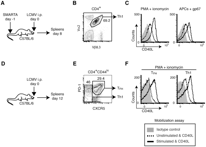
To further characterize the involvement of pCD40L in vivo, we examined effector CD4+ T cell subsets generated in vivo. SMARTA CD4+ T cells, which have a transgenic TCR specific for an LCMV epitope [39], were transferred into normal mice followed by infection with LCMV (Fig. 2A ). Th1 differentiation of these cells was confirmed by intracellular staining of IFN-γ upon in vitro stimulation with PMA plus ionomycin (60–70% IFN-γ+, data not shown). The mobilization assay shows that in vivo-generated Th1 SMARTA (CD4+Vα2+Vβ8.3+, Fig. 2B ) cells mobilize pCD40L in an antigen-specific manner (Fig. 2C ). SMARTA cells expanded during LCMV infection were reported to contain both Th1 and TFH cells [40]. To rule out the possibility of preferential pCD40L expression in TFH cells rather than Th1 cells, mobilization of pCD40L was assessed in endogenous polyclonal TFH cells (CD4+CD44hiCXCR5hiPD-1hi) and Th1 cells (CD4+CD44hiCXCR5lowPD-1low) from spleens of LCMV-infected mice (Figs. 2D, 2E ). The result shows that both Th1 and TFH cells mobilize pCD40L upon stimulation (Fig. 2F ).
Figure 2. In vivo-generated Th1 and TFH cells possess and mobilize pCD40L.
A, Generation of antigen-specific Th1 cells. LCMV-glycoprotein-specific, TCR-transgenic SMARTA CD4+ T cells were recovered from LCMV-infected mice on day 8 post-infection. B, Gating strategy for SMARTA cells (Vα2+, Vβ8.3+). C, Mobilization of pCD40L by SMARTA cells in response to PMA plus ionomycin or the LCMV peptide gp67-pulsed APC. D, Polyclonal Th1 and TFH cells. Spleen cells were obtained from LCMV-infected mice on day 12 post-infection. E, Gating strategy for TFH (CD4+CD44hiCXCR5hiPD-1hi) and Th1 (CD4+CD44hiCXCR5lowPD-1low) cells. F, Mobilization of pCD40L by TFH and Th1 cells in response to PMA plus ionomycin. Data are representative of two independent experiments.
In vivo-generated Th2 cells possess and mobilize pCD40L upon antigenic stimulation
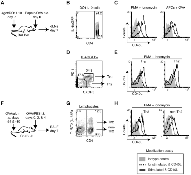
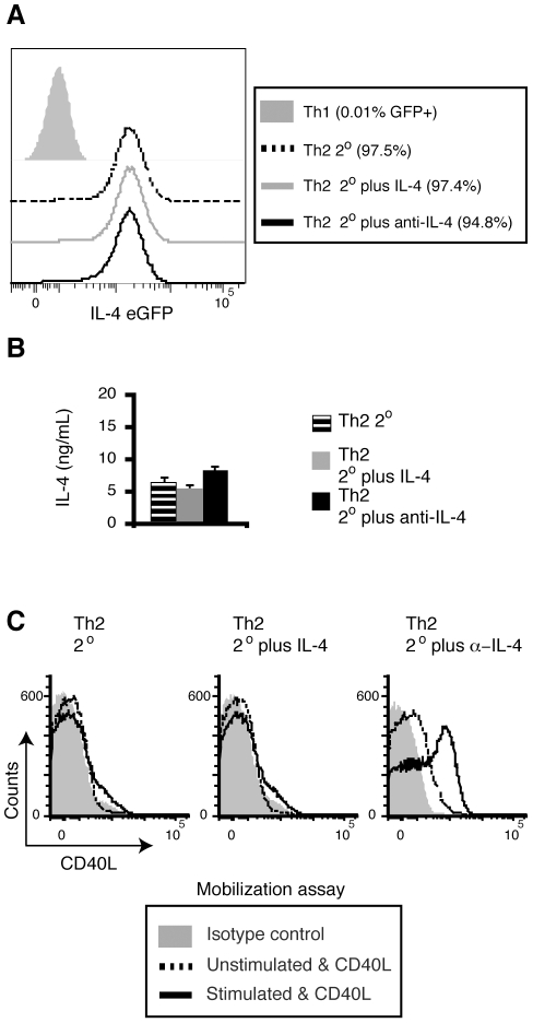
Next, we tested in vivo-generated Th2 cells by transferring CD4+ T cells from 4get/DO11.10 TCR transgenic mice into normal mice and immunizing recipients with papain plus OVA protein, which induces a robust Th2 response in vivo [30] (Fig. 3A ). A week after immunization, 20 to 40% of the DO11.10 CD4+ T cells became IL-4/eGFP positive Th2 cells (Fig. 3B and data not shown). In contrast to what we observed with in vitro-generated Th2 cells, in vivo-generated Th2 cells mobilize a substantial amount of pCD40L upon cognate interactions with APCs (Fig. 3C ). To rule out the possibility of preferential pCD40L expression in TFH cells rather than in Th2 cells, we conducted the mobilization assay for pCD40L followed by staining of CXCR5 and PD-1 [26], [41]. The mobilization assay shows that both TFH (IL-4/eGFP+CXCR5hiPD-1hi) and Th2 (IL-4/eGFP+CXCR5lowPD-1low) DO11.10 cells mobilize pCD40L (Figs. 3D, 3E ). To further verify these findings, we analyzed endogenous, polyclonal Th2 cells in an asthma model (Fig. 3F ). We observed mobilization of pCD40L in polyclonal Th2 cells, which were identified by a Th2 marker (T1/ST2, IL-33R), in BALF from sensitized and challenged animals (Figs. 3G, 3H ) [42]. These results were surprising since in vitro-generated Th2 cells stored and mobilized very little pCD40L. To determine the root of this discrepancy, we considered cell-extrinsic factors that might downregulate pCD40L in Th2 cells generated in vitro. Because IL-4 has been shown to repress the late phase of de novo CD40L expression by stimulated naïve CD4+ T cells [43], we tested whether exogenous or accumulated IL-4 in the culture is responsible for the diminished pCD40L expression in Th2 cells. In vitro-generated Th2 cells were prepared as in Fig. 1 and then restimulated with antigen-pulsed APCs either in the absence or presence of recombinant IL-4 or neutralizing anti-IL-4 for 4 days. After the two rounds of stimulation, all groups of Th2 cells maintained their stable Th2 phenotype as measured by IL-4/eGFP expression (Fig. 4A ) and IL-4 protein secretion upon PMA plus ionomycin stimulation (Fig. 4B ). When the three groups of Th2 cells were analyzed, we found that only Th2 cells that underwent the second round of stimulation in the presence of anti-IL-4 mobilized pCD40L upon stimulation (Fig. 4C ). These data indicate that a non-physiological level of IL-4 in the in vitro culture causes downregulation of pCD40L. IL-4 levels in situ in lymph nodes undergoing an intense Th2 response were estimated to be 15–500 pg/ml [44], whereas the culture media in this experiment may contain 5–100 ng/ml of IL-4. We conclude that Th2 cells do store and mobilize pCD40L except in the presence of high levels of exogenous or accumulated IL-4.
Figure 3. In vivo-generated Th2 cells store and mobilize pCD40L.
A, BALB/c mice that had received 4get/DO11.10 CD4+ T cells were subcutaneously immunized with papain plus OVA protein. Seven days later, cells from dLNs were analyzed by the mobilization assay. B, Gating strategy for antigen-specific IL-4/eGFP+ DO11.10 cells (CD4+IL-4/eGFP+KJ1-26+). C, Mobilization of pCD40L by IL-4/eGFP+ DO11.10 cells in response to PMA plus ionomycin or OVA peptide-pulsed APC. D, Those cells were further differentiated as TFH (CXCR5hiPD-1hi) and Th2 (CXCR5lowPD-1low) cells. E, Mobilization of pCD40L by TFH and Th2 cells in response to PMA plus ionomycin. F, To obtain polyclonal Th2 cells, mice were sensitized with OVA/alum followed by intratracheal challenge with OVA/PBS. G, The gating strategy for Th2 cells (CD4+T1/ST2+) and non-Th2 cells in BALF. H, Mobilization of pCD40L by polyclonal Th2 and non-Th2 cells in response to PMA plus ionomycin. Data are representative of two to three independent experiments.
Figure 4. Excess IL-4 downregulates pCD40L levels in Th2 cells generated in vitro.
Recovery of pCD40L in Th2 cells by neutralizing IL-4. A–C, Differentiated Th2 cells from 4get/DO11.10 spleen cells were restimulated with peptide-pulsed splenic APCs alone (Th2 2°) or in the presence of exogenous IL-4 (Th2 2° plus IL-4) or anti-IL-4 (Th2 2° plus anti-IL-4) for 4 days. A, The levels of IL-4/eGFP in each group of Th2 cells as well as Th1 negative control cells. B, IL-4 production upon PMA plus ionomycin stimulation by each group of Th2 cells was analyzed by ELISA. Each bar represents the mean ± standard deviation for triplicates. C, Mobilization of pCD40L by each group of Th2 cells in response to PMA plus ionomycin. Data are representative of two independent experiments.
In vivo-generated Th17 cells possess and mobilize pCD40L upon antigenic stimulation
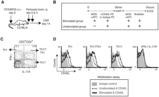
We also tested in vivo-generated Th17 cells for pCD40L using an EAE model (Fig. 5A ). To distinguish Th17 cells from Th17/Th1 and Th1 cells in the mobilization assay, cells from the CNS of symptomatic EAE animals were analyzed with a combination of intracellular cytokine staining and the mobilization assay (Fig. 5B ). Endogenous Th17 cells, as well as Th17/Th1 and Th1 cells, but not antigen non-specific (IFN-γ−IL-17A−) CD4+ T cells from EAE lesions mobilize pCD40L upon antigen recognition (Figs. 5C, 5D). Together, the findings above show that pCD40L is widely shared among effector CD4+ T cells.
Figure 5. In vivo-generated Th17 and Th17/Th1 cells store and mobilize pCD40L.
A, CNS infiltrating leukocytes were obtained from brains and spinal cords of EAE animals induced by immunization with CFA plus MOG peptide and pertussis toxin. B, CNS infiltrating leukocytes were analyzed by the CD40L mobilization assay followed by intracellular cytokine staining. C, Intracellular staining of IL-17A and IFN-γ in CNS CD4+ T cells. D, CD40L mobilization in Th1 (IL-17A−IFN-γ+), Th17 (IL-17A+IFN-γ−), Th17/Th1 (IL-17A+IFN-γ+), and antigen non-specific (IL-17A−IFN-γ−) CD4+ T cells upon antigenic stimulation. Data are representative of three independent experiments.
Treg cells possess little or no pCD40L
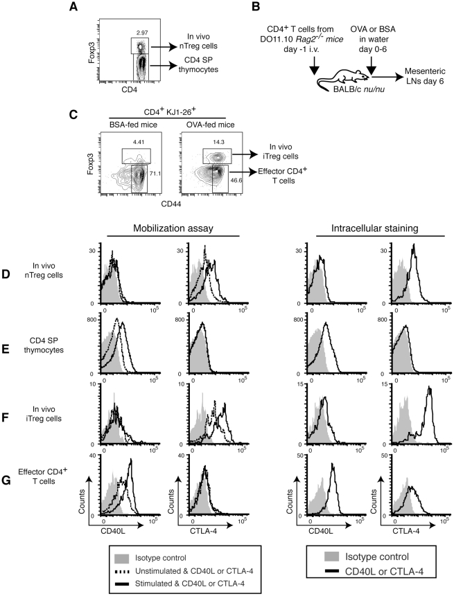
Although Treg cells are reported to accumulate low levels of surface CD40L in the steady state in CD40−/− mice [45], we observed little or no pCD40L in iTreg cells generated in vitro (Fig. 1). We observed differences in pCD40L level between in vitro and in vivo Th2 cells (Figs. 1, 3, and 4), leading us to examine pCD40L expression and mobilization in Treg cells obtained in vivo. We were unable to detect unambiguous pCD40L expression in either thymic natural Treg (nTreg) cells or splenic Treg cells from unmanipulated mice, although they both express and mobilize CTLA-4 (Figs. 6A, 6D and Figs. S2 A, S2B). In accord with this notion, exclusion of CD44hi nTreg cells from memory phenotype (MP) CD4+ T cells resulted in higher pCD40L mobilization than previously reported [17] (Figs. S2 A, S2C). Although we previously concluded that naïve CD4+ T cells do not have pCD40L [17], we reproducibly detected a very low level of pCD40L in naïve CD4+ T cells (Figs. S2 A, S2D). Low level pCD40L expression in naïve CD4+ T cells has also been reported by others [28].
Figure 6. CD4 SP thymocytes, but not Treg cells, store and mobilize pCD40L.
A, Gating strategy for thymic nTreg cells and CD4 SP thymocytes. B, Generation of in vivo iTreg cells. DO11.10 CD4+ T cells were recovered from OVA- or BSA-fed recipient mice on day 6. C, Gating strategy for in vivo iTreg cells and effector CD4+ T cells. A BSA-fed mouse is shown as a negative control. D, thymic nTreg cells; E, CD4 SP thymocytes; F, in vivo iTreg cells; and G, effector CD4+ T cells, are analyzed by the mobilization assay and intracellular staining for pCD40L and CTLA-4. Data are representative of three (D and E) or two (F and G) independent experiments.
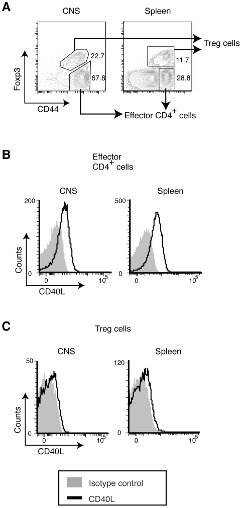
Next, we tested antigen-driven, in vivo-generated iTreg cells induced in an oral tolerance model with OVA [33] (Figs. 6B, 6C ). Although it has been shown that CD40L is required for the induction of oral tolerance [46], we found that in vivo-generated iTreg cells nearly lack pCD40L expression (Fig. 6F ), while the CD44hi effector CD4+ T cells in the same model had a high level of pCD40L (Fig. 6G ). Because this finding was obtained in a tolerogenic environment, we next examined whether Treg cells might acquire pCD40L in an inflammatory environment. In EAE, most Treg cells in inflamed CNS tissue are nTreg cells of thymic origin [47]. Even though almost all Treg cells in the CNS upregulate CD44 (Fig. 7A ) and effector CD4+ T cells acquire abundant pCD40L (Fig. 7B ), Treg cells from inflamed CNS as well as spleens lacked detectable pCD40L (Fig. 7C ). Taken together, these data indicate that pCD40L expression in Treg cells is tightly suppressed.
Figure 7. Severe inflammation does not compromise the lack of expression of pCD40L by Treg cells.
CNS infiltrating leukocytes and splenocytes were obtained from EAE animals induced as Fig. 5A . A, Gating strategy for cells from CNS and spleen. B & C, Intracellular CD40L levels for effector CD4+ T cells (B) and Treg cells (C). Data are representative of two independent experiments.
CD4 SP thymocytes and iNKT cells possess pCD40L
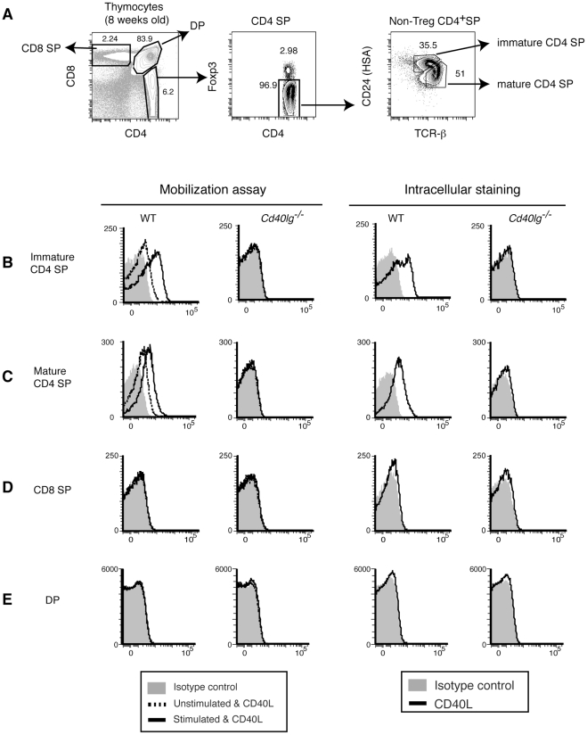
We were surprised to see low level pCD40L expression in peripheral naïve CD4+ T cells. This suggested to us that there might be a developmental role for pCD40L, so we decided to examine pCD40L in thymic T cell populations. When we looked in the thymus, we found that CD4 SP (CD4+CD8−Foxp3−) thymocytes express and mobilize pCD40L (Fig. 6E ). Further analysis showed that immature CD4 SP thymocytes have more pCD40L than mature CD4 SP thymocytes (Figs. 8A, B, C ). We could not detect any CD40L staining in CD4−CD8+ (CD8 SP) and CD4+CD8+ double-positive (DP) thymocytes (Figs. 8D, 8E ). These results suggest that immature CD4 SP thymocytes acquire pCD40L, and then decrease pCD40L to lower levels as they mature.
Figure 8. CD4 SP thymocytes, but not CD8 SP or DP thymocytes, store and mobilize pCD40L.
A, Gating strategy for identifying CD8 SP, DP, and immature and mature CD4 SP thymocytes. B–E, Mobilization of pCD40L for immature (B) and mature (C) CD4 SP thymocytes, CD8 SP (D), and DP (E) thymocytes. Cd40lg−/− : CD40L-deficient mouse. Data are representative of at least five independent experiments.
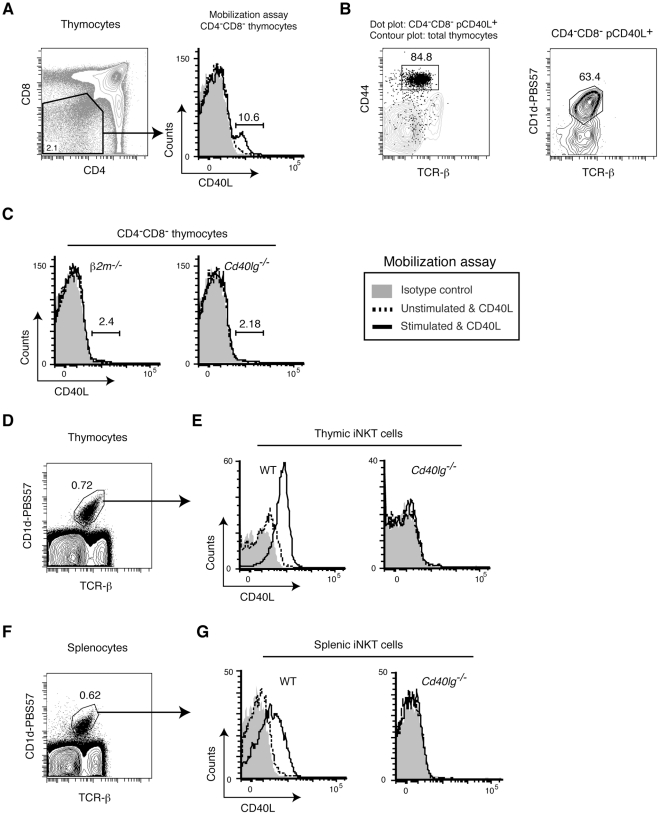
We also detected mobilization of pCD40L in a small fraction of the CD4−CD8− double-negative (DN) population (Fig. 9A ). Further analysis showed that those cells were CD44hi, TCR-βint, and CD1d-PBS57 tetramer positive iNKT cells (Fig. 9B ). In fact, in β2m-deficient (β2m−/−) mice, which lack both CD8+ T cells and iNKT cells, the pCD40L positive population in DN thymocytes is absent (Fig. 9C ). Direct assessment of thymic iNKT cells shows clear and uniform mobilization of pCD40L (Figs. 9D, 9E ). Splenic iNKT had somewhat reduced pCD40L (Figs. 9F, 9G ). We found no detectable pCD40L in NK cells and CD8+ T cells (Figs. S3 A, S3B) [17], [48].
Figure 9. iNKT cells possess and mobilize pCD40L.
A, A fraction of the DN thymocyte population mobilizes pCD40L upon stimulation with PMA plus ionomycin. B, pCD40L positive DN thymocytes are iNKT cells. C, iNKT deficient β2m−/− mice lack the pCD40L positive DN thymocyte population. D and F, Gating strategy for thymic (D) and splenic (F) iNKT cells. E and G, Mobilization of pCD40L in CD1d-tetramer-positive thymic (E) or splenic (G) iNKT cells upon stimulation with PMA plus ionomycin. Data are representative of two independent experiments.
Discussion
Recent two-photon microscopy studies indicate that interactions between effector CD4+ T cells and APCs in vivo are surprisingly brief [12], [13], [14], [15], necessitating a reassessment of our ideas about how CD4+ T cells deliver their effector functions. The dominant idea is directional secretion of newly synthesized cytokines toward antigen-bearing APCs [49], but de novo cytokine synthesis requires more time than is provided by these short in vivo interactions. On the other hand, preformed effector molecules stored in T cells can be delivered by regulated secretion in minutes as opposed to hours. As a membrane-bound cytokine, CD40L is designed for delivery by cell contact, and is necessary for cognate help for B cells and licensing of DC [1], [2], so regulated surface expression of pCD40L could explain antigen-specific activation of APC in brief interactions with CD4+ T cells in vivo. pCD40L has been reported in CD4+ T cells [16], [17], [24], [25], [26], [27], [28], but it is not yet known which functions of CD40L require de novo CD40L synthesis and which can be supplied by pCD40L. Our recent study showed that pCD40L is sufficient for the activation of DCs and selective activation of antigen-bearing B cells in vitro [22]. In this paper, we assessed possession and surface mobilization of pCD40L among the newly expanded classification of CD4+ T cell subsets by signature cytokines [23] to determine whether pCD40L is restricted to specific subsets with certain functions, for example, TFH or Th1 cells. We found instead that pCD40L is expressed and mobilized to the cell surface in all tested effector CD4+ T cell subsets, as well as CD4 SP thymocytes and iNKT cells. However, neither nTreg nor iTreg cells possess easily detectable pCD40L. Taken together, our recent findings indicate that pCD40L may be involved in T cell development and function broadly in innate and adaptive immunity.
pCD40L may also contribute to the pathology of inflammatory diseases because pCD40L is not limited to the primary and secondary lymphoid organs. Th2 cells recovered from the airway of a mouse asthma model and Th1 and Th17 cells from CNS disease lesions of EAE animals possess and mobilize pCD40L. Similar findings were reported in effector CD4+ T cells recovered from synovial fluid of human rheumatoid arthritis patients [24]. It has been shown that anti-CD40L treatment not only blocks the induction phase but also ameliorates the effector phase of EAE [50], [51]. Two-photon microscopy shows predominantly brief interactions of effector memory CD4+ T cells with APCs in target tissues [14]. Together, these findings suggest that pCD40L may function during the effector phase of inflammation through the promotion of cytokine secretion by APCs.
Strikingly, we found that only Treg cells lack reproducibly detectable pCD40L among all CD4+ T cells, further suggesting that activation of APCs is the primary role of pCD40L. In contrast, it has been reported that a fraction of Treg cells express de novo CD40L upon activation [52] and elicit CD8 T cell responses in a CD40L-dependent manner in vivo [53]. These cases can be seen as functional reprogramming of Treg cells to manifest helper/effector activity [54]. Therefore, one might imagine that aberrant pCD40L expression in Treg cells could be observed in severe inflammation. However, Treg cells defined by Foxp3 expression had barely detectable pCD40L expression even in the presence of pathologic inflammation caused by Th1 and Th17 cells in EAE lesions, in keeping with a recent report that showed an extremely stable phenotype of Treg cells [55]. Alternatively, ex-Treg cells that have lost Foxp3 expression might gain pCD40L after conversion to aggressive effector CD4+ T cells [56]. In fact, converted ex-nTreg cells in the gut preferentially become TFH cells and express CD40L [57]. Based on the above findings, it would be interesting to test whether engineering Treg cell-specific expression of pCD40L could redirect their activity from regulation to activation of APCs.
There are several non-mutually exclusive mechanisms that could explain the lack of pCD40L in Treg cells. One possibility is transcriptional repression. It was shown that Foxp3 can form a heterodimer with NFAT1, and that the NFAT1:Foxp3 complex prevents the NFAT1:AP-1 complex from binding to the IL-2 promoter, resulting in repression of IL-2 mRNA transcription [58]. The CD40L promoter has a consensus sequence (5′-GGAANNNNTGTTT-3′) for the NFAT1:Foxp3 complex [58], suggesting repression of CD40L mRNA transcription by the NFAT1:Foxp3 complex. In fact, a reduced level of CD40L mRNA was found in Treg cells compared to naïve CD4+ T cells [45]. CD4+ T cells from Foxp3 transgenic mice also showed reduced CD40L expression upon anti-CD3 plus anti-CD28 stimulation [59]. Independent of Foxp3, anergic CD4+ T cells produced by TCR stimulation without costimulation, presumably through NFAT in the absence of AP-1 [60], also lack CD40L expression [61], [62]. Our preliminary data indicate that type 1 regulatory T (Tr1) cells [63] lack pCD40L, strengthening the link between diminished pCD40L and an anergic phenotype (Y. Koguchi, D.C. Parker, unpublished data). Post-transcriptional regulation of CD40L mRNA may be different between effector CD4+ T cells and Treg cells because CD40L mRNA is stabilized in response to Ag recognition in effector T cells [64]. Another possibility is post-translational modification of pCD40L. It is reported that GRAIL (gene related to anergy in lymphocytes) directly downregulates CD40L through its E3 ubiquitin ligase activity [65], although this finding is controversial since GRAIL-deficient mice from two different groups did not show any evidence of CD40L overexpression [66], [67]. Investigation of the cause(s) of the lack of pCD40L in Treg cells could shed light on how effector CD4+ T cells acquire and regulate pCD40L.
The selective expression of pCD40L in CD4 SP thymocytes implies a role in T cell development. Through a careful examination of pCD40L in thymocytes, we found that CD4 SP thymocytes have pCD40L and immature CD4 SP thymocytes have more pCD40L than mature CD4 SP thymocytes. CD40L is necessary for thymic negative selection to endogenous superantigens [68] and contributes to the maintenance of medullary thymic epithelial cells (mTECs) [69], [70], [71]. Unregulated expression of surface CD40L on thymocytes in CD40L transgenic mice caused hyper-proliferation of mTECs [6]. Therefore, it seems likely that regulated provision of pCD40L by CD4 SP thymocytes plays an important role in homeostasis of mTECs. Provision of pCD40L by CD4 SP thymocytes may also be important for homeostasis of Treg cells by promoting IL-2 production from DCs [72].
iNKT cells, as innate immune cells, produce proinflammatory and immunoregulatory cytokines and deliver effector functions that depend on perforin and granzyme B [73]. Although de novo CD40L expression in iNKT cells has been reported [74], [75], [76], this is the first report of the presence of pCD40L in iNKT cells. The contribution of CD40L to iNKT cell development has not been studied. Although it is still a matter of debate, CD1d expressing thymic DCs may mediate negative selection of iNKT cells [77], in which case, pCD40L may play a role in that process. In the periphery, iNKT cells can mediate acute hepatitis in response to Con A in a CD40L-dependent manner [78], and provide cognate help for iNKT glycolipid-pulsed B cells via delivery of CD40L [79], [80]. iNKT cells can license DCs and myeloid-derived suppressor cells via delivery of CD40L for optimal CD4+ and CD8+ T cell responses [81], [82], [83]. A recent study using two-photon microscopy showed that cognate interactions between lymph node macrophages and iNKT cells are relatively stable (only 20% show short interaction less than 20 min) [84] allowing ample time for de novo CD40L production. Whether cognate interactions of iNKT cells with DCs and B cells are stable or transient is currently unknown. Further studies are required to address whether iNKT cells mobilize pCD40L upon stimulation with glycolipid-pulsed APCs and whether any of the CD40L-dependent functions of iNKT cells are owing to pCD40L.
The ability of pCD40L to be rapidly mobilized to the cell surface upon antigen recognition provides a mechanism for T cells to activate cognate APC during transient, antigen-specific interactions in vivo. The broad distribution of pCD40L among CD4+ T effector cell subsets and iNKT cells indicates that use of pCD40L may be widespread in T cell development and innate and adaptive immune responses. Future research to define the molecular machinery that regulates the formation of the pCD40L secretory compartment and its delivery to the cell surface will allow studies in vivo to determine which of the many known functions of CD40L are owing to pCD40L, and provide new targets for therapeutic intervention in inflammatory diseases.
Supporting Information
Schematic explanation of the mobilization assay. In the mobilization assay, fluorochrome-labeled anti-CD40L mAb is included in the culture during the activation of cells at 37°C. Compared to the “snap shot” nature of conventional staining at 4°C after completion of stimulation, the mobilization assay captures CD40L that has been delivered to the cell surface during stimulation while blocking CD40-dependent internalization, thereby providing the “long exposure” view of CD40L surface expression. By limiting the stimulation period to 30 minutes, we were able to exclude surface expression of de-novo CD40L made following stimulation (Koguchi, 2007). Intracellular staining can be seen as an “x-ray”, and is useful to distinguish defective mobilization of pCD40L from absence of stored pCD40L in cases where no mobilization of pCD40L is observed.
(TIF)
pCD40L expression in splenic CD4+ T cell subsets. A, Gating strategy for Treg cells, memory phenotype (MP) and naive CD4+ T cells. B–D, Mobilization of pCD40L upon stimulation with PMA plus ionomycin and intracellular staining of Treg cells (B), MP (C) and naive (D) CD4+ T cells. Data are representative of seven independent experiments.
(TIF)
NK cells and CD8+ T cells do not have pCD40L. Gating strategies and the data for the mobilization of pCD40L following stimulation with PMA plus ionomycin and intracellular staining of pCD40L for NK cells (A) and CD8+CD44hi T cells (B) are shown. Data are representative of two independent experiments.
(TIF)
Acknowledgments
We thank Dr. Susan Murray for reviewing this manuscript. We thank Dr. J. Lindsay Whitton, The Scripps Research Institute, for providing SMARTA mice.
We also thank Katelynne Gardner Toren and Fanny Polesso for excellent technical assistance.
Footnotes
Competing Interests: The authors have declared that no competing interests exist.
Funding: This study was supported by National Institutes of Health grants ROI AI050823, R01 AI070934, and R21 AI077032 to DCP, AI054458 to MKS, HL061013, HL071795, and AI075064 to DBJ, and Oregon National Primate Research Center grant (RR000163) to MKS. JLG and TJT have been supported as trainees on an institutional training grant from the NIH (T32 AI078903). The funders had no role in study design, data collection and analysis, decision to publish, or preparation of the manuscript.
References
- 1.McHeyzer-Williams LJ, McHeyzer-Williams MG. Antigen-specific memory B cell development. Annu Rev Immunol. 2005;23:487–513. doi: 10.1146/annurev.immunol.23.021704.115732. [DOI] [PubMed] [Google Scholar]
- 2.Williams MA, Bevan MJ. Effector and memory CTL differentiation. Annu Rev Immunol. 2007;25:171–192. doi: 10.1146/annurev.immunol.25.022106.141548. [DOI] [PubMed] [Google Scholar]
- 3.Parker DC. T cell-dependent B cell activation. Annu Rev Immunol. 1993;11:331–360. doi: 10.1146/annurev.iy.11.040193.001555. [DOI] [PubMed] [Google Scholar]
- 4.Feau S, Arens R, Togher S, Schoenberger SP. Autocrine IL-2 is required for secondary population expansion of CD8(+) memory T cells. Nat Immunol. 2011;12:908–913. doi: 10.1038/ni.2079. [DOI] [PMC free article] [PubMed] [Google Scholar]
- 5.van Kooten C, Banchereau J. CD40-CD40 ligand. J Leukoc Biol. 2000;67:2–17. doi: 10.1002/jlb.67.1.2. [DOI] [PubMed] [Google Scholar]
- 6.Clegg CH, Rulffes JT, Haugen HS, Hoggatt IH, Aruffo A, et al. Thymus dysfunction and chronic inflammatory disease in gp39 transgenic mice. Int Immunol. 1997;9:1111–1122. doi: 10.1093/intimm/9.8.1111. [DOI] [PubMed] [Google Scholar]
- 7.Erickson LD, Durell BG, Vogel LA, O'Connor BP, Cascalho M, et al. Short-circuiting long-lived humoral immunity by the heightened engagement of CD40. J Clin Invest. 2002;109:613–620. doi: 10.1172/JCI14110. [DOI] [PMC free article] [PubMed] [Google Scholar]
- 8.Pham LV, Tamayo AT, Yoshimura LC, Lo P, Terry N, et al. A CD40 Signalosome anchored in lipid rafts leads to constitutive activation of NF-kappaB and autonomous cell growth in B cell lymphomas. Immunity. 2002;16:37–50. doi: 10.1016/s1074-7613(01)00258-8. [DOI] [PubMed] [Google Scholar]
- 9.Bolduc A, Long E, Stapler D, Cascalho M, Tsubata T, et al. Constitutive CD40L expression on B cells prematurely terminates germinal center response and leads to augmented plasma cell production in T cell areas. J Immunol. 2010;185:220–230. doi: 10.4049/jimmunol.0901689. [DOI] [PMC free article] [PubMed] [Google Scholar]
- 10.Jain A, Kovacs JA, Nelson DL, Migueles SA, Pittaluga S, et al. Partial immune reconstitution of X-linked hyper IgM syndrome with recombinant CD40 ligand. Blood. 2011;118:3811–3817. doi: 10.1182/blood-2011-04-351254. [DOI] [PMC free article] [PubMed] [Google Scholar]
- 11.Murphy KM, Travers P, Walport M. Janeway's Immunobiology. New York, NY: Garland Science, Taylor & Francis Group, LLC; 2008. 384 [Google Scholar]
- 12.Allen CD, Okada T, Tang HL, Cyster JG. Imaging of germinal center selection events during affinity maturation. Science. 2007;315:528–531. doi: 10.1126/science.1136736. [DOI] [PubMed] [Google Scholar]
- 13.Qi H, Cannons JL, Klauschen F, Schwartzberg PL, Germain RN. SAP-controlled T-B cell interactions underlie germinal centre formation. Nature. 2008;455:764–769. doi: 10.1038/nature07345. [DOI] [PMC free article] [PubMed] [Google Scholar]
- 14.Matheu MP, Beeton C, Garcia A, Chi V, Rangaraju S, et al. Imaging of effector memory T cells during a delayed-type hypersensitivity reaction and suppression by Kv1.3 channel block. Immunity. 2008;29:602–614. doi: 10.1016/j.immuni.2008.07.015. [DOI] [PMC free article] [PubMed] [Google Scholar]
- 15.Cannons JL, Qi H, Lu KT, Dutta M, Gomez-Rodriguez J, et al. Optimal germinal center responses require a multistage T cell:B cell adhesion process involving integrins, SLAM-associated protein, and CD84. Immunity. 2010;32:253–265. doi: 10.1016/j.immuni.2010.01.010. [DOI] [PMC free article] [PubMed] [Google Scholar]
- 16.Casamayor-Palleja M, Khan M, MacLennan IC. A subset of CD4+ memory T cells contains preformed CD40 ligand that is rapidly but transiently expressed on their surface after activation through the T cell receptor complex. J Exp Med. 1995;181:1293–1301. doi: 10.1084/jem.181.4.1293. [DOI] [PMC free article] [PubMed] [Google Scholar]
- 17.Koguchi Y, Thauland TJ, Slifka MK, Parker DC. Preformed CD40 ligand exists in secretory lysosomes in effector and memory CD4+ T cells and is quickly expressed on the cell surface in an antigen-specific manner. Blood. 2007;110:2520–2527. doi: 10.1182/blood-2007-03-081299. [DOI] [PMC free article] [PubMed] [Google Scholar]
- 18.Blott EJ, Griffiths GM. Secretory lysosomes. Nat Rev Mol Cell Biol. 2002;3:122–131. doi: 10.1038/nrm732. [DOI] [PubMed] [Google Scholar]
- 19.Heller KN, Gurer C, Munz C. Virus-specific CD4+ T cells: ready for direct attack. J Exp Med. 2006;203:805–808. doi: 10.1084/jem.20060215. [DOI] [PMC free article] [PubMed] [Google Scholar]
- 20.van de Berg PJ, van Leeuwen EM, ten Berge IJ, van Lier R. Cytotoxic human CD4(+) T cells. Curr Opin Immunol. 2008;20:339–343. doi: 10.1016/j.coi.2008.03.007. [DOI] [PubMed] [Google Scholar]
- 21.Beal AM, Anikeeva N, Varma R, Cameron TO, Vasiliver-Shamis G, et al. Kinetics of early T cell receptor signaling regulate the pathway of lytic granule delivery to the secretory domain. Immunity. 2009;31:632–642. doi: 10.1016/j.immuni.2009.09.004. [DOI] [PMC free article] [PubMed] [Google Scholar]
- 22.Koguchi Y, Gardell JL, Thauland TJ, Parker DC. Cyclosporine-resistant, Rab27a-independent mobilization of intracellular preformed CD40 ligand mediates antigen-specific T cell help in vitro. J Immunol. 2011;187:626–634. doi: 10.4049/jimmunol.1004083. [DOI] [PMC free article] [PubMed] [Google Scholar]
- 23.King C, Tangye SG, Mackay CR. T follicular helper (TFH) cells in normal and dysregulated immune responses. Annu Rev Immunol. 2008;26:741–766. doi: 10.1146/annurev.immunol.26.021607.090344. [DOI] [PubMed] [Google Scholar]
- 24.MacDonald KP, Nishioka Y, Lipsky PE, Thomas R. Functional CD40 ligand is expressed by T cells in rheumatoid arthritis. J Clin Invest. 1997;100:2404–2414. doi: 10.1172/JCI119781. [DOI] [PMC free article] [PubMed] [Google Scholar]
- 25.Lettesjo H, Burd GP, Mageed RA. CD4+ T lymphocytes with constitutive CD40 ligand in preautoimmune (NZB×NZW)F1 lupus-prone mice: phenotype and possible role in autoreactivity. J Immunol. 2000;165:4095–4104. doi: 10.4049/jimmunol.165.7.4095. [DOI] [PubMed] [Google Scholar]
- 26.Breitfeld D, Ohl L, Kremmer E, Ellwart J, Sallusto F, et al. Follicular B helper T cells express CXC chemokine receptor 5, localize to B cell follicles, and support immunoglobulin production. J Exp Med. 2000;192:1545–1552. doi: 10.1084/jem.192.11.1545. [DOI] [PMC free article] [PubMed] [Google Scholar]
- 27.Campbell DJ, Kim CH, Butcher EC. Separable effector T cell populations specialized for B cell help or tissue inflammation. Nat Immunol. 2001;2:876–881. doi: 10.1038/ni0901-876. [DOI] [PubMed] [Google Scholar]
- 28.Martin-Fontecha A, Baumjohann D, Guarda G, Reboldi A, Hons M, et al. CD40L+ CD4+ memory T cells migrate in a CD62P-dependent fashion into reactive lymph nodes and license dendritic cells for T cell priming. J Exp Med. 2008;205:2561–2574. doi: 10.1084/jem.20081212. [DOI] [PMC free article] [PubMed] [Google Scholar]
- 29.Benson MJ, Pino-Lagos K, Rosemblatt M, Noelle RJ. All-trans retinoic acid mediates enhanced T reg cell growth, differentiation, and gut homing in the face of high levels of co-stimulation. J Exp Med. 2007;204:1765–1774. doi: 10.1084/jem.20070719. [DOI] [PMC free article] [PubMed] [Google Scholar]
- 30.Sokol CL, Barton GM, Farr AG, Medzhitov R. A mechanism for the initiation of allergen-induced T helper type 2 responses. Nat Immunol. 2008;9:310–318. doi: 10.1038/ni1558. [DOI] [PMC free article] [PubMed] [Google Scholar]
- 31.Keane-Myers A, Gause WC, Linsley PS, Chen SJ, Wills-Karp M. B7-CD28/CTLA-4 costimulatory pathways are required for the development of T helper cell 2-mediated allergic airway responses to inhaled antigens. J Immunol. 1997;158:2042–2049. [PubMed] [Google Scholar]
- 32.Buenafe AC, Bourdette DN. Lipopolysaccharide pretreatment modulates the disease course in experimental autoimmune encephalomyelitis. J Neuroimmunol. 2007;182:32–40. doi: 10.1016/j.jneuroim.2006.09.004. [DOI] [PubMed] [Google Scholar]
- 33.Coombes JL, Siddiqui KR, Arancibia-Carcamo CV, Hall J, Sun CM, et al. A functionally specialized population of mucosal CD103+ DCs induces Foxp3+ regulatory T cells via a TGF-beta and retinoic acid-dependent mechanism. J Exp Med. 2007;204:1757–1764. doi: 10.1084/jem.20070590. [DOI] [PMC free article] [PubMed] [Google Scholar]
- 34.Yellin MJ, Sippel K, Inghirami G, Covey LR, Lee JJ, et al. CD40 molecules induce down-modulation and endocytosis of T cell surface T cell-B cell activating molecule/CD40-L. Potential role in regulating helper effector function. J Immunol. 1994;152:598–608. [PubMed] [Google Scholar]
- 35.Roy M, Aruffo A, Ledbetter J, Linsley P, Kehry M, et al. Studies on the interdependence of gp39 and B7 expression and function during antigen-specific immune responses. European journal of immunology. 1995;25:596–603. doi: 10.1002/eji.1830250243. [DOI] [PubMed] [Google Scholar]
- 36.Cohen GB, Kaur A, Johnson RP. Isolation of viable antigen-specific CD4 T cells by CD40L surface trapping. Journal of immunological methods. 2005;302:103–115. doi: 10.1016/j.jim.2005.05.002. [DOI] [PubMed] [Google Scholar]
- 37.Chattopadhyay PK, Yu J, Roederer M. A live-cell assay to detect antigen-specific CD4+ T cells with diverse cytokine profiles. Nature medicine. 2005;11:1113–1117. doi: 10.1038/nm1293. [DOI] [PubMed] [Google Scholar]
- 38.Vavassori S, Covey LR. Post-transcriptional regulation in lymphocytes: the case of CD154. RNA biology. 2009;6:259–265. doi: 10.4161/rna.6.3.8581. [DOI] [PMC free article] [PubMed] [Google Scholar]
- 39.Oxenius A, Bachmann MF, Zinkernagel RM, Hengartner H. Virus-specific MHC-class II-restricted TCR-transgenic mice: effects on humoral and cellular immune responses after viral infection. Eur J Immunol. 1998;28:390–400. doi: 10.1002/(SICI)1521-4141(199801)28:01<390::AID-IMMU390>3.0.CO;2-O. [DOI] [PubMed] [Google Scholar]
- 40.Johnston RJ, Poholek AC, DiToro D, Yusuf I, Eto D, et al. Bcl6 and Blimp-1 are reciprocal and antagonistic regulators of T follicular helper cell differentiation. Science. 2009;325:1006–1010. doi: 10.1126/science.1175870. [DOI] [PMC free article] [PubMed] [Google Scholar]
- 41.Zaretsky AG, Taylor JJ, King IL, Marshall FA, Mohrs M, et al. T follicular helper cells differentiate from Th2 cells in response to helminth antigens. J Exp Med. 2009;206:991–999. doi: 10.1084/jem.20090303. [DOI] [PMC free article] [PubMed] [Google Scholar]
- 42.Lohning M, Stroehmann A, Coyle AJ, Grogan JL, Lin S, et al. T1/ST2 is preferentially expressed on murine Th2 cells, independent of interleukin 4, interleukin 5, and interleukin 10, and important for Th2 effector function. Proc Natl Acad Sci U S A. 1998;95:6930–6935. doi: 10.1073/pnas.95.12.6930. [DOI] [PMC free article] [PubMed] [Google Scholar]
- 43.Lee BO, Haynes L, Eaton SM, Swain SL, Randall TD. The biological outcome of CD40 signaling is dependent on the duration of CD40 ligand expression: reciprocal regulation by interleukin (IL)-4 and IL-12. J Exp Med. 2002;196:693–704. doi: 10.1084/jem.20020845. [DOI] [PMC free article] [PubMed] [Google Scholar]
- 44.Perona-Wright G, Mohrs K, Mohrs M. Sustained signaling by canonical helper T cell cytokines throughout the reactive lymph node. Nat Immunol. 2010;11:520–526. doi: 10.1038/ni.1866. [DOI] [PMC free article] [PubMed] [Google Scholar]
- 45.Lesley R, Kelly LM, Xu Y, Cyster JG. Naive CD4 T cells constitutively express CD40L and augment autoreactive B cell survival. Proc Natl Acad Sci U S A. 2006;103:10717–10722. doi: 10.1073/pnas.0601539103. [DOI] [PMC free article] [PubMed] [Google Scholar]
- 46.Kweon MN, Fujihashi K, Wakatsuki Y, Koga T, Yamamoto M, et al. Mucosally induced systemic T cell unresponsiveness to ovalbumin requires CD40 ligand-CD40 interactions. J Immunol. 1999;162:1904–1909. [PubMed] [Google Scholar]
- 47.Korn T, Reddy J, Gao W, Bettelli E, Awasthi A, et al. Myelin-specific regulatory T cells accumulate in the CNS but fail to control autoimmune inflammation. Nat Med. 2007;13:423–431. doi: 10.1038/nm1564. [DOI] [PMC free article] [PubMed] [Google Scholar]
- 48.Beadling C, Slifka MK. Differential regulation of virus-specific T-cell effector functions following activation by peptide or innate cytokines. Blood. 2005;105:1179–1186. doi: 10.1182/blood-2004-07-2833. [DOI] [PubMed] [Google Scholar]
- 49.Huse M, Quann EJ, Davis MM. Shouts, whispers and the kiss of death: directional secretion in T cells. Nat Immunol. 2008;9:1105–1111. doi: 10.1038/ni.f.215. [DOI] [PMC free article] [PubMed] [Google Scholar]
- 50.Gerritse K, Laman JD, Noelle RJ, Aruffo A, Ledbetter JA, et al. CD40-CD40 ligand interactions in experimental allergic encephalomyelitis and multiple sclerosis. Proc Natl Acad Sci U S A. 1996;93:2499–2504. doi: 10.1073/pnas.93.6.2499. [DOI] [PMC free article] [PubMed] [Google Scholar]
- 51.Becher B, Durell BG, Miga AV, Hickey WF, Noelle RJ. The clinical course of experimental autoimmune encephalomyelitis and inflammation is controlled by the expression of CD40 within the central nervous system. J Exp Med. 2001;193:967–974. doi: 10.1084/jem.193.8.967. [DOI] [PMC free article] [PubMed] [Google Scholar]
- 52.Li W, Carlson TL, Green WR. Stimulation-dependent induction of CD154 on a subset of CD4+ FoxP3+ T-regulatory cells. International immunopharmacology. 2011;11:1205–1210. doi: 10.1016/j.intimp.2011.03.021. [DOI] [PMC free article] [PubMed] [Google Scholar]
- 53.Sharma MD, Hou DY, Baban B, Koni PA, He Y, et al. Reprogrammed foxp3(+) regulatory T cells provide essential help to support cross-presentation and CD8(+) T cell priming in naive mice. Immunity. 2010;33:942–954. doi: 10.1016/j.immuni.2010.11.022. [DOI] [PMC free article] [PubMed] [Google Scholar]
- 54.Mellor AL, Munn DH. Physiologic control of the functional status of Foxp3+ regulatory T cells. Journal of immunology (Baltimore, Md. 2011;186:4535–4540. doi: 10.4049/jimmunol.1002937. [DOI] [PMC free article] [PubMed] [Google Scholar]
- 55.Rubtsov YP, Niec RE, Josefowicz S, Li L, Darce J, et al. Stability of the regulatory T cell lineage in vivo. Science. 2010;329:1667–1671. doi: 10.1126/science.1191996. [DOI] [PMC free article] [PubMed] [Google Scholar]
- 56.Zhou X, Bailey-Bucktrout SL, Jeker LT, Penaranda C, Martinez-Llordella M, et al. Instability of the transcription factor Foxp3 leads to the generation of pathogenic memory T cells in vivo. Nat Immunol. 2009;10:1000–1007. doi: 10.1038/ni.1774. [DOI] [PMC free article] [PubMed] [Google Scholar]
- 57.Tsuji M, Komatsu N, Kawamoto S, Suzuki K, Kanagawa O, et al. Preferential generation of follicular B helper T cells from Foxp3+ T cells in gut Peyer's patches. Science. 2009;323:1488–1492. doi: 10.1126/science.1169152. [DOI] [PubMed] [Google Scholar]
- 58.Wu Y, Borde M, Heissmeyer V, Feuerer M, Lapan AD, et al. FOXP3 controls regulatory T cell function through cooperation with NFAT. Cell. 2006;126:375–387. doi: 10.1016/j.cell.2006.05.042. [DOI] [PubMed] [Google Scholar]
- 59.Kasprowicz DJ, Smallwood PS, Tyznik AJ, Ziegler SF. Scurfin (FoxP3) controls T-dependent immune responses in vivo through regulation of CD4+ T cell effector function. J Immunol. 2003;171:1216–1223. doi: 10.4049/jimmunol.171.3.1216. [DOI] [PubMed] [Google Scholar]
- 60.Fathman CG, Lineberry NB. Molecular mechanisms of CD4+ T-cell anergy. Nat Rev Immunol. 2007;7:599–609. doi: 10.1038/nri2131. [DOI] [PubMed] [Google Scholar]
- 61.Bowen F, Haluskey J, Quill H. Altered CD40 ligand induction in tolerant T lymphocytes. Eur J Immunol. 1995;25:2830–2834. doi: 10.1002/eji.1830251018. [DOI] [PubMed] [Google Scholar]
- 62.Yi Y, McNerney M, Datta SK. Regulatory defects in Cbl and mitogen-activated protein kinase (extracellular signal-related kinase) pathways cause persistent hyperexpression of CD40 ligand in human lupus T cells. J Immunol. 2000;165:6627–6634. doi: 10.4049/jimmunol.165.11.6627. [DOI] [PubMed] [Google Scholar]
- 63.Roncarolo MG, Gregori S, Battaglia M, Bacchetta R, Fleischhauer K, et al. Interleukin-10-secreting type 1 regulatory T cells in rodents and humans. Immunol Rev. 2006;212:28–50. doi: 10.1111/j.0105-2896.2006.00420.x. [DOI] [PubMed] [Google Scholar]
- 64.Vavassori S, Shi Y, Chen CC, Ron Y, Covey LR. In vivo post-transcriptional regulation of CD154 in mouse CD4+ T cells. Eur J Immunol. 2009;39:2224–2232. doi: 10.1002/eji.200839163. [DOI] [PMC free article] [PubMed] [Google Scholar]
- 65.Lineberry NB, Su LL, Lin JT, Coffey GP, Seroogy CM, et al. Cutting edge: The transmembrane E3 ligase GRAIL ubiquitinates the costimulatory molecule CD40 ligand during the induction of T cell anergy. J Immunol. 2008;181:1622–1626. doi: 10.4049/jimmunol.181.3.1622. [DOI] [PMC free article] [PubMed] [Google Scholar]
- 66.Kriegel MA, Rathinam C, Flavell RA. E3 ubiquitin ligase GRAIL controls primary T cell activation and oral tolerance. Proc Natl Acad Sci U S A. 2009;106:16770–16775. doi: 10.1073/pnas.0908957106. [DOI] [PMC free article] [PubMed] [Google Scholar]
- 67.Nurieva RI, Zheng S, Jin W, Chung Y, Zhang Y, et al. The E3 ubiquitin ligase GRAIL regulates T cell tolerance and regulatory T cell function by mediating T cell receptor-CD3 degradation. Immunity. 2010;32:670–680. doi: 10.1016/j.immuni.2010.05.002. [DOI] [PMC free article] [PubMed] [Google Scholar]
- 68.Foy TM, Page DM, Waldschmidt TJ, Schoneveld A, Laman JD, et al. An essential role for gp39, the ligand for CD40, in thymic selection. J Exp Med. 1995;182:1377–1388. doi: 10.1084/jem.182.5.1377. [DOI] [PMC free article] [PubMed] [Google Scholar]
- 69.Gray DH, Seach N, Ueno T, Milton MK, Liston A, et al. Developmental kinetics, turnover, and stimulatory capacity of thymic epithelial cells. Blood. 2006;108:3777–3785. doi: 10.1182/blood-2006-02-004531. [DOI] [PubMed] [Google Scholar]
- 70.Akiyama T, Shimo Y, Yanai H, Qin J, Ohshima D, et al. The tumor necrosis factor family receptors RANK and CD40 cooperatively establish the thymic medullary microenvironment and self-tolerance. Immunity. 2008;29:423–437. doi: 10.1016/j.immuni.2008.06.015. [DOI] [PubMed] [Google Scholar]
- 71.Irla M, Hugues S, Gill J, Nitta T, Hikosaka Y, et al. Autoantigen-specific interactions with CD4+ thymocytes control mature medullary thymic epithelial cell cellularity. Immunity. 2008;29:451–463. doi: 10.1016/j.immuni.2008.08.007. [DOI] [PubMed] [Google Scholar]
- 72.Guiducci C, Valzasina B, Dislich H, Colombo MP. CD40/CD40L interaction regulates CD4+CD25+ T reg homeostasis through dendritic cell-produced IL-2. Eur J Immunol. 2005;35:557–567. doi: 10.1002/eji.200425810. [DOI] [PubMed] [Google Scholar]
- 73.Bendelac A, Savage PB, Teyton L. The biology of NKT cells. Annu Rev Immunol. 2007;25:297–336. doi: 10.1146/annurev.immunol.25.022106.141711. [DOI] [PubMed] [Google Scholar]
- 74.Kawano T, Cui J, Koezuka Y, Toura I, Kaneko Y, et al. CD1d-restricted and TCR-mediated activation of valpha14 NKT cells by glycosylceramides. Science. 1997;278:1626–1629. doi: 10.1126/science.278.5343.1626. [DOI] [PubMed] [Google Scholar]
- 75.Kitamura H, Iwakabe K, Yahata T, Nishimura S, Ohta A, et al. The natural killer T (NKT) cell ligand alpha-galactosylceramide demonstrates its immunopotentiating effect by inducing interleukin (IL)-12 production by dendritic cells and IL-12 receptor expression on NKT cells. J Exp Med. 1999;189:1121–1128. doi: 10.1084/jem.189.7.1121. [DOI] [PMC free article] [PubMed] [Google Scholar]
- 76.Yoshimoto T, Min B, Sugimoto T, Hayashi N, Ishikawa Y, et al. Nonredundant roles for CD1d-restricted natural killer T cells and conventional CD4+ T cells in the induction of immunoglobulin E antibodies in response to interleukin 18 treatment of mice. J Exp Med. 2003;197:997–1005. doi: 10.1084/jem.20021701. [DOI] [PMC free article] [PubMed] [Google Scholar]
- 77.Chun T, Page MJ, Gapin L, Matsuda JL, Xu H, et al. CD1d-expressing dendritic cells but not thymic epithelial cells can mediate negative selection of NKT cells. J Exp Med. 2003;197:907–918. doi: 10.1084/jem.20021366. [DOI] [PMC free article] [PubMed] [Google Scholar]
- 78.Zhou F, Ajuebor MN, Beck PL, Le T, Hogaboam CM, et al. CD154-CD40 interactions drive hepatocyte apoptosis in murine fulminant hepatitis. Hepatology. 2005;42:372–380. doi: 10.1002/hep.20802. [DOI] [PubMed] [Google Scholar]
- 79.Galli G, Pittoni P, Tonti E, Malzone C, Uematsu Y, et al. Invariant NKT cells sustain specific B cell responses and memory. Proc Natl Acad Sci U S A. 2007;104:3984–3989. doi: 10.1073/pnas.0700191104. [DOI] [PMC free article] [PubMed] [Google Scholar]
- 80.Leadbetter EA, Brigl M, Illarionov P, Cohen N, Luteran MC, et al. NK T cells provide lipid antigen-specific cognate help for B cells. Proc Natl Acad Sci U S A. 2008;105:8339–8344. doi: 10.1073/pnas.0801375105. [DOI] [PMC free article] [PubMed] [Google Scholar]
- 81.Fujii S, Liu K, Smith C, Bonito AJ, Steinman RM. The linkage of innate to adaptive immunity via maturing dendritic cells in vivo requires CD40 ligation in addition to antigen presentation and CD80/86 costimulation. J Exp Med. 2004;199:1607–1618. doi: 10.1084/jem.20040317. [DOI] [PMC free article] [PubMed] [Google Scholar]
- 82.Taraban VY, Martin S, Attfield KE, Glennie MJ, Elliott T, et al. Invariant NKT cells promote CD8+ cytotoxic T cell responses by inducing CD70 expression on dendritic cells. J Immunol. 2008;180:4615–4620. doi: 10.4049/jimmunol.180.7.4615. [DOI] [PubMed] [Google Scholar]
- 83.De Santo C, Salio M, Masri SH, Lee LY, Dong T, et al. Invariant NKT cells reduce the immunosuppressive activity of influenza A virus-induced myeloid-derived suppressor cells in mice and humans. J Clin Invest. 2008;118:4036–4048. doi: 10.1172/JCI36264. [DOI] [PMC free article] [PubMed] [Google Scholar]
- 84.Barral P, Polzella P, Bruckbauer A, van Rooijen N, Besra GS, et al. CD169(+) macrophages present lipid antigens to mediate early activation of iNKT cells in lymph nodes. Nat Immunol. 2010;11:303–312. doi: 10.1038/ni.1853. [DOI] [PMC free article] [PubMed] [Google Scholar]
Associated Data
This section collects any data citations, data availability statements, or supplementary materials included in this article.
Supplementary Materials
Schematic explanation of the mobilization assay. In the mobilization assay, fluorochrome-labeled anti-CD40L mAb is included in the culture during the activation of cells at 37°C. Compared to the “snap shot” nature of conventional staining at 4°C after completion of stimulation, the mobilization assay captures CD40L that has been delivered to the cell surface during stimulation while blocking CD40-dependent internalization, thereby providing the “long exposure” view of CD40L surface expression. By limiting the stimulation period to 30 minutes, we were able to exclude surface expression of de-novo CD40L made following stimulation (Koguchi, 2007). Intracellular staining can be seen as an “x-ray”, and is useful to distinguish defective mobilization of pCD40L from absence of stored pCD40L in cases where no mobilization of pCD40L is observed.
(TIF)
pCD40L expression in splenic CD4+ T cell subsets. A, Gating strategy for Treg cells, memory phenotype (MP) and naive CD4+ T cells. B–D, Mobilization of pCD40L upon stimulation with PMA plus ionomycin and intracellular staining of Treg cells (B), MP (C) and naive (D) CD4+ T cells. Data are representative of seven independent experiments.
(TIF)
NK cells and CD8+ T cells do not have pCD40L. Gating strategies and the data for the mobilization of pCD40L following stimulation with PMA plus ionomycin and intracellular staining of pCD40L for NK cells (A) and CD8+CD44hi T cells (B) are shown. Data are representative of two independent experiments.
(TIF)


