Abstract
A beautiful smile definitely enhances the personality of an individual and reveals self-confidence. The harmony of the smile is determined not only by the shape, position, and color of the teeth but also by the gingival tissues. Gingival pigmentation results from melanin granules which are produced by melanoblasts. Although melanin pigmentation of the gingiva is a completely benign condition and does not pose any medical problem, complaints of “black gums” are common particularly in patients having a very high smile line. The different treatment modalities that have been reported for depigmentation are bur abrasion, partial thickness flap, cryotherapy, electrosurgery, and lasers. In this paper we have compared the results of electrosurgery and scalpel technique, i.e., partial thickness flap.
Keywords: Depigmentation, electrosurgery, scalpel technique, pigmented gingiva
INTRODUCTION
Melanin pigmentation of the gingiva occurs in all races.[1] Melanin, a brown pigment, is the most common natural pigment contributing to endogenous pigmentation of gingiva and the gingiva is also the most predominant site of pigmentation on the mucosa. Melanin pigmentation is the result of melanin granules produced by melanoblasts intertwined between epithelial cells at the basal layer of gingival epithelium.[2]
Gingival hyperpigmentation is seen as a genetic trait in some populations irrespective of age and gender; hence it is termed physiologic or racial gingival pigmentation.[1,3] The degree of pigmentation varies from one individual to another which is mainly dictated by the melanoblastic activity.[4] Melanin pigmentation of gingiva is symmetric and persistent and it does not alter normal gingival architecture. Melanosis of gingiva is frequently encountered among dark-skinned ethnic groups, as well as in medical conditions such as Addison's syndrome, Peutz-jegher's syndrome, and Von Recklinghausen's disease (neurofibromatosis).[5]
Earlier studies have shown that no significant difference exists in the density of distribution of melanocytes between light-skinned, dark-skinned, and black individuals. However, melanocytes of dark-skinned and black individuals are uniformly highly reactive than in light-skinned individuals.[6]
Demand for cosmetic therapy is made, especially by fair-skinned people with moderate or severe gingival pigmentation.[7] Gingival depigmentation is a periodontal plastic surgical procedure whereby the gingival hyperpigmentation is removed or reduced by various techniques. The first and foremost indication for depigmentation is patient demand for improved esthetics. Various depigmentation techniques have been employed with similar results. Selection of the technique should be based on clinical experiences and individual preferences. One of the first and still popular techniques to be employed is the surgical removal of undesirable pigmentation using scalpels.[8] There is only limited information in the literature on depigmentation using surgical techniques. The procedure essentially involves surgical removal of gingival epithelium along with a layer of the underlying connective tissue and allowing the denuded connective tissue to heal by secondary intention. The new epithelium that forms is devoid of melanin pigmentation.[8]
Electrosurgery unit
A radiosurgical electrosurgical unit was used for the procedure. It is based upon the biterminal principle which grounds the patient in the circuit.
Surgical procedure
Surgical gingival de-epithelization can be performed by the following techniques.
Scalpel technique[9]
Gingival abrasion technique using diamond bur[10]
Combination of both the scalpel and bur
We have used the scalpel technique in the following cases.
CASE REPORTS
Case 1
A healthy 21-year-old male patient reported to the Department of Periodontology, Yerala Dental College, with the chief complaint of “blackish gums” which esthetically interfered with his smile [Figure 1]. The patient requested for any cosmetic therapy which would eventually enhance the esthetics on smiling. The patient's history revealed that the blackish discoloration of gingiva was present since birth suggestive of physiologic melanin pigmentation. His medical history was noncontributory. On intraoral examination, generalized diffused blackish pigmentation of gingiva was observed on the labial surface of both maxillary and mandibular arches; however it was healthy and completely free of any inflammation.
Figure 1.
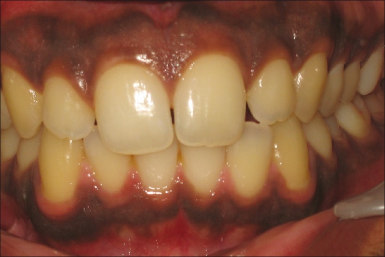
Pre-operative view – case 1
Considering the patient's concern, a surgical gingival de-epithelization procedure was planned using electrosurgery unit for the right side and scalpel technique for the left side of the maxillary arch.
For the scalpel technique, following the administration of a local anesthetic solution, a partial split thickness flap was raised with the help of blade no. 15 with the Bard Parker handle to scrape the epithelium. Bleeding was controlled using pressure pack with sterile gauze. Care was taken that excessive tissue was not removed thereby avoiding any bone exposure [Figure 2].
Figure 2.
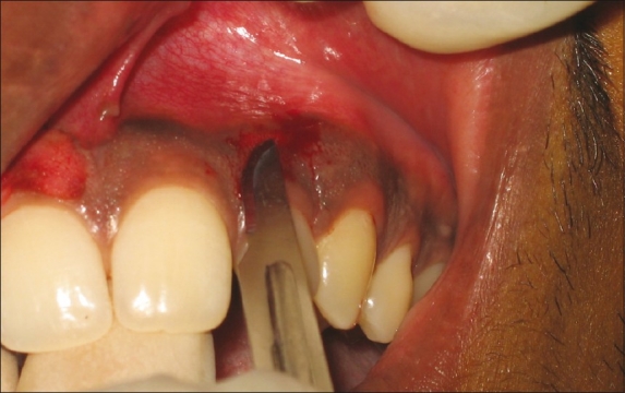
De-epithelisation using scalpel techique
Similarly epithelial excision was done on the right side with the electrosurgery unit using the loop electrode [Figure 3]. Again care was taken not to expose the bone on the attached gingiva and not to remove excessive tissue on the marginal gingiva thereby disturbing the gingival harmony [Figure 4].
Figure 3.
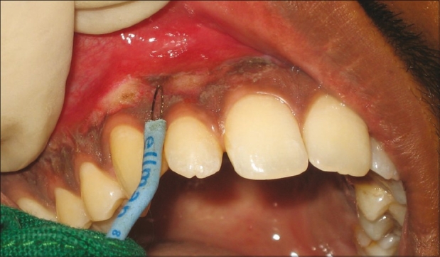
De-epithelisation using the electrosurgery unit
Figure 4.
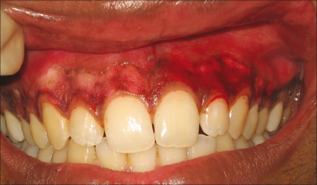
Immediately after the surgery
The surgical area was covered with a periodontal pack and postoperative instructions were given. Analgesic was prescribed for the management of pain. After 1 week, the pack was removed and the surgical area was examined. The healing was uneventful without any post-surgical complications. The gingiva appeared pink, healthy, and firm giving a normal appearance [Figure 5]. The patient was very impressed with such a pleasing esthetic outcome.
Figure 5.
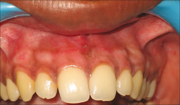
Post-operative view after 7 days
Case 2 and 3
A 24-year-old female patient [Figures 6 and 7] and a 19-year-old male patient [Figures 8and 9] reported with similar findings and underwent the same procedure
Figure 6.
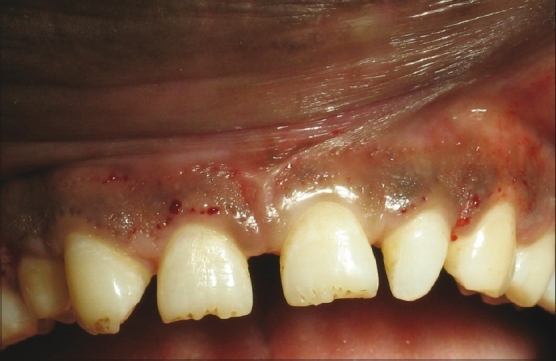
Pre-operative view – case 2
Figure 7.
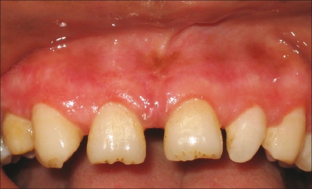
Post-operative view – case 2
Figure 8.
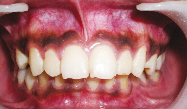
Pre-operative view Case 3
Figure 9.
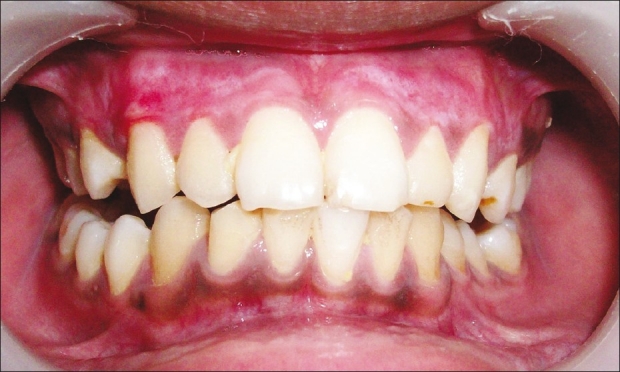
Post-operative view Case 3
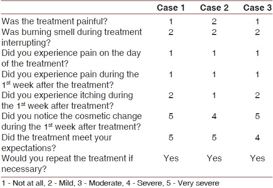
Patients were recalled after 7 days and the following was recorded:
Healing – absence of pain, bleeding, and infection
Postoperative pain and itching if any
Inflammation and color change
The McGill Pain Questionnaire[11]
The McGill Pain Questionnaire:
DISCUSSION
There are wide variations in gingival color in normal healthy persons. The degree of vascularization, the thickness of the keratinized layer, and the amount of the pigment containing cells will determine the color of the gingiva.[12] There are few studies published so far regarding clinical methods of treatment of pigmented gingiva. The techniques that were tried in the past to treat gingival pigmentation include chemical cauterization,[13] gingivectomy,[14] scalpel scraping procedure,[9] and abrasion of gingiva.[10] The recent techniques of gingival depigmentation in practice are cryotherapy,[7] free gingival autograft,[15] and laser therapy[16] and these have achieved satisfactory results.
The use of the scalpel technique for the depigmentation is most economical compared to other techniques, which require more advanced armamentarium. However, scalpel surgery causes unpleasant bleeding during and after the operation, and it is necessary to cover the surgical site with periodontal dressing for 7–10 days. Electrosurgery has its own limitations in that its repeated and prolonged use induces heat accumulation and undesired tissue destruction.
We found the scalpel and abrasion technique relatively simple and versatile and it required minimum time and effort. No sophisticated and expensive armamentarium was required; only blade and bur were sufficient.
Though the initial result of the depigmentation surgery is highly encouraging, repigmentation is a common problem. The exact mechanism of repigmentation is not known. Different studies show variation in the timing for early repigmentation. To return to the full clinical baseline repigmentation it takes about 1.5–3 years.[12] This variation may be due to the different techniques performed or due to the patient's race. Thus, the gingival depigmentation procedure, if performed primarily for cosmetic reason, will not be of permanent value, because pigmentation tends to return to baseline values.[12]
In future, even if gingival repigmentation occurs in this patient, the same procedure could be repeated in the same region. Therefore, the scalpel surgical technique and gingival abrasion technique are highly recommended in consideration of the equipment constraints in developing countries. It is simple, easy to perform, cost effective, and above all provides minimum discomfort to the patient and esthetically pleasing results.
ACKNOWLEDGMENTS
I would like to thank our Chairman, Dr. G.D. Pol, Dean Dr. Sharat Kokate, my staff members, and PG students for helping me to carry out this study in our institution.
Footnotes
Source of Support: Nil
Conflict of Interest: None declared.
REFERENCES
- 1.Dummett CO. Oral pigmentation. First symposium of oral pigmentation. J Periodontol. 1960;31:356–60. [Google Scholar]
- 2.Cicek Y, Ertas U. The normal and pathological pigmentation of oral mucous membrane: A review. J Contemp Dent Pract. 2003;4:76–86. [PubMed] [Google Scholar]
- 3.Dummet CO, Barens G. Oromucosal pigmentation: An updated literary review. J Periodontol. 1971;42:726–36. doi: 10.1902/jop.1971.42.11.726. [DOI] [PubMed] [Google Scholar]
- 4.Perlmutter S, Tal H. Repigmentation of gingival following injury. J Periodontal. 1986;57:48–50. doi: 10.1902/jop.1986.57.1.48. [DOI] [PubMed] [Google Scholar]
- 5.Shafer WG, Hine MK, Levy BM. Philadelphia: WB Saunders Co; 1984. Text Book of Oral Pathology; pp. 89–136. [Google Scholar]
- 6.Szako G, Gerald SB, Pathak MA, Fitz Patrick TB. Racial differences in the fate of melanosomes in human epidermis. Nature. 1969;222:1081. doi: 10.1038/2221081a0. [DOI] [PubMed] [Google Scholar]
- 7.Tal H, Landsberg J, Koztovsky A. Cryosurgical depigmentation of the gingiva - A case report. J Clin Periodontol. 1987;14:614–7. doi: 10.1111/j.1600-051x.1987.tb01525.x. [DOI] [PubMed] [Google Scholar]
- 8.Roshna T, Nandakumar K. Anterior esthetic gingival depigmentation and crown lengthening: Report of a case. J Contemp Dent Pract. 2005;6:139–47. [PubMed] [Google Scholar]
- 9.Almas K, Sadig W. Surgical treatment of melanin pigmented gingiva: An esthetic approach. Indian J Dent Res. 2002;13:70–3. [PubMed] [Google Scholar]
- 10.Pal TK, Kapoor KK, Parel CC, Mukharjee K. Gingival melanin pigmentation- A study on its removal for esthetics. J Indian Soc Periodontol. 1994;3:52–4. [Google Scholar]
- 11.Melzack R. The McGill pain questionnaire: Major properties and scoring methods. Pain. 1975;1:277–99. doi: 10.1016/0304-3959(75)90044-5. [DOI] [PubMed] [Google Scholar]
- 12.Begamaschi O, Kon S, Doine AI, Ruben MP. Melanin repigmentation after gingivectomy: A five year clinical and transmission electron microscopic study in humans. Int J Periodontics Restorative Dent. 1993;13:85–92. [PubMed] [Google Scholar]
- 13.Hirschfeld I, Hirschfeld L. Oral pigmentation and method of removing it. Oral Surg Oral Med Oral Pathol. 1951;4:1012. doi: 10.1016/0030-4220(51)90448-3. [DOI] [PubMed] [Google Scholar]
- 14.Dummet CO, Bolden TE. Post surgical repigmentation of the gingiva. Oral Surg Oral Med Oral Pathol. 1963;16:353–6. [Google Scholar]
- 15.Tamizi M, Taheri M. Treatment of severe physiologic gingival pigmentation with free gingival autograft. Quintessence Int. 1996;27:555–8. [PubMed] [Google Scholar]
- 16.Trelles MA, Verkruysse W, Segui JM, Udaeta A. Treatment of melanotic spots in the gingiva by Argon laser. J Oral Maxillofac Surg. 1993;51:759–61. doi: 10.1016/s0278-2391(10)80416-1. [DOI] [PubMed] [Google Scholar]


