Abstract
The Phase I clinical study was designed to assess the safety and feasibility of a dose escalating intracoronary infusion of autologous bone marrow (BM)-derived CD133+ stem cell therapy to the patients with chronic total occlusion (CTO) and ischemia. Nine patients were received CD133+ cells into epicardial vessels supplying collateral flow to areas of viable ischemic myocardium in the distribution of the CTO. There were no major adverse cardiac events (MACE), revascularization, re-admission to the hospital secondary to angina, or acute myocardial infarction (AMI) for the 24-month period following cellular infusion. In addition, there were no periprocedural infusion-related complications including malignant arrhythmias, loss of normal coronary blood flow or acute neurologic events. Cardiac enzymes were negative in all patients. There was an improvement in the degree of ischemic myocardium, which was accompanied by a trend towards reduction in anginal symptoms. Intracoronary infusion of autologous CD133+ marrow-derived cells is safe and feasible. Cellular therapy with CD133+ cells to reduce anginal symptoms and to improve ischemia in patients with CTO awaits clinical investigation in Phase II/III trials.
Keywords: Safety, Efficacy, Autologous, Bone marrow, CD133, Chronic, Myocardial, Ischemia, Therapy
2. INTRODUCTION
Therapies for coronary ischemia and subsequent myocardial necrosis include acute revascularization, coronary artery bypass grafting (CABG) and pharmacologic interventions targeted at reducing ischemia and ultimately preserving myocardium and prolonging life. Morbidity and mortality related to myocardial necrosis regardless of etiology has been reduced dramatically with the institution of these therapies. Others and we reported that hematopoietic stem cells have the promise in reducing total ischemia via neovascularization in various animal models (1–4). Researchers and clinicians are interested to develop improved therapeutic regime for the treatment of ischemia and ultimately facilitate improvement of symptoms for the betterment of patient’s life. Specifically, patients with chronic total occlusion (CTO) provide a therapeutic challenge as they experience stable chronic angina, which is difficult to control and or ameliorate (5). Percutaneous coronary intervention (PCI) to CTO account for approximately 12–16% of all interventions in the United States (6). The Occluded Artery Trial recently revealed that PCI does not reduce MACE and may in fact provoke myocardial ischemia with increase reinfarction rates in clinically stable patients with persistent total occlusion (7). Many of these patients have areas of viable, yet jeopardized areas of myocardium. Development of collateral growth is an important compensatory mechanism with respect to CTO. Collateralization may provide adequate augmentation of blood flow to ischemic tissue protecting patients with CTO from angina. However, collateralization may not provide this protection and further augmentation of collateral growth may be necessary to generate reduction or cessation of symptoms.
Hematopoietic stem cells, which express CD34 and CD133 surface markers have been shown in models of acute and chronic ischemia to augment blood flow and prevent myocardial necrosis (4, 8–16). Postulated mechanisms for this process include paracrine effects, which may provide substrate for promoting myocardial healing and/or angiogenesis (17, 18). Recently, a number of clinical trials have attempted to determine the role of stem cells in promoting an in situ reparative milieu (19–29). The studies utilized autologous cellular products, primarily whole mononuclear cell preparations. In addition, different delivery techniques were attempted and a unified cell dose was not used. We postulate that a selected cell population may have a therapeutic advantage over whole cell preparations because it provides a pure potent stem cell fraction, which were reportedly having the potential for neovascularization and differentiation ultimately resulted to reduction of ischemia. Moreover, all of the current studies have illustrated safety with single dose applications without an attempt at titration with respect to safety. Therefore, we aimed to determine if infusion with increasing cell dose of autologous CD133+ selected stem cells was safe and feasible in patients with CTO.
3. METHODS
3.1. Patient selection
A total of nine patients were enrolled between January and June 2006 and followed for a period of 24 months after the date of the procedure. Patients underwent screening for enrollment within 30 days of therapy. This Phase I, single center study enrolled patients of at least 18 years of age who experienced class II–IV angina (Canadian Cardiovascular Society classification). Identification by nuclear imaging of at least one region of chronically ischemic or viable (hibernating) myocardium formerly perfused by a non-revascularizable totally occluded coronary artery was required for inclusion. In addition, well-established collateral vessels of at least 1.5-mm in luminal diameter to the viable myocardium at the time of diagnostic coronary angiography must have been present to be included in the trial. Patients included had also a left ventricular (LV) ejection fraction of greater than 45%, as measured by echocardiography Patients with coronary lesions amenable to PCI including brachytherapy, or where CABG was indicated was excluded. Any contraindication for cardiac catheterization, PCI and BM aspiration as per institutional guidelines, patients with an AMI within the previous 3 months and/or New York Heart Association (NYHA) class III or IV congestive heart failure were also excluded. Patients with baseline electrocardiogram (ECG) abnormalities that would hinder interpretation of baseline ECG un-interpretable for ischemia (e.g., left bundle branch block, LV hypertrophy with strain pattern, Wolff-Parkinson-White syndrome) were excluded. Hematologic abnormalities including a documented bleeding diathesis, anemia with a hemoglobin concentration of < 8 mg/dl, a platelet count < 100,000 and known malignancy involving the hematopoietic or lymphoid system excluded entry into this study. Moreover, the presence of severe co-morbidities including renal and hepatic failure was additionally excluded. Informed consent was obtained from those patients that fulfilled these criteria.
3.2. Study design and parameters of safety
The study design and protocol was approved by the institutional review board of Case Western Reserve University and University Hospitals of Cleveland. Informed consent was obtained from all participants. The primary endpoint of the study was to assess the safety and feasibility of a dose-escalating injection of autologous BM derived CD133+ hematopoietic stem cells in chronic ischemic patients with a staged twenty-four months follow up. Secondary endpoints included reduction in the area of ischemic myocardium, improvement in LV function and myocardial viability and reduction of symptoms. Preenrollment procedures included coronary angiogram, two-dimensional echocardiogram, pharmacologic stress test with nuclear imaging, ECG and 24 hour Holter monitor, laboratory studies and completion of a Seattle Angina Questionnaire. After the infusion, all patients were monitored for 16–24 hours on telemetry and had vital signs per routine until discharge. Cardiac enzymes and basic metabolic panels were collected at 8, 16, and 24 hours following the procedure. An ECG was completed at 24 hours post-injection or at the time of discharge. Staged follow-up began immediately following infusion and subsequently at 7, 14, 30, 90, 180 days, 12 months, 18 months and 24 months. At each visit the following assessments were performed: (1) Focused cardiovascular history and angina questionnaire, (2) Limited physical exam, (3) Review of medication list, (4) ECG, (5) Evaluation of MACE as defined as death (cardiac and noncardiac), PCI or CABG, readmission to the hospital secondary to angina and AMI. Laboratory and other testing including cardiac enzymes, basic metabolic panel, complete blood count and coagulation parameters (7, 30, 90, 180 days, 12 months, 18 months and 24 months), 24 hour Holter monitoring (7 and 30 days) and temperature log (at every visit) were carried out. At the 1, 6, 12 and 18-month follow-ups, all of the above items were completed in addition to an echocardiogram.
3.3. Cell preparation and delivery
Cellular preparation was carried out on the morning of the day of infusion. BM aspiration was performed in a sterile fashion. The posterior iliac crest was prepped with betadine and a 25-gauge needle introduced with 1% lidocaine to provide local anesthesia. A 4-inch, 11-gauge Jamsheedi biopsy needle was introduced into the marrow cavity and 150–200 ml of BM was removed. Following isolation of BM, mononuclear cells were isolated by density gradient separation using Ficoll-Paque solution. Selection of CD133+ cells was achieved by initial magnetic labeling of CD133+ cells with CD133 microbeads (Miltenyi, Bergisch Gladbach, Germany) and subsequent separation of these cells from CD133− cells in the same marrow aliquot by passing all cells through a positive selection column, which is placed in a magnetic field. The magnetically-labeled CD133+ cells were retained in the column while the unlabeled CD133− cells pass through. After removal of the column from the magnetic field, the magnetically retained CD133+ cells were eluted as the positively selected fraction. The final cell harvest was centrifuged prior to final formulation and three washes in buffering solution were performed. The final product was brought up to five ml in buffering solution at a concentration corresponding to the appropriate dose.
Cellular aliquots were subjected to flow cytometry to evaluate the expression of cellular markers prior to and after cell selection. Cellular markers evaluated included phycoerythrin (PE) conjugated CD133 and fluoroscein isothiocyanate-conjugated CD45. Other lineage markers to determine other contaminating cells included PE conjugated CD3, CD13, CD19 (Invitrogen, Carlsbad, California), CD33, CD41, CD61, CD71, and CD56 (Becton Dickinson, Franklin Lakes, New Jersey). These surface phenotype analyses served to innumerate and characterize the purity of CD133+ cells and included identification and quantification of any contaminating cells. Sterility was assessed by gram stain and assays for mycoplasma and endotoxin as well as bacterial and fungal cultures. The minimum acceptable total number of infused CD133+ cells was 1 × 10⁶ (range 0.5 – 1.5 × 10⁶ cells), 2 × 10⁶ (range 1.5 – 2.5 × 10⁶ cells) and 3 × 10⁶ (range 2.5 – 3.5 × 10⁶ cells). A minimum purity and viability of ≥ 70 % CD45+ CD133+ cells was deemed acceptable for infusion. Mononuclear cells were assessed for colony-forming units per manufacturer protocol (StemCell Technologies, Vancouver, British Columbia). The cells were transported to the catheterization laboratory at ambient temperature for infusion within 8 hours after final formulation.
An intracoronary perfusion over the wire dilation catheter (Maverick, Boston Scientific, Natick, Massachusetts) was utilized for cellular infusion. The epicardial coronary artery supplying collateral vessels to the viable myocardium was identified via angiography. Equipment was passed through an introducer sheath placed in a peripheral vessel for the index PCI. The target parent vessel was cannulated with an infusion catheter through a guide catheter that was placed in the ostium of the appropriate coronary artery in the sinus of Valsalva. Patients received a cell dose of CD133+ cells by manual injection over five minutes followed by a flush as indicated by the respective cohort (e.g., 1 × 10⁶, 2 × 10⁶, or 3 × 10⁶). The infusion was performed by an independent operator without knowledge of the dosing strategy. At the end of the treatment, a selected coronary angiogram of the vessel was performed to assess Thrombolysis in Myocardial Infarction (TIMI) flow and to evaluate the integrity of the vessel wall.
3.4. Evaluation of safety and feasibility
A data safety and monitoring board was established and comprised of independent experts in relevant biomedical fields and managed through the independent assistance of the Harvard Clinical Research Institute. The board reviewed the protocol prior to study initiation and thereafter periodically monitored progress, serious adverse events, data, outcomes, toxicity, safety and other confidential data from this trial.
3.5. Pharmacologic stress test and 99mTc-sestamibi assessments
At baseline, 1, 6, 12 and 18-month follow-up a pharmacologic stress test was done on each patient. Reduction in the area of ischemia was evaluated by 99mTc-sestamibi imaging coupled with the pharmacologic study. A standard scintillation camera was used to assess viable myocardium under ischemia. Planar views were captured at rest and stress with identification of a reversible defect corresponding to the appropriate vascular territory. Ischemic burden was quantified on the four (short axis, vertical long axis, horizontal long axis and transaxial) tomographic projections. A straight anterior projection was used to determine LV function following radiotracer injection.
3.6. Angina assessment
The Seattle Angina Questionnaire was given to patients at 7, 30, 90, 180 days, 12 months and 24 months. The questionnaire consisted of eleven questions designed to measure cardiovascular clinical outcomes. It assessed physical limitation, anginal stability and frequency, treatment satisfaction and disease perception as a measure of the impact of coronary artery disease on a patient’s quality of life.
3.7. Statistics
A paired Student’s t test was used to evaluate the p-value. A p-value less than 0.05 is considered statistically significant.
4. Results
4.1. Baseline characteristics
Table 1 illustrates the patient demographics including clinical variables. The majority of patients were white males with an average age of 65 years. They experienced primarily class I and II anginal symptoms and had preserved LV ejection fractions. Clinical histories were similar and consistent with respect to the study population. All patients were discharged on aspirin and a beta-blocker. ACE inhibitor and statins were used widely.
Table 1.
Baseline characteristics
| n | % | |
|---|---|---|
| Demographic Variables | ||
| Age, yrs | 65.2 ± 8.1 | |
| Gender | ||
| Male | 7 | |
| Female | 2 | |
| Race | ||
| White | 9 | |
| Black | 0 | |
| Clinical History | ||
| Diabetes | 5 | |
| Hypertension | 8 | |
| Smoking | 5 | |
| Dyslipidemia | 8 | |
| CAD | 5 | |
| CVA or TIA | 0 | |
| Ejection fraction | 57.8 ± 6.44 | |
| Myocardial infarction | 6 | |
| PCI | 5 | |
| CHF | 0 | |
| Prior CABG | 5 | |
| Angina | ||
| Class 1 or 2 | 6 | |
| Class 3 or 4 | 3 | |
| Sestamibi Grade | ||
| Enrollment | ||
| Mild | 7 | |
| Moderate | 2 | |
| 6 months | ||
| Absent | 6 | |
| Mild | 3 | |
| Cell Therapy | ||
| Target vessel | ||
| LAD | 1 | |
| RCA | 6 | |
| LCX | 2 | |
| CD133 purity | 85.0 ± 7.37 | |
| Cell viability | 84.1 ± 10.0 | |
| Colony-forming units/well | 0.87 ± 0.81 | |
| Discharge medications | ||
| Aspirin (%) | 9 | 100 |
| Clopidogrel (%) | 3 | 33 |
| ACE inhibitor (%) | 6 | 67 |
| Beta-blocker (%) | 9 | 100 |
| Statin (%) | 8 | 89 |
Values are expressed as mean ± SD. CAD = coronary artery disease; CVA = cerebral vascular accident; TIA = transient ischemic attack; PCI = percutaneous coronary intervention; CHF = congestive heart failure; CABG = coronary artery bypass grafting; ACE = angiotensin-converting enzyme; LAD = left anterior descending artery; LCX = left circumflex; RCA = right coronary artery.
4.2. Procedural and cellular characteristics
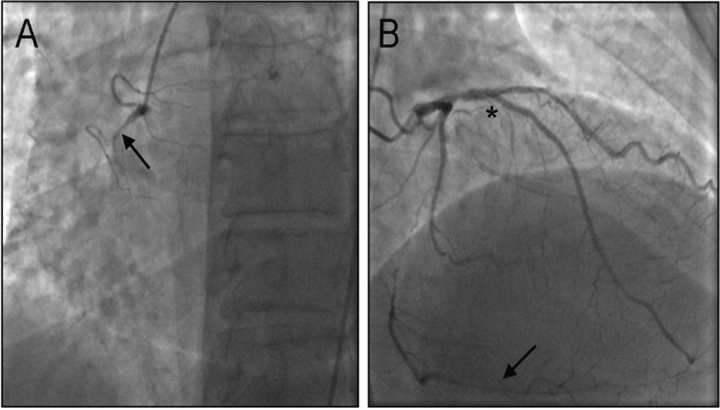
Parent vessel anatomy was consistent with population studies of CTO with the RCA being the most prevalent vessel occluded (Table 2). Marrow aspirations were carried out without complication. Aspirated volumes were adequate and similar among patients. Mononuclear cell preparations and subsequent selection of CD133+ cells were variable in number ranging from 8–48 × 106 and yielded substantially more cells than required for infusion. Purity fell within the range of acceptable as defined in the protocol for all patients but was also highly variable (77–97%). In vitro analysis of the whole cell preparations from all patients produced few and in some cases no colony-forming units. Analysis of the CD133+ selected cells as a percent of CD45+ cells (hematopoietic cells) is expressed in Table 3. All hematopoietic lineages were represented in the contaminating population, the largest percentage of which was myeloid precursors and megakaryocytes. Each of the three dosing tiers was tolerated and then triggered an additional dose escalation (doubling of cell number). Cells were held, transported and infused to the target vessel with timely success. There was a lack of infusion related morbidity including acute obstruction or dissection and no change in TIMI flow of the target vessel (Table 4). Figure 1 is a representative angiogram and illustrates collateral vessel directed infusion of cells. Cardiac biomarkers remained within normal limits for all patients at time points up to 24 hours following infusion. There were no detected arrhythmias or ECG abnormalities during the post-procedural period. Moreover, patients experienced no symptoms consistent with angina following infusion.
Table 2.
Patient infusion data
| Patient | Age | Marrow volume (ml) | Number of selected CD133+ (106) | CD133+ purity (%) | Target vessel | CD133+ infused |
|---|---|---|---|---|---|---|
| 1 | 65 | 200 | 23.2 | 97.0 | LCX | 1 × 10⁶ |
| 2 | 76 | 200 | 48.5 | 77.0 | RCA | 1 × 10⁶ |
| 3 | 76 | 185 | 11.6 | 77.3 | RCA | 1 × 10⁶ |
| 4 | 65 | 170 | 8.0 | 80.8 | RCA | 2 × 10⁶ |
| 5 | 71 | 200 | 11.8 | 91.0 | OM2 | 2 × 10⁶ |
| 6 | 61 | 200 | 30.2 | 94.5 | RCA | 2 × 10⁶ |
| 7 | 63 | 200 | 19.5 | 78.0 | LAD | 3 × 10⁶ |
| 8 | 51 | 200 | 13.2 | 88.3 | RCA | 3 × 10⁶ |
| 9 | 59 | 200 | 17.8 | 80.8 | RCA | 3 × 10⁶ |
| Summary | 65.2 ± 7.67 | 195 ±10.0 | 20.4 ± 11.8 | 85.0 ± 7.37 |
Values are expressed as mean ± SD.
Table 3.
CD133+ contamination characteristics
| Patient | CD3 | CD3neg/CD56+ | CD13+/SSC high | CD14 | CD19 immature | CD19 mature | CD61 | CD71 |
|---|---|---|---|---|---|---|---|---|
| 1 | 2.8 | 0.06 | 4.7 | 0.68 | 4.5 | 1.9 | 4.6 | 0.21 |
| 2 | 7.7 | 0.43 | 7.7 | 0.59 | 1.5 | 0.26 | 0.5 | 0.78 |
| 3 | 1.3 | 0.01 | 3.8 | 0.23 | 0.55 | 0.17 | 2.9 | 0.58 |
| 4 | 1.4 | 0.17 | 6.3 | 0.49 | 2.6 | 0.47 | 22.8 | 0.28 |
| 5 | 0.95 | 0.05 | 11.1 | 0.61 | 5.4 | 0.2 | 0.13 | 2.2 |
| 6 | -- | -- | -- | -- | -- | -- | 3.1 | 0.15 |
| 7 | 0.57 | 0.05 | 15.1 | 0.09 | 1.2 | 0.02 | 1.3 | 1.7 |
| 8 | 2.6 | 0.03 | 6.2 | 0.36 | 3.7 | 0.88 | 2.0 | 1.5 |
| 9 | 10.9 | 0.15 | 5.2 | 0.99 | 3.2 | 1.9 | 9.9 | 0.3 |
| Summary | 3.53 ± 3.50 | 0.12 ± 0.13 | 7.51 ± 3.55 | 0.51 ± 0.26 | 2.83 ± 1.58 | 0.73 ± 0.72 | 5.25 ±6.79 | 0.86 ± 0.71 |
Absolute values are expressed as percent. Summary values are expressed as mean ± SD
Table 4.
Clinical safety markers
| Procedural complications * | 0 |
|---|---|
| Creatinine kinase (U/l) † | |
| Baseline | 80.5 ± 39.4 |
| 6–8 h after cell therapy | 180.1 ± 78.9 |
| 12–16 h after cell therapy | 162.9 ± 58.8 |
| 20–24 after cell therapy | 145.1 ± 58.6 |
| Creatinine kinase -MB (ng/ml) ‡ | |
| Baseline | 1.11 ± 0.57 |
| 6–8 h after cell therapy | 1.33 ± 0.67 |
| 12–16 h after cell therapy | 1.33 ± 1.05 |
| 20–24 after cell therapy | 1.22 ± 1.03 |
Values are expressed as mean ± SD.
Reduction in TIMI flow, dissection, aneurysm and thrombosis.
upper normal limit = 232.
upper normal limit = 7
Figure 1.
(A) Representative coronary angiograms illustrating a chronic total occlusion (CTO) of the right coronary artery (RCA) and (B*) target left anterior descending artery (LAD) during directed cellular infusion. Arrowheads indicate the regions of occlusion.
4.3. Outcomes
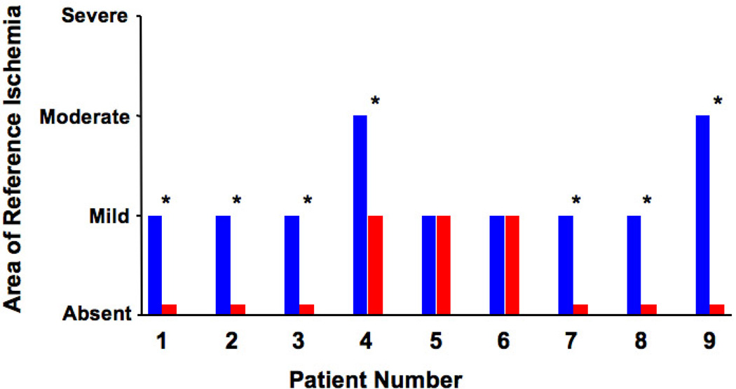
In our single center study of nine patients the isolation and infusion of autologous CD133+ cells was feasible. No in-hospital events were recorded including death, AMI and ventricular arrhythmias. No patients were lost to follow-up and all clinical and ischemic functional assessments were completed. The primary outcome of safety over the course of the study was met. No MACE was incurred during the twenty-four months follow up period. There were no arrhythmias detected by 24 hour Holter monitoring and no laboratory abnormalities attributable to infusion. With respect to secondary outcomes, seven of the nine patients had a reduction in the area of reference ischemia (Figure 2). There was suggestion of an increase in anginal stability and quality of life (Table 5). However, there appeared to be no change in treatment satisfaction, anginal frequency or improvement in physical limitation as assessed by the Seattle Angina Questionnaire.
Figure 2.
Comparison of the area of reference ischemia, measured by 99mTc-sestamibi. Columns in the left (blue) and in the right (red) represent the degree of ischemia at the time of enrollment and at 12 months, respectively for each patient. (*) indicates statistical significance (p<0.001).
Table 5.
12-Month change in seattle angina questionnaire score
| Scale of the Seattle Angina Questionnaire | Baseline Mean Value | 12-Month Mean Value | Mean Difference | |
|---|---|---|---|---|
| Physical limitation | 71.35 | 76.79 | 5.43 | |
| Angina stability | 50.00 | 65.63 | 15.63 | |
| Angina frequency | 77.50 | 83.75 | 6.25 | |
| Treatment satisfaction | 85.94 | 91.41 | 5.47 | |
| Disease perception | 53.13 | 67.71 | 14.58 |
5. DISCUSSION
In this phase I pilot study we demonstrated that coronary infusion of autologous BM derived CD133+ hematopoietic stem cells is feasible and safe in patients with CTO. This safety was achieved with a step-wise dose escalation approach.
Recent clinical trials in humans have illustrated reasonable safety and feasibility. However, there remain additional questions regarding efficacy, timing of delivery, cell potential and dosing. As with other pharmaceutical agents dosing kinetics and toxicity are critical. This interplay has not been addressed in previous studies with cellular products. The largest trials to date have generated varying results with respect to efficacy and those that have shown benefit reflect nominal responses (20, 24, 25). In addition, these studies employed large numbers of heterogeneous cell populations. Heterogeneity of cell populations prohibits analysis of potency because the functionally effective cell is not specifically quantified. Additionally, there are no studies to date addressing the question of cell dose and the relationship with clinical safety.
In our study no procedural complications were encountered and there were no subsequent post-procedural arrhythmias, ischemia or immediate acceleration of angina in any patients. Each dose was tolerated and triggered an additional two increases in cell dose without acute clinical consequences. There were no patients lost to follow up and all patients completed the components of the clinical follow up protocol. During the twenty-four months monitoring period following cell infusion there were no documented arrhythmias or MACE.
There were no logistical issues related to harvesting and selecting adequate numbers of a relatively pure fraction of hematopoietic stem cells. Isolation of distinct populations of stem cells is possible in most large clinical centers with appropriate core facilities as this is done regularly for bone marrow transplantation. Large volume bone marrow aspirates generated significantly more CD133+ cells than required for infusion and cell preparations were free of infectious agents after processing. The wide range of cell numbers retrieved may represent the health of the patient’s bone marrow. Although adequate numbers of cells were selected, there was a range of purity of these cells. The variability in purity reflects small numbers of contaminating precursor cells and may be related to antibody specificity. However, because this fraction represents a minute portion of the total numbers of cells infused we don’t believe they are significantly involved in mediating toxicity or the potential for efficacy. Further investigation of this cell fraction is ongoing to determine the exact role of these cells and their functional capacity.
Previous trials have utilized a cell source, which potentially may have reduced potential as a number of studies suggest that stem cell dysfunction may be inherent to patients with chronic disease (30–32). Functional quantification was carried out in this study utilizing a colony-forming assay with cells selected from each patient. None of the patient samples yielded significant numbers of colony-forming units suggesting a possible diminution of function.
The patients enrolled in this study were required to have an LV ejection fraction greater than 45% and thus represent a cohort without significant impairment of myocardial function. Additionally, six of nine patients had symptoms that did not dramatically impact their function (i.e. class I and II angina). There was an overall trend towards reduction in ischemic myocardium.
5.1. Limitations
Although a number of products have been utilized with varying success it remains unclear as to the efficacy of stem cells and the potential role in myocardial ischemia whether acute or chronic. Many of the patients in this trial may have received benefit from the infusion however the study was not powered to answer this provocative question. Overall myocardial function was initially good with established collateralization in this mostly functional population and thus represents a population difficult to detect functional or symptomatic improvement.
6. CONCLUSION
Cellular therapy for myocardial ischemia and subsequent functional deficiency has been shown to be beneficial in preclinical studies. Furthermore, application of cellular therapy in vivo can enhance repair via angiogenesis and possibly regenerate myocardium. Although improvement has been achieved in animal studies it remains unclear as to whether this form of therapy is effective in humans. There is a paucity of therapeutic options for patients with CTO and perhaps augmentation of the compensatory response with stem cells may be effective. Our study provides a novel approach of collateral vessel targeting for stem cell delivery in patients with CTO and appears to be safe. We believe this study paves the way for future trials powered to answer questions of efficacy of a selected hematopoietic stem cell in this difficult to treat population of patients.
ACKNOWLEDGEMENTS
The authors wish to acknowledge the extensive efforts of our clinical trial nurses involved in this study, without them this study would not have been possible. This study was supported by the National Institutes of Health NHLBI 4R42 HL080856-02. Dr. Pompili is equity-holders in Arteriocyte, Inc.
REFERENCES
- 1.Das H, Abdulhameed N, Joseph M, Sakthivel R, Mao HQ, Pompili VJ. Ex vivo nanofiber expansion and genetic modification of human cord blood-derived progenitor/stem cells enhances vasculogenesis. Cell Transplant. 2009;18(3):305–318. doi: 10.3727/096368909788534870. [DOI] [PMC free article] [PubMed] [Google Scholar]
- 2.Das H, George JC, Joseph M, Das M, Abdulhameed N, Blitz A, Khan M, Sakthivel R, Mao HQ, Hoit BD, Kuppusamy P, Pompili VJ. Stem cell therapy with overexpressed VEGF and PDGF genes improves cardiac function in a rat infarct model. PLoS One. 2009;4(10):e7325. doi: 10.1371/journal.pone.0007325. [DOI] [PMC free article] [PubMed] [Google Scholar]
- 3.Aggarwal R, Pompili VJ, Das H. Genetic modification of ex-vivo expanded stem cells for clinical application. Front Biosci. 2010;15:854–871. doi: 10.2741/3650. [DOI] [PMC free article] [PubMed] [Google Scholar]
- 4.Finney MR, Greco NJ, Haynesworth SE, Martin JM, Hedrick DP, Swan JZ, Winter DG, Kadereit S, Joseph ME, Fu P, Pompili VJ, Laughlin MJ. Direct comparison of umbilical cord blood versus bone marrow-derived endothelial precursor cells in mediating neovascularization in response to vascular ischemia. Biol Blood Marrow Transplant. 2006;12(5):585–593. doi: 10.1016/j.bbmt.2005.12.037. [DOI] [PubMed] [Google Scholar]
- 5.Prasad A, Rihal CS, Lennon RJ, Wiste HJ, Singh M, Holmes DR., Jr Trends in outcomes after percutaneous coronary intervention for chronic total occlusions: a 25-year experience from the Mayo Clinic. J Am Coll Cardiol. 2007;49(15):1611–1618. doi: 10.1016/j.jacc.2006.12.040. [DOI] [PubMed] [Google Scholar]
- 6.Stone GW, Kandzari DE, Mehran R, Colombo A, Schwartz RS, Bailey S, Moussa I, Teirstein PS, Dangas G, Baim DS, Selmon M, Strauss BH, Tamai H, Suzuki T, Mitsudo K, Katoh O, Cox DA, Hoye A, Mintz GS, Grube E, Cannon LA, Reifart NJ, Reisman M, Abizaid A, Moses JW, Leon MB, Serruys PW. Percutaneous recanalization of chronically occluded coronary arteries: a consensus document: part I. Circulation. 2005;112(15):2364–2372. doi: 10.1161/CIRCULATIONAHA.104.481283. [DOI] [PubMed] [Google Scholar]
- 7.Hochman JS, Lamas GA, Buller CE, Dzavik V, Reynolds HR, Abramsky SJ, Forman S, Ruzyllo W, Maggioni AP, White H, Sadowski Z, Carvalho AC, Rankin JM, Renkin JP, Steg PG, Mascette AM, Sopko G, Pfisterer ME, Leor J, Fridrich V, Mark DB, Knatterud GL. Coronary intervention for persistent occlusion after myocardial infarction. N Engl J Med. 2006;355(23):2395–2407. doi: 10.1056/NEJMoa066139. [DOI] [PMC free article] [PubMed] [Google Scholar]
- 8.Agbulut O, Vandervelde S, Al Attar N, Larghero J, Ghostine S, Leobon B, Robidel E, Borsani P, Le Lorc'h M, Bissery A, Chomienne C, Bruneval P, Marolleau JP, Vilquin JT, Hagege A, Samuel JL, Menasche P. Comparison of human skeletal myoblasts and bone marrow-derived CD133+ progenitors for the repair of infarcted myocardium. J Am Coll Cardiol. 2004;44(2):458–463. doi: 10.1016/j.jacc.2004.03.083. [DOI] [PubMed] [Google Scholar]
- 9.Botta R, Gao E, Stassi G, Bonci D, Pelosi E, Zwas D, Patti M, Colonna L, Baiocchi M, Coppola S, Ma X, Condorelli G, Peschle C. Heart infarct in NOD-SCID mice: therapeutic vasculogenesis by transplantation of human CD34+ cells and low dose CD34+KDR+ cells. Faseb J. 2004;18(12):1392–1394. doi: 10.1096/fj.03-0879fje. [DOI] [PubMed] [Google Scholar]
- 10.Chen HK, Hung HF, Shyu KG, Wang BW, Sheu JR, Liang YJ, Chang CC, Kuan P. Combined cord blood stem cells and gene therapy enhances angiogenesis and improves cardiac performance in mouse after acute myocardial infarction. Eur J Clin Invest. 2005;35(11):677–686. doi: 10.1111/j.1365-2362.2005.01565.x. [DOI] [PubMed] [Google Scholar]
- 11.Friedrich EB, Walenta K, Scharlau J, Nickenig G, Werner N. CD34−/CD133+/VEGFR-2+ endothelial progenitor cell subpopulation with potent vasoregenerative capacities. Circ Res. 2006;98(3):e20–e25. doi: 10.1161/01.RES.0000205765.28940.93. [DOI] [PubMed] [Google Scholar]
- 12.Leor J, Guetta E, Feinberg MS, Galski H, Bar I, Holbova R, Miller L, Zarin P, Castel D, Barbash IM, Nagler A. Human umbilical cord blood-derived CD133+ cells enhance function and repair of the infarcted myocardium. Stem Cells. 2006;24(3):772–780. doi: 10.1634/stemcells.2005-0212. [DOI] [PubMed] [Google Scholar]
- 13.Ma N, Ladilov Y, Moebius JM, Ong L, Piechaczek C, David A, Kaminski A, Choi YH, Li W, Egger D, Stamm C, Steinhoff G. Intramyocardial delivery of human CD133+ cells in a SCID mouse cryoinjury model: Bone marrow vs. cord blood-derived cells. Cardiovasc Res. 2006;71(1):158–169. doi: 10.1016/j.cardiores.2006.03.020. [DOI] [PubMed] [Google Scholar]
- 14.Ott I, Keller U, Knoedler M, Gotze KS, Doss K, Fischer P, Urlbauer K, Debus G, von Bubnoff N, Rudelius M, Schomig A, Peschel C, Oostendorp RA. Endothelial-like cells expanded from CD34+ blood cells improve left ventricular function after experimental myocardial infarction. Faseb J. 2005;19(8):992–994. doi: 10.1096/fj.04-3219fje. [DOI] [PubMed] [Google Scholar]
- 15.Suuronen EJ, Veinot JP, Wong S, Kapila V, Price J, Griffith M, Mesana TG, Ruel M. Tissue-engineered injectable collagen-based matrices for improved cell delivery and vascularization of ischemic tissue using CD133+ progenitors expanded from the peripheral blood. Circulation. 2006;114(1 Suppl):I138–I144. doi: 10.1161/CIRCULATIONAHA.105.001081. [DOI] [PubMed] [Google Scholar]
- 16.Orlic D, Kajstura J, Chimenti S, Jakoniuk I, Anderson SM, Li B, Pickel J, McKay R, Nadal-Ginard B, Bodine DM, Leri A, Anversa P. Bone marrow cells regenerate infarcted myocardium. Nature. 2001;410(6829):701–705. doi: 10.1038/35070587. [DOI] [PubMed] [Google Scholar]
- 17.Gnecchi M, He H, Noiseux N, Liang OD, Zhang L, Morello F, Mu H, Melo LG, Pratt RE, Ingwall JS, Dzau VJ. Evidence supporting paracrine hypothesis for Akt-modified mesenchymal stem cell-mediated cardiac protection and functional improvement. Faseb J. 2006;20(6):661–669. doi: 10.1096/fj.05-5211com. [DOI] [PubMed] [Google Scholar]
- 18.Gnecchi M, He H, Liang OD, Melo LG, Morello F, Mu H, Noiseux N, Zhang L, Pratt RE, Ingwall JS, Dzau VJ. Paracrine action accounts for marked protection of ischemic heart by Akt-modified mesenchymal stem cells. Nat Med. 2005;11(4):367–368. doi: 10.1038/nm0405-367. [DOI] [PubMed] [Google Scholar]
- 19.Assmus B, Schachinger V, Teupe C, Britten M, Lehmann R, Dobert N, Grunwald F, Aicher A, Urbich C, Martin H, Hoelzer D, Dimmeler S, Zeiher AM. Transplantation of Progenitor Cells and Regeneration Enhancement in Acute Myocardial Infarction (TOPCARE-AMI) Circulation. 2002;106(24):3009–3017. doi: 10.1161/01.cir.0000043246.74879.cd. [DOI] [PubMed] [Google Scholar]
- 20.Assmus B, Honold J, Schachinger V, Britten MB, Fischer-Rasokat U, Lehmann R, Teupe C, Pistorius K, Martin H, Abolmaali ND, Tonn T, Dimmeler S, Zeiher AM. Transcoronary transplantation of progenitor cells after myocardial infarction. N Engl J Med. 2006;355(12):1222–1232. doi: 10.1056/NEJMoa051779. [DOI] [PubMed] [Google Scholar]
- 21.Bartunek J, Vanderheyden M, Vandekerckhove B, Mansour S, De Bruyne B, De Bondt P, Van Haute I, Lootens N, Heyndrickx G, Wijns W. Intracoronary injection of CD133-positive enriched bone marrow progenitor cells promotes cardiac recovery after recent myocardial infarction: feasibility and safety. Circulation. 2005;112(9 Suppl):I178–I183. doi: 10.1161/CIRCULATIONAHA.104.522292. [DOI] [PubMed] [Google Scholar]
- 22.Kocher AA, Schuster MD, Szabolcs MJ, Takuma S, Burkhoff D, Wang J, Homma S, Edwards NM, Itescu S. Neovascularization of ischemic myocardium by human bone-marrow-derived angioblasts prevents cardiomyocyte apoptosis, reduces remodeling and improves cardiac function. Nat Med. 2001;7(4):430–436. doi: 10.1038/86498. [DOI] [PubMed] [Google Scholar]
- 23.Lunde K, Solheim S, Aakhus S, Arnesen H, Abdelnoor M, Forfang K. Autologous stem cell transplantation in acute myocardial infarction: The ASTAMI randomized controlled trial. Intracoronary transplantation of autologous mononuclear bone marrow cells, study design and safety aspects. Scand Cardiovasc J. 2005;39(3):150–158. doi: 10.1080/14017430510009131. [DOI] [PubMed] [Google Scholar]
- 24.Lunde K, Solheim S, Aakhus S, Arnesen H, Abdelnoor M, Egeland T, Endresen K, Ilebekk A, Mangschau A, Fjeld JG, Smith HJ, Taraldsrud E, Grogaard HK, Bjornerheim R, Brekke M, Muller C, Hopp E, Ragnarsson A, Brinchmann JE, Forfang K. Intracoronary injection of mononuclear bone marrow cells in acute myocardial infarction. N Engl J Med. 2006;355(12):1199–1209. doi: 10.1056/NEJMoa055706. [DOI] [PubMed] [Google Scholar]
- 25.Schachinger V, Erbs S, Elsasser A, Haberbosch W, Hambrecht R, Holschermann H, Yu J, Corti R, Mathey DG, Hamm CW, Suselbeck T, Assmus B, Tonn T, Dimmeler S, Zeiher AM. Intracoronary bone marrow-derived progenitor cells in acute myocardial infarction. N Engl J Med. 2006;355(12):1210–1221. doi: 10.1056/NEJMoa060186. [DOI] [PubMed] [Google Scholar]
- 26.Strauer BE, Brehm M, Zeus T, Kostering M, Hernandez A, Sorg RV, Kogler G, Wernet P. Repair of infarcted myocardium by autologous intracoronary mononuclear bone marrow cell transplantation in humans. Circulation. 2002;106(15):1913–1918. doi: 10.1161/01.cir.0000034046.87607.1c. [DOI] [PubMed] [Google Scholar]
- 27.Stamm C, Westphal B, Kleine HD, Petzsch M, Kittner C, Klinge H, Schumichen C, Nienaber CA, Freund M, Steinhoff G. Autologous bone-marrow stemcell transplantation for myocardial regeneration. Lancet. 2003;361(9351):45–46. doi: 10.1016/S0140-6736(03)12110-1. [DOI] [PubMed] [Google Scholar]
- 28.Wollert KC, Meyer GP, Lotz J, Ringes-Lichtenberg S, Lippolt P, Breidenbach C, Fichtner S, Korte T, Hornig B, Messinger D, Arseniev L, Hertenstein B, Ganser A, Drexler H. Intracoronary autologous bone-marrow cell transfer after myocardial infarction: the BOOST randomised controlled clinical trial. Lancet. 2004;364(9429):141–148. doi: 10.1016/S0140-6736(04)16626-9. [DOI] [PubMed] [Google Scholar]
- 29.Janssens S, Dubois C, Bogaert J, Theunissen K, Deroose C, Desmet W, Kalantzi M, Herbots L, Sinnaeve P, Dens J, Maertens J, Rademakers F, Dymarkowski S, Gheysens O, Van Cleemput J, Bormans G, Nuyts J, Belmans A, Mortelmans L, Boogaerts M, Van de Werf F. Autologous bone marrow-derived stem-cell transfer in patients with ST-segment elevation myocardial infarction: double-blind, randomised controlled trial. Lancet. 2006;367(9505):113–121. doi: 10.1016/S0140-6736(05)67861-0. [DOI] [PubMed] [Google Scholar]
- 30.Werner N, Kosiol S, Schiegl T, Ahlers P, Walenta K, Link A, Bohm M, Nickenig G. Circulating endothelial progenitor cells and cardiovascular outcomes. N Engl J Med. 2005;353(10):999–1007. doi: 10.1056/NEJMoa043814. [DOI] [PubMed] [Google Scholar]
- 31.Hill JM, Zalos G, Halcox JP, Schenke WH, Waclawiw MA, Quyyumi AA, Finkel T. Circulating endothelial progenitor cells, vascular function, and cardiovascular risk. N Engl J Med. 2003;348(7):593–600. doi: 10.1056/NEJMoa022287. [DOI] [PubMed] [Google Scholar]
- 32.Urbich C, Dimmeler S. Risk factors for coronary artery disease, circulating endothelial progenitor cells, and the role of HMG-CoA reductase inhibitors. Kidney Int. 2005;67(5):1672–1676. doi: 10.1111/j.1523-1755.2005.00261.x. [DOI] [PubMed] [Google Scholar]


