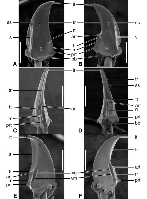
Figure 3.
SEM photographs of mandibles of Pelophila rudis LeConte. A, C, E left mandible, dorsal, occlusal, ventral aspects, respectively; B, D, F right mandible, dorsal, occlusal, ventral aspects, respectively. Legend: see Table 2. Scale bars = 0.5 mm.

