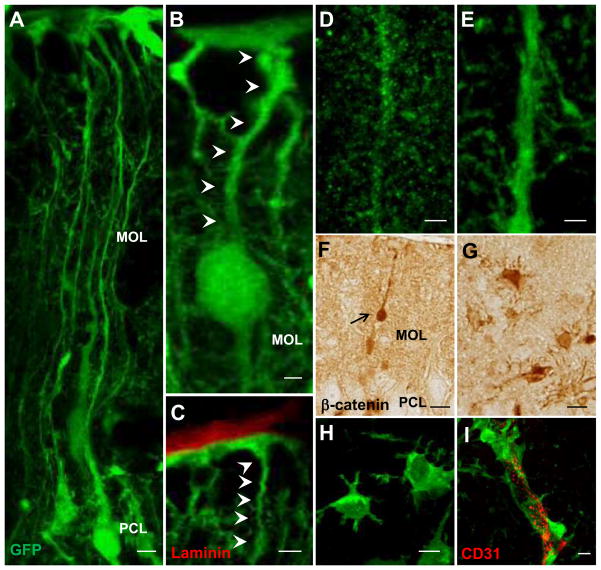
Figure 6.
Morphological transformation of APC-deficient Bergmann glia. Representative images of the reporter GFP-positive cells in the cerebellar cortex of APC-CKO reporter mice. At P14, many GFP-positive Bergmann glia situate in the PCL and their radial fibers span the MOL to the pial surface (A). A portion of GFP-positive cells still retain radial glia morphology as well as the contact with the pial surface (B, C), but their fibers shorten and the cell bodies translocate into the MOL with beta-catenin accumulation (B, F). The radial fibers of APC-deficient Bergmann glia are thick and smooth with a marked reduction of collateral branches (E) in comparison to those of control reporter mice (D). At P21, the majority of GFP-positive cells loses the pial contact and transforms into a stellate morphology (G, H) with the prominent perivascular endfeet (I). Scale bar: A, C, F, G, H, 5μm; B, D, E, I, 2 μm. P, postnatal day; PCL, Purkinje cell layer; MOL, molecular layer; GCL, granule cell layer.

