Abstract
Background
The purpose of this study is to evaluate the efficacy of computed tomography (CT) scans compared with plain radiographs on detecting the involvement of the sigmoid notch.
Methods
This study involved 121 cases diagnosed as the intra-articular distal radius fracture and performed post-reduction CT scans. We determined the presence of the sigmoid notch involvement with both plain radiographs and CT scans and compared findings of plain radiographs with CT scans about the incidence and the pattern of injuries. And the differences of results between arbeitsgemeinschaft für osteosyntheses (AO) type C2 and C3 were compared.
Results
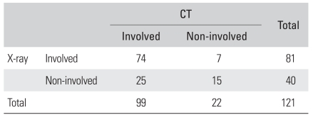
The incidences of sigmoid notch involvement detected in plain radiographs were 81 cases (66.9%), whereas CT scans were 99 cases (81.9%). The sensitivity of plain radiographs compared with CT scans was 74.7%, the specificity was 68.2%, the positive predictive value was 91.4%, the negative predictive value was 37.5%, the false negative value was 25.3%, and the false positive value was 31.8%. In comparison between AO type C2 and C3, the incidence of sigmoid notch involvement was not a significant difference, but the displacement of fracture fragment showed a significant difference.
Conclusions
The intra-articular distal radius fracture usually accompanies the sigmoid notch involvement. Considering that the evaluation of sigmoid notch involvement by plain radiography often results in misinterpretation or underestimation, performing CT scan in intra-articular distal radius fracture is thought to be beneficial.
Keywords: Distal radius, Intra-articular fracture, Sigmoid notch, Computed tomography
The intra-articular distal radius fracture is very common fracture which occupies 10% to 12% of whole fractures and its characteristics are known to be diverse and complications also frequently seen.1) And various treatment methods are used such as closed reduction and cast immobilization, percutaneous K-wire fixation, external fixation, intramedullary fixation, open reduction and internal fixation. Choosing an appropriate treatment method up on the characteristics of the fracture is vital for precise reduction and satisfactory functional recovery and the characteristics of the fracture have to be accurately understood to be able to achieve this.2) Computed tomography (CT) is commonly used to understand the precise characteristics of the intra-articular distal radius fracture along with plain X-ray.3-5) That is known to be more beneficial to understand the precise characteristics of the intra-articular distal radius fracture than performing only plain X-ray and that also aids to decide the direction of its treatment.6-8) However, the frequency and the type of the sigmoid notch following intra-articular distal radius fracture with advanced diagnostic methods such as computed tomography have been very rarely researched.9,10) For this reason, this study aimed to understand the involvement of sigmoid notch following intra-articular distal radius fracture by reconstructing the sagittal, axial and coronal CT images and to evaluate its benefits.
METHODS
The subjects were 119 patients (121 cases) underwent CT scan after being diagnosed with intra-articular distal radius fracture by plain X-ray between March 2007 and January 2010. The cases diagnosed as the intra-articular fracture of distal radius by plain X-ray and CT scan were included in this study. The cases which suspected on plain X-ray but not confirmed in CT scan were excluded. Also, the cases which only distal radio-ulnar joint was involved were excluded. There were 46 male patients and 75 female patients and their mean age was 56.4 years (range, 16 to 97 years) old. The fracture of right wrist was 67 cases and left was 52 cases and bilateral was 2 cases. The reduction of the fracture was completed by manual traction and all the patients underwent anterio-posterior, lateral and oblique view of plain X-ray of the wrist prior and post to the reduction. CT scan was performed after wearing sugar tongs splint in a neutral rotation of forearm and the CT machine used was Mx8000IDT (Philips, Eindhoven, Netherlands). One millimeter interval of sectioned images were obtained and reconstructed to the sagittal, axial and coronal images using MxView program (version 3.5).
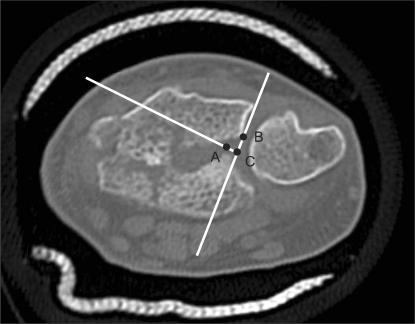
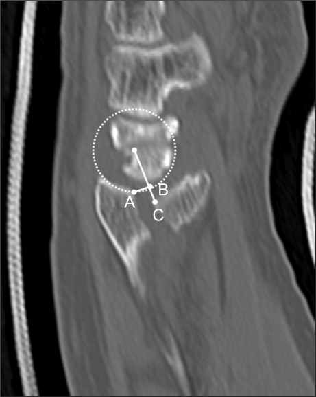
Radiological analysis was done by two different doctors (one hand surgery specialist and one orthopedic resident). The plain X-ray reading and CT reading were performed in two weeks interval and each reading was repeated twice. The intra-articular distal radius fracture was classified by arbeitsgemeinschaft für osteosyntheses (AO) classification and the presence of sigmoid notch involvement was evaluated in pre- and post-reduction plain X-ray. The sigmoid notch involvement on CT images was classified by the method of Rozental et al.10) Type 1 was identified as the intra-articular distal radius fracture without extension into sigmoid notch. And it was subdivided into type 1a if there were non-displaced fracture of distal radius, otherwise, it was into type 1b. Type 2 was as the extension of fracture into sigmoid notch that not displaced of fragments. Type 3 was as the extension of fracture into sigmoid notch with displacement of fragments. And it was subdivided into type 3a when there was rotation and/or translation of one of volar and dorsal fracture fragments and type 3b when were rotation and/or translation of both fragments. The sensitivity, specificity, positive predictive value, negative predictive value, false positive value and false negative value of plain X-ray to recognize sigmoid notch involvement were investigated by comparing the findings of plain X-ray and CT. The gap and the step-off of displaced fragments were measured on axial images and sagittal images when sigmoid notch involvement was found on CT scan. The method of Rozental et al.10) was used in the axial measurements and the gap and the step-off were measured on the images showed subchondral bone (Fig. 1). The method of Cole et al.6) was quoted in the sagittal measurements and the images which was closer to the sigmoid notch and both palmar and dorsal fragments were seen on were chosen for the measurement (Fig. 2). Moreover, the degrees of sigmoid notch involvement between AO type C2 and C3 were compared.
Fig. 1.
Axial computed tomography image of the distal radius showing a displaced fracture of the sigmoid notch (SN). Point A and B were marked at subchondral fracture margins of SN. First line was drawn between the dorsal corner of SN and point B. Second line through point A was perpendicular to first line. The intersection of two lines was point C. Step-off of SN was measured as the distance between point A and C. Gap displacement was measured as the distance between point B and C.
Fig. 2.
Sagittal computed tomography image of the radiocarpal joint. Point A and C were marked at subchondral fracture margin of lunate facet of distal radius. A circle matching curvature of remaining articular surface of distal radius was traced and passed through point A. First line was drawn from center of circle to point C. The intersection between circle and first line was point B. Step displacement is measured as the distance between points B and C. Gap displacement is measured as the distance between points A and B.
The results of each group were compared with paired t-test and chi-square test. And intra-class correlation coefficients (ICCs) were used to evaluate the reproducibility within each assessors and the reliability between the assessors.11) It was referred to be poor when the value of ICCs was between 0.00-0.39, to be average when it was between 0.40-0.74 and to be excellent when it was between 0.75-1.00. Statistical analysis was done by SPSS ver. 16.0 (SPSS Inc., Chicago, IL, USA).
RESULTS
The Incidences of Sigmoid Notch Involvement
Total 81 cases (66.9%) of sigmoid notches were detected on plain X-ray image. Distal radius fractures were classified as the followings: type B1 was 5 cases, type B3 was 4, type C1 was 2, type C2 was 62, and type C3 was 48. On the other hand, total 99 cases (81.9%) were detected on CT scan. Classifying subjects with the CT images, Rozental type 1a was 12 cases, type 1b was 22, type 2 was 39, type 3a was 27, and type 3b was 21. Especially, the sigmoid notches involvement in cases of AO type C were 2 cases from 2 cases of C1, 45 (72.6%) from 62 of C2 and 34 (70.8%) from 48 of C3 on plain X-ray and 2 from 2 of C1, 51 (82.3%) from 62 of C2 and 42 (87.5%) from 48 of C3.
On plain X-ray analysis, the reproducibility of the first observer was 0.822 and the reproducibility of the second observer was 0.895. And, the values of ICCs for the reliability between observers were 0.628 and 0.742. In terms of the reading agreement between plain X-ray and CT scan, the initial value of the first observer was 0.616 and the initial value of the second observer was 0.683. And the reading agreement was increased in the second measurements (ICCs of the first observer, 0.733; the second observer, 0.762).
The Evaluation of Sigmoid Notch Involvement by CT Scan
There was a statistically significant difference between sigmoid notch findings between plain X-ray and CT scan (p = 0.001) (Table 1). Among 99 cases which sigmoid notch was identified on CT images, 25 cases, that were 17 from C2 and 8 from C3 of AO classification, were missed on the plain X-ray. Eight among 25 cases had greater than 1 mm displaced fracture fragment on CT scan but it was failed to identify (Fig. 3). Nine of them didn't show any evidence of displaced fracture fragment. Five cases were identified the sigmoid notch involvement on sagittal CT images. And it was difficult to diagnose in the other 3 cases because the sigmoid notch had a feature of posterior thin cortical fracture accompanying dorsal metaphyseal comminution. On the other hand, among 22 cases which were ruled out by CT scan, 7 (2 of C3 and 5 of C3) cases were diagnosed with sigmoid notch involvement by the plain X-ray and were judged to be misdiagnosed by CT scan. Four among 7 cases were intra-articular fracture of radio-carpal joint located near the sigmoid notch (Fig. 4) and the other 3 were misdiagnosed due to the impact of severely comminuted fragment of metaphysis.
Table 1.
Comparison of Plain X-ray Results and Computed Tomography (CT) Results
Sensitivity: 74.7%, Specificity: 68.2%.
Positive predictive value: 91.4%, Negative predictive value: 37.5%.
False negative value: 25.3%, False positive value: 31.8%.
Fig. 3.
Radiographs of 57-year-old woman with left distal radius fracture. Initial radiographs (A-C) were showing intra-articular fracture line (black arrow on image (A)) and metaphyseal comminution. But, authors misinterpreted the involvement of the sigmoid notch presented because of overlaps of ulnar head and lunate facet on lateral view (B) or dorsal-ulnar corner of distal radius on oblique view (C). The fracture of the sigmoid notch with displaced fragment was confirmed on axial computed tomography image (D).
Fig. 4.
Initial radiographs (A-C) of 79-year-old man were showing intra-articular fracture of left distal radius fracture. The authors interpreted the disruption of the sigmoid notch with displacement of fracture fragment on image A-C. But, the sigmoid notch was intact and intra-articular fracture line was located near the sigmoid notch on axial computed tomography image (D).
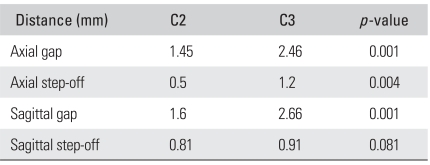
The displacement of fragments in 99 cases with sigmoid notch involvement on CT images was measured. The average gap of displaced fragments were 1.98 mm (range, 0 to 4.9 mm) on axial images and 2.17 mm (range, 0 to 4.6 mm) on sagittal images and the average step-off were 0.82 mm (range, 0 to 4.1 mm) and 0.87 mm (range, 0 to 4.6 mm), respectively. Especially, the average gap of 51 sigmoid notch cases from C2 were 1.45 mm (range, 0 to 3.2 mm) on axial images and 1.61 mm (range, 0 to 4.3 mm) on sagittal images and the average step-off were 0.5 mm (range, 0 to 2.4 mm) and 0.81 mm (range, 0 to 2.9 mm), respectively. The average gap of 42 sigmoid notch cases from C3 were 2.46 mm (range, 0 to 4.9 mm) on axial images and 2.67 mm (range, 0 to 4.5 mm) on sagittal images and the average step-off were 1.2 mm (range, 0 to 4.1 mm) and 0.91 mm (range, 0 to 4.6 mm), respectively. The gap and step-off of above 1 mm were identified in 15 (29.4%) from 51 cases of C2 and 23 (54.8%) from 42 cases of C3. There was no difference in the incidences of sigmoid notch involvement between C2 and C3 (p = 0.84). However, there was a significant difference in the level of displacement of fragments between C2 and C3 (Table 2).
Table 2.
Mean Gap and Step-off of AO Type C2 and C3 Measured by Computed Tomography
AO: arbeitsgemeinschaft für osteosyntheses.
The Additional Type of Fractures to be Considered
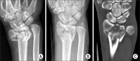
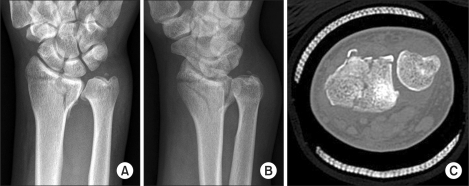
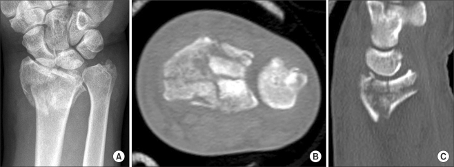
First of all, axial fracture line of distal radius fracture invaded to sigmoid notch was observed in 17 cases among 62 cases of AO type C2 and 12 cases among 48 cases of C3. This damage aided the diagnosis of AP images of plain X-ray or coronal image of CT scan (Fig. 5). Secondly, posterior thin cortical fracture invaded to sigmoid notch was observed in 9 cases among 62 cases of C2 and 4 cases among 48 cases of C3 and the fragments with less than 2 mm thickness on coronal CT images showing subchondral bone were also included (Fig. 6). This type of fracture was difficult to identify with plain X-ray and they were confirmed on the reconstructed coronal CT images. Thirdly, there were cases of comminuted fracture on the sigmoid notch. It was shown in 3 cases of type C3 and they were all identified on the coronal CT images (Fig. 7).
Fig. 5.
Distal radius fracture involved the articular surface of radiocarpal and distal radioulnar joint. This case was classified as arbeitsgemeinschaft für osteosyntheses type C3 and Ronzental type 3a. Axial-plane fracture line of the sigmoid notch can be easily found in coronal computed tomography image (C) than pre-reduction (A) or post-reduction (B) radiographs.
Fig. 6.
Initial radiographs (A and B) and axial CT image (C) showing intra-articular distal radius fracture. This case was classified as arbeitsgemeinschaft für osteosyntheses type C2 and Rozental type 1b. Dorso-ulnar cortical corner fracture of the distal radius was not found on postero-anterior view (A) but oblique view (B). And minimal involvement of the sigmoid notch was confirmed on axial computed tomography image (C).
Fig. 7.
Comminuted distal radius fracture was classified as arbeitsgemeinschaft für osteosyntheses type C3 and Rozental type 3b. An accurate assessment is difficult about sigmoid notch in plane radiograph (A). Axial (B) and sagittal (C) computed tomography image shows comminuted fracture of articular surface of the sigmoid notch.
DISCUSSION
There have been a number of changes to manage distal radius fracture and successful anatomical reduction is vital to achieve satisfactory outcomes. Intra-articular fracture in distal radius fracture is commonly seen and closed or open reduction depending on the level of displaced articular surface is required. It is widely known that the prevalence of traumatic osteoarthritis is high when the displacement of radiocarpal articular surface is greater than 2 mm.1,12,13) There is a literature recommending less than 1 mm reduction of articular surface in recent years.8) Therefore, it is important to judge the type of fracture at early stage after trauma and the use of CT scan is expected to beneficial because it is difficult to do this with only plain X-ray.3-5) Cole et al.6) reported that plain X-ray failed to identify the displacement of articular surface greater than 2 mm in 24% and there was a huge difference between findings from plain X-ray and CT scan. Pruitt et al.7) claimed that CT was more useful to understand the invasion of distal radioulnar joint and the level of depression and comminution of articular surface although there was no significant difference between plain X-ray and CT scan in the fracture of radiocarpal joint. Trumble et al.8) reported that there were significant correlations between the postoperative gap of fracture fragments, the formation of step-off, the incongruency of articular surface and the shortening of radius. And the gap among those was identified as the most important factor and they stated that a decrease in the function could be expected when it was greater than 1 mm. Reviewing these research results, CT is beneficial for the accurate judgment to manage the fracture either conservatively or surgically.
One of commonly complained symptoms after distal radius fracture is reported to be the pain around distal radioulnar joint.3,9,12,14) Instability of the joint, incongruency of the joint, pain secondary to ulnar impaction syndrome and limited rotation are also commonly expressed and the former 2 symptoms can cause irreversible damages to articular cartilage leading to degenerative conditions. Hence, appropriate treatment should be initiated during the acute period and accurate understanding of the fracture is essential in the early stage of trauma. It was easy to detect sigmoid notch involvement in AO type B2 or B3 distal radius fracture but it was difficult to judge in type C with plain X-ray. Generally, sigmoid notch involvement may be difficult to be detected on plain X-ray image and the increased anterioposterior length of distal radius sometimes indicates the fracture of sigmoid notch and the displacement of fracture fragment.2) The authors in this study found the oblique image helpful to confirm sigmoid notch but it was difficult to understand the level of damage and the displacement with single plain X-ray. Moreover, the shadow of the cast to maintain the reduction of the fracture interrupted the observation of fracture line. Pruitt et al.7) compared the plain X-ray reading to the CT reading of 18 cases of distal radioulnar fracture. They failed to identify 6 cases of distal radioulnar fracture on plain X-ray images and CT images of one case were interpreted as distal radioulnar fracture although it wasn't observed on the image. Rozental et al.,10) also, reported that the fracture was identified in 7 cases among 20 cases (35%) with plain X-ray but 13 cases were identified with CT scan (65%). Katz et al.3) reported that more than 63% of distal radioulnar fracture confirmed by CT scan was underestimated on the finding of plain X-ray. The incidences of sigmoid notch involvement identified in this study were 67% on plain X-ray and 82% on CT scan. And 32 cases (26.4%) were misdiagnosed with sigmoid notch involvement by plain X-ray. The reason for the high incidences of sigmoid notch involvement in this study was thought to be that most of the subjects were AO C2 or C3 type fracture accompanying severe level of metaphysis or articular surface comminution. In addition to this, the misinterpretation of the radiological investigation was either confused with the intra-articular fracture line or comminuted metaphyseal fragments or it was difficult to identify because fracture fragments were not displaced on plain X-ray. Therefore, it has been acknowledged that CT is more useful to understand the damages of distal radioulnar joint in distal radius fracture than plain X-ray.
Instability of distal radioulnar joint can be caused by the malalignment of distal radius fracture but it can be rectified by osteotomy. However, distal radioulnar joint osteoarthritis is difficult to treat and salvage operations are required in most cases.15) Generally, the description of sigmoid notch involvement was hardly seen although Frykman16) reported that 19% of distal radius fracture accompanied with distal radioulnar joint damages. Geissler et al.9) classified the distal radioulnar joint damages in distal radius fracture and it was said to be type IIIA when there was the sigmoid notch involvement. They explained that this type of fracture carried a high risk of distal radioulnar joint osteoarthritis and limited forearm rotation when sufficient reduction was not achieved because the joint is extremely unstable. They also highlighted the importance of accurate understanding of the damage during the early stage of trauma in order to reduce these complications.
Rozental et al.10) first classified and evaluated the sigmoid notch involvement using CT. Among 20 cases in this study, sigmoid notch involvement was identified in 7 cases (35%) with plain X-ray and 13 cases (65%) with CT scan and the undetected interval of sigmoid notch (9 cases) and step-off formation (7 cases) by plain X-ray were confirmed on CT images. Nevertheless, they evaluated with sigmoid notch involvement only axial images and they didn't mention about the results of treatments. The authors in this study used axial images and sagittal images to evaluate the displacement of the fracture. As it was mentioned in the results, the displacement of the fracture was observed in the sagittal images as it was done in the axial images. And the greater displacement on both axial images and sagittal images was observed as the comminution of articular surface was severer.
It is difficult to understand the reduction status in surgical management to treat sigmoid notch involvement and distal radius fracture accompanying displacement. Either the attempt for arthroscopic reduction or confirmation of lunate-lunate facet ratio17) is known to be beneficial. The authors confirmed the anteroposterior distance of distal radius from the standard lateral image and lunate facet of distal radius articular surface from the 10° lateral projection image using radiography after the fixation of the fracture.2) Furthermore, the displacement during the insertion of screws for the plates fixation was prevented by pressing the posterior fragment of the sigmoid notch with fingers and the screws that were shorter than actual measurements were used to avoid the displacement of posterior fragment induced by screws. Postoperative CT might have helped but wasn't able to perform due to the cost and the failure to obtain consent.
The authors were able to observe other types of sigmoid notch involvement which wasn't mentioned by Rozental et al.10) by reading CT images but they were described in the results because it was difficult to include them under any categories. First of all, sigmoid notch damage induced by axial fracture was found in 29 cases (26%) among 112 cases of AO type C. This damage was easily seen in the AO type A3 which was excluded in this study because it didn't presented with intra-articular distal radius fracture and it was confirmed in 10 cases (38%) among 28 cases of A3. Radial displacement is commonly occurred in the distal area of the fracture when this type of fracture is accompanied and precise reduction is expected to aid to reduce the complications of distal radioulnar joint. Secondly, minimally invaded sigmoid notch induced by posterior thin cortical fracture of distal radius was observed in 13 cases (12%) among 112 cases of AO type C. Limited extension of wrist joint can be occurred when this fragments are displaced to distal area and healed and ulnar displacement can cause chronic distal radioulnar joint pain induced by the collision to the head of ulnar during rotation. Besides, the accurate judgment during the early stage of radiography is vital because posterior thin cortical fracture is often accompanied with posterior subluxation of distal radioulnar joint.18) Thirdly, precise anatomical reduction was difficult in the comminuted fracture on articular surface of sigmoid notch since it was generated from a high energy impact and accompanied with severe comminuted fracture of distal radius.
In conclusions, sigmoid notch involvement is commonly seen in intra-articular distal radius fracture and inappropriate treatment can cause pain around the wrist. Therefore, performing CT scan prior to surgical managements is beneficial because the diagnosis and the evaluation of sigmoid notch involvement in intra-articular distal radius fracture is insufficient.
Footnotes
No potential conflict of interest relevant to this article was reported.
References
- 1.Turner RG, Faber KJ, Athwal GS. Complications of distal radius fractures. Hand Clin. 2010;26(1):85–96. doi: 10.1016/j.hcl.2009.08.005. [DOI] [PubMed] [Google Scholar]
- 2.Medoff RJ. Essential radiographic evaluation for distal radius fractures. Hand Clin. 2005;21(3):279–288. doi: 10.1016/j.hcl.2005.02.008. [DOI] [PubMed] [Google Scholar]
- 3.Katz MA, Beredjiklian PK, Bozentka DJ, Steinberg DR. Computed tomography scanning of intra-articular distal radius fractures: does it influence treatment. J Hand Surg Am. 2001;26(3):415–421. doi: 10.1053/jhsu.2001.22930a. [DOI] [PubMed] [Google Scholar]
- 4.Johnston GH, Friedman L, Kriegler JC. Computerized tomographic evaluation of acute distal radial fractures. J Hand Surg Am. 1992;17(4):738–744. doi: 10.1016/0363-5023(92)90326-k. [DOI] [PubMed] [Google Scholar]
- 5.Catalano LW, 3rd, Barron OA, Glickel SZ. Assessment of articular displacement of distal radius fractures. Clin Orthop Relat Res. 2004;(423):79–84. doi: 10.1097/01.blo.0000132884.51311.28. [DOI] [PubMed] [Google Scholar]
- 6.Cole RJ, Bindra RR, Evanoff BA, Gilula LA, Yamaguchi K, Gelberman RH. Radiographic evaluation of osseous displacement following intra-articular fractures of the distal radius: reliability of plain radiography versus computed tomography. J Hand Surg Am. 1997;22(5):792–800. doi: 10.1016/s0363-5023(97)80071-8. [DOI] [PubMed] [Google Scholar]
- 7.Pruitt DL, Gilula LA, Manske PR, Vannier MW. Computed tomography scanning with image reconstruction in evaluation of distal radius fractures. J Hand Surg Am. 1994;19(5):720–727. doi: 10.1016/0363-5023(94)90174-0. [DOI] [PubMed] [Google Scholar]
- 8.Trumble TE, Schmitt SR, Vedder NB. Factors affecting functional outcome of displaced intra-articular distal radius fractures. J Hand Surg Am. 1994;19(2):325–340. doi: 10.1016/0363-5023(94)90028-0. [DOI] [PubMed] [Google Scholar]
- 9.Geissler WB, Fernandez DL, Lamey DM. Distal radioulnar joint injuries associated with fractures of the distal radius. Clin Orthop Relat Res. 1996;(327):135–146. doi: 10.1097/00003086-199606000-00018. [DOI] [PubMed] [Google Scholar]
- 10.Rozental TD, Bozentka DJ, Katz MA, Steinberg DR, Beredjiklian PK. Evaluation of the sigmoid notch with computed tomography following intra-articular distal radius fracture. J Hand Surg Am. 2001;26(2):244–251. doi: 10.1053/jhsu.2001.22930. [DOI] [PubMed] [Google Scholar]
- 11.Shrout PE, Fleiss JL. Intraclass correlations: uses in assessing rater reliability. Psychol Bull. 1979;86(2):420–428. doi: 10.1037//0033-2909.86.2.420. [DOI] [PubMed] [Google Scholar]
- 12.Knirk JL, Jupiter JB. Intra-articular fractures of the distal end of the radius in young adults. J Bone Joint Surg Am. 1986;68(5):647–659. [PubMed] [Google Scholar]
- 13.Taras JS, Ladd AL, Kalainov DM, Ruch DS, Ring DC. New concepts in the treatment of distal radius fractures. Instr Course Lect. 2010;59:313–332. [PubMed] [Google Scholar]
- 14.Kihara H, Palmer AK, Werner FW, Short WH, Fortino MD. The effect of dorsally angulated distal radius fractures on distal radioulnar joint congruency and forearm rotation. J Hand Surg Am. 1996;21(1):40–47. doi: 10.1016/S0363-5023(96)80152-3. [DOI] [PubMed] [Google Scholar]
- 15.Nagy L. Salvage of post-traumatic arthritis following distal radius fracture. Hand Clin. 2005;21(3):489–498. doi: 10.1016/j.hcl.2005.03.005. [DOI] [PubMed] [Google Scholar]
- 16.Frykman G. Fracture of the distal radius including sequelae--shoulder-hand-finger syndrome, disturbance in the distal radio-ulnar joint and impairment of nerve function: a clinical and experimental study. Acta Orthop Scand. 1967;(Suppl 108):3+. doi: 10.3109/ort.1967.38.suppl-108.01. [DOI] [PubMed] [Google Scholar]
- 17.Harreld KL, Apel P, Koman LA, Li Z. Lunate-lunate facet ratio and variance to predict articular gap after distal radius fracture. J Hand Surg Am. 2009;34(9):1625–1632. doi: 10.1016/j.jhsa.2009.06.008. [DOI] [PubMed] [Google Scholar]
- 18.Lozano-Calderon SA, Doornberg J, Ring D. Fractures of the dorsal articular margin of the distal part of the radius with dorsal radiocarpal subluxation. J Bone Joint Surg Am. 2006;88(7):1486–1493. doi: 10.2106/JBJS.E.00930. [DOI] [PubMed] [Google Scholar]


