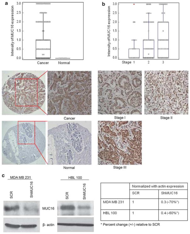
Figure 1.
Expression of MUC16 in breast cancer tissues and non-neoplastic breast tissues. Immunohistochemical analysis was carried out with CA125 antibody in breast cancer tissue. (a) Shows high immunoreactivity for MUC16 was observed in breast cancer tissues compared with non-neoplastic breast tissues. (b) Shows the comparison of MUC16 expression between various stages of breast carcinoma. Stable knockdown of MUC16 in two different breast cancer cell lines MDA MB 231 and HBL 100. Endogenously expressed MUC16 was stably knocked down using a MUC16 shRNA construct (pSUPER-Retro-MUC16-sh) in highly aggressive MDA MB 231 and HBL 100 breast cancer cells. (c) Western blot analysis confirms the decreased expression of MUC16 in MDA MB 231 and HBL 100 cells that were stably transfected with the MUC16 shRNA construct in comparison with empty-vector control cells.

