Abstract
The circumsporozoite protein (CSP) plays a key role in malaria sporozoite infection of both mosquito salivary glands and the vertebrate host. The conserved Regions I and II have been well studied but little is known about the immunogenic central repeat region and the N-terminal region of the protein. Rodent malaria Plasmodium berghei parasites, in which the endogenous CS gene has been replaced with the avian Plasmodium gallinaceum CS (PgCS) sequence, develop normally in the A. stephensi mosquito midgut but the sporozoites are not infectious. We therefore generated P. berghei transgenic parasites carrying the PgCS gene, in which the repeat region was replaced with the homologous region of P. berghei CS (PbCS). A further line, in which both the N-terminal region and repeat region were replaced with the homologous regions of PbCS, was also generated. Introduction of the PbCS repeat region alone, into the PgCS gene, did not rescue sporozoite species-specific infectivity. However, the introduction of both the PbCS repeat region and the N-terminal region into the PgCS gene completely rescued infectivity, in both the mosquito vector and the mammalian host. Immunofluorescence experiments and western blot analysis revealed correct localization and proteolytic processing of CSP in the chimeric parasites. The results demonstrate, in vivo, that the repeat region of P. berghei CSP, alone, is unable to mediate sporozoite infectivity in either the mosquito or the mammalian host, but suggest an important role for the N-terminal region in sporozoite host cell invasion.
Introduction
The circumsporozoite protein (CSP) is the predominant surface antigen of Plasmodium sporozoites and is highly immunogenic being one of the key targets recognised by the host immune system. Pre-erythrocytic malaria vaccines have been primarily based on CSP [1], [2]. In particular, the central repeat region of P. falciparum CSP, which contains an immunodominant B cell epitope, represented the target of the first two vaccine trials [3], [4]. Recently, the results of a phase III trial of the RTS,S vaccine, based on both the repeat region and T-cell epitopes from the C-terminal region, provided evidence for protection against both clinical and severe malaria in African children [5].
CSP performs numerous functions for the sporozoite at different stages in the life cycle. The protein is first detected at high levels in the oocyst and has been shown to be vital in the process of sporogenesis [6], [7]. CSP is likely to be important in sporozoite gliding motility although the precise role that the protein plays in gliding remains unknown. Antibodies against CSP inhibit gliding motility [8] and sporozoites leave behind trails of CSP that correspond to their pattern of movement [9]. CSP has long been known to be involved in sporozoite infectivity [10], [11]. Specifically, it appears to be important in the binding of the sporozoite to both mosquito salivary glands [12], [13], [14] and vertebrate host hepatocytes [15], [16], [17].
Comparison of the deduced amino acid sequences of CS proteins from all species of Plasmodium shows that they have a similar overall structure. They all contain a central repeat region, whose amino acid sequence is species specific, and two conserved regions: a five amino acid sequence called Region I (RI), immediately upstream of the repeats, and a known cell-adhesive sequence with similarity to the type I repeat of thrombospondin called Region II (RII), downstream of the repeat region. CSP has a canonical glycosylphosphatidylinositol (GPI) anchor addition sequence in its C-terminus. Much evidence has been gathered on the functions of the conserved Regions I and II of CSP, as well as on residues outside of RI and RII, within the N- and C-terminal portions of the protein, which have been implicated in host binding [18], [19], [20], [21]. However, little evidence exists to date on the role of the CSP repeat region in the parasite life cycle.
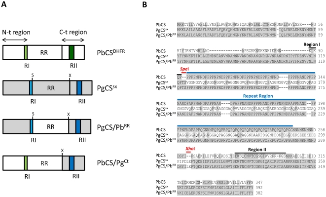
The RI core is a five amino acid sequence (KLKQP), conserved in almost all Plasmodium species with the exception of the avian malaria parasite P. gallinaceum, in which there are two amino acid changes in the core (NLNQP) (Figure 1). RI has been strongly implicated in the binding of salivary glands [13], [22]. A peptide encompassing RI was shown to inhibit binding of both recombinant CSP and sporozoites to the salivary glands. However, the RI core alone could not achieve this inhibition and upstream residues were also required [13], [22]. The sequence upstream of RI contains stretches of positively charged residues which are implicated, together with RI, in the interaction with liver heparan sulphate proteoglycans (HSPGs) [19], [21]. RI and RII peptides inhibited P. falciparum CSP (PfCSP) binding to HepG2 cell HSPGs to a similar extent, suggesting they both contribute to the interaction [23]. RII is a common motif present in the proteins of a wide range of organisms and is also found in another sporozoite surface protein, the thrombospondin-related adhesive protein (TRAP). It is an 18 amino acid region and part of a larger type I repeat of human thrombospondin (TSR) domain which acts as an adhesive module and binds with high affinity to heparin and certain sulphated glycoconjugates (Figure 1). The rapid and specific homing to the liver by sporozoites of mammalian Plasmodium spp. has been ascribed to the interaction between RII and glycosaminoglycan (GAG) side chains on HSPGs, located on the basolateral surface of hepatocytes [15], [16], [23], [24], [25]. The only difference between the P. gallinaceum RII and the RII consensus, present in all Plasmodium species, is a single amino acid substitution, despite the fact that P. gallinaceum does not infect hepatocytes.
Figure 1. Schematic representation and alignment of wild type and transgenic P.berghei circumsporozoite proteins.
(A) Schematic representation of the CS proteins present in each of the four transgenic P. berghei parasite lines generated. The corresponding names of the transgenic parasite lines are shown on the right. PbCSDHFR parasites carry the wildtype PbCS coding sequence (white boxes, RI and RII indicated with light and dark green respectively) and, similar to all transgenic lines, contain the T. gondii DHFR drug selectable marker inserted in the CS locus. PgCSSX parasites carry the full PgCS coding sequence (grey boxes, RI and RII indicated with light and dark blue respectively), but with the SpeI (S) and XhoI (X) restriction endonuclease sites inserted on either side of the repeat region. PgCS/PbRR parasites contain the PgCS N-terminal and C-terminal regions (grey boxes) and the PbCS repeat region (white box). PbCS/PgCT parasites carry the PbCS N-terminal and repeat regions (white boxes) and the PgCS C-terminal region (grey box). (B) Alignment of the wild type PbCS and transgenic PgCSSX and PgCS/PbRR amino acid sequences. Shaded boxes represent areas of amino acid identity. Regions I and II (black lines), the repeat region (blue line) and the SpeI and XhoI restriction sites (red lines) are labelled. The SpeI and XhoI sites were introduced into the PgCS sequence to mediate exchange of the PgCS repeat region with the PbCS repeat region.
The repeat region is species-specific, immunodominant and constitutes about one half of the molecule. It contains multiple copies of tandemly arrayed repeat peptides (Figure 1) and has no obvious sequence homology to any known protein. The repeats contain B-cell immunodominant epitopes and antibodies specific to this region can protect against malaria by blocking sporozoite invasion of host hepatocytes [26], [27]. The function of the CSP repeat region within the parasite life cycle has so far not been investigated. However, there are reasons to believe that this large domain of CSP could perform perhaps multiple functions for the parasite. The repeat peptide structure might lend itself well to high avidity interactions with host receptors, for example GAGs which are composed of repeating sugar units. Multivalency is able to enhance the affinity of protein-carbohydrate interactions, particularly those involving GAGs [28], and the repeats could provide multivalent binding sites. In addition, the species-specific nature of the region may indicate a role in maintaining the host specificity of infection.
P. berghei parasites, in which the entire CS gene (PbCS) was replaced with its P. gallinaceum orthologue, failed to both invade salivary glands and to infect mice [29], although sporozoite development and CSP expression appeared unaffected. P. gallinaceum is carried by the mosquito vector Aedes aegypti whereas P. berghei is carried by anopheline vectors. The lack of infectivity with the PgCS replacement parasites could be due to the inability of PgCSP to bind to A. stephensi salivary gland receptors and rodent liver receptors.
To assess whether the repeat region has a role in salivary gland or vertebrate infectivity, we generated transgenic P. berghei parasites carrying the PgCS gene, in which the repeat region was replaced with the PbCS repeat region (Figure 1), in order to determine if introduction of the wild-type sequence rescues infectivity. Another parasite line containing the full-length PbCS N-terminal sequence (up to RI), the PbCS repeat region and the PgCS C-terminal sequence was also generated and allowed further analysis of the role of the N- and C-terminal regions (Figure 1).
Results
Generation of transgenic parasites
Transgenic P. berghei parasites were generated that carried chimeric P. gallinaceum - P. berghei CS genes. In the first transgenic parasite line, the endogenous PbCS gene was substituted with the PgCS gene in which the repeat region was replaced with the homologous region of P. berghei: line PgCS/PbRR (Figure 1A). A second line contained both the N-terminal region and repeat region of P. berghei, while the C-terminal region was replaced with the homologous sequence of P. gallinaceum: line PbCS/PgCT (Figure 1A). As controls, we generated: i) a transgenic line carrying the wild type PbCS gene and the drug selectable marker inserted in the PbCS locus: line PbCSDHFR and ii) a transgenic line carrying the PgCS gene in which the SpeI and XhoI restriction sites had been introduced on either side of the repeat region: line PgCSSX (Figure 1A). A schematic representation and alignment of PbCSP and the CS proteins expressed in the transgenic parasites, and their corresponding names, are presented in Figure 1A and 1B respectively.
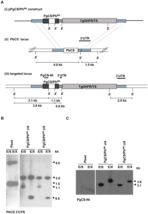
The targeting constructs were designed to direct the double cross-over event between the 1.13 kb sequence of the PbCS 5′ untranslated region (UTR) and the 0.85 kb sequence of the PbCS 3′ UTR in the linearized plasmid and their corresponding sequences in the PbCS locus (Figure 2A and Figure S1A). Two targeting constructs, pPgCS/PbRR and pPgCSSX were then used to transfect P. berghei wild type (Pbwt) parasites (Figure 2A and Figure S1A respectively).
Figure 2. Generation and southern blot analysis of transgenic PgCS/PbRR parasite lines.
(A) Schematic representation of (i) the PgCS/PbRR targeting construct, (ii) the wt PbCS locus and (iii) the targeted locus after recombination between the PbCS 5′UTR and 3′UTR sequences. The vertically dashed box indicates the 1.13 kb 5′UTR sequence used in the construct and the horizontally dashed boxes indicate the 0.3 kb and 0.85 kb 3′UTR sequences between which the TgDHFR-TS selectable marker cassette (light grey) was inserted in the construct. The PgCS/PbRR chimeric gene contains the PgCS N- and C-terminal regions (dark grey boxes) and the PbCS repeat region (white box) flanked by the SpeI (S) and XhoI (X) sites. In the wt PbCS locus the white box indicates the full PbCS gene. Thick black lines indicate the probes used in southern blots. E: EcoRV site. (B) Southern blot of EcoRV/SpeI (E/S) and EcoRV/XhoI (E/X) digested genomic DNA from Pbwt parasites and transgenic PgCS/PbRR parasites (clones 4 and 5), hybridised with the PbCS 3′UTR probe. Two bands of 4.0 and 1.5 kb were present in Pbwt DNA in both digestions. E/S digested DNA from the PgCS/PbRR clones revealed two bands of 1.1 and 2.0 kb, while E/X digested DNA revealed two bands of 0.6 and 2.0 kb, demonstrating the replacement of the endogenous PbCS gene with the targeted construct. (C) The same membrane was then hybridised with the PgCS N-terminal probe (PgCS-Nt), encompassing the entire N-terminal region of PgCS. No band was present in Pbwt DNA. However, a band of 3.1 kb or 3.6 kb was present in the E/S or E/X digested transgenic DNA, respectively.
In construct pPgCSSX (Figure S1A), the SpeI and XhoI restriction sites had been introduced by site-directed mutagenesis into the PgCS sequence in order to mediate exchange of the PgCS repeat region with the homologous sequence of P. berghei. The SpeI site was inserted immediately downstream of the PgCS RI core sequence, NLNQP (Figure 1). The XhoI restriction site was inserted after the PgCS repeat region and before RII (Figure 1). Correct insertion of the construct, to generate the control transgenic parasite line PgCSSX (clones 1 and 2), was confirmed by Southern blot analysis (Figure S1).
A second construct was generated by replacing the PgCS repeat region in pPgCSSX with the PbCS repeat region, which had been amplified from genomic DNA. The resulting construct, pPgCS/PbRR, was then tranfected into Pbwt parasites to create the transgenic line PgCS/PbRR (clones 4 and 5), and correct integration was confirmed by southern blot (Figure 2).
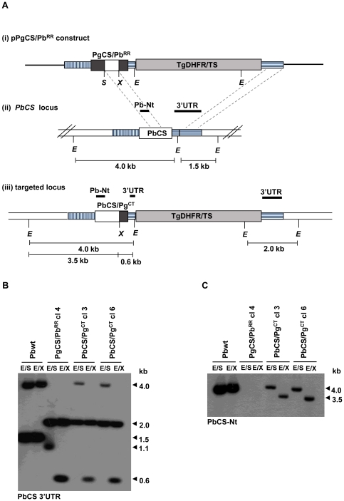
Other integration events were also observed after transfection with the pPgCS/PbRR vector, as a consequence of cross-over events that took place between sequences present in the internal part of the construct and the homologous sequences in the PbCS locus. This resulted in the generation of two further transgenic parasite lines, PbCS/PgCT and PbCSDHFR (Figure 3 and Figure S2). PbCS/PgCT clones 3 and 6 originated from cross-over events at the 0.5 kb PbCS repeat region and the 3′ UTR sequence (Figure 3A). The PbCS/PgCT parasite line generated therefore contained a chimeric CS gene where both the N-terminal sequence and the repeat region were from P. berghei, while the C-terminal sequence was from P. gallinaceum, as demonstrated by Southern blot analysis (Figure 3).
Figure 3. Generation and southern blot analysis of transgenic PbCS/PgCT parasite lines.
(A) Schematic representation of (i) the PgCS/PbRR targeting construct, (ii) the wt PbCS locus and (iii) the targeted locus after recombination between the PbCS repeat region (white box) and the PbCS 3′UTR (horizontally dashed box). The PbCS/PgCT chimeric gene contains both the PbCS N- terminal and repeat regions (white boxes) and the PgCS C-terminal region (dark grey box). The light grey box indicates the TgDHFR-TS selectable marker cassette. Thick black lines indicate the probes used in southern blots. E = EcoRV site, S = SpeI site and X = XhoI site. (B) Southern blot of EcoRV/SpeI (E/S) and EcoRV/XhoI (E/X) digested genomic DNA from Pbwt parasites, transgenic PgCS/PbRR clone 4 parasites and PbCS/PgCT parasites (clones 3 and 6), hybridised with the PbCS 3′UTR probe. PgCS/PbRR parasites acted as a control for the presence of both the SpeI and XhoI sites in the CS chimeric gene. Both PbCS/PgCT clones contained a gene with the XhoI site but lacked the SpeI site, as demonstrated by the presence of the 4.0 and 2.0 kb bands after E/S digestion and the 2.0 and 0.6 kb bands after E/X digestion, indicating the 5′ cross-over event had occurred between the repeat regions. (C) Southern blot of E/S and E/X digested genomic DNA, hybridised with the PbCS N-terminal region probe. A band of 4.0 kb was detected in both the Pbwt DNA and E/S digested DNA from the PbCS/PgCT clones, demonstrating the presence of the PbCS N-terminal sequence in the transgenic parasites. Genomic DNA from the PbCS/PgCT clones, digested with E/X, revealed a band of 3.5 kb, indicating the presence of the XhoI site. No bands were visible in PgCS/PbRR transgenic parasite DNA due to the absence of the PbCS N-terminal sequence.
Moreover, just 300 bp of the PbCS 3′ UTR, present between the chimeric CS gene and the TgDHFR/TS cassette in the construct pPgCS/PbRR, was sufficient to direct recombination, generating the PbCSDHFR transgenic line (Figure S2A). Double cross-over recombination, between the two PbCS 3′ UTR regions present in the construct and the homologous regions in the PbCS locus, resulted in the insertion of only the TgDHFR/TS cassette, in the 3′ UTR region of the PbCS locus, leaving the endogenous PbCS gene intact. The transgenic PbCSDHFR clones 9 and 10 generated (Figure S2) were used as a control for any phenotypic effects arising from the presence of the selectable marker cassette within the CS locus.
The insertion events described, and the presence of the correct CS gene sequences in the transgenic parasites lines, was further confirmed by sequencing the CS genes, which had been PCR amplified from genomic DNA.
Development of the transgenic parasites in the mosquito vector
Blood stage parasites from all transgenic lines replicated normally in mice and in vitro generated gametocytes that developed into fertile gametes. The development of the transgenic parasites in the mosquito was monitored in Anopheles stephensi fed on infected mice. Mosquito midguts infected with each of the transgenic clones, and dissected at 14 and 18 days post-infection (p.i.), revealed normal oocyst development compared to mosquitoes infected with Pbwt parasites. All transgenic oocysts had the same morphology as Pbwt oocysts. Interestingly, melanisation (observed in mosquitoes infected with PgCS-replacement P. berghei parasites [29]) was only seen in midguts infected with transgenic lines containing PgCS sequences. However, it occurred with a very low frequency both in terms of the proportion of infected midguts and also the number of melanised oocysts per midgut.
Midguts dissected on day 14 p.i. showed no significant differences in oocyst and sporozoite numbers between any of the transgenic parasite lines and Pbwt parasites (Table 1), indicating that both heterologous PgCSP and the chimeric proteins function normally during sporozoite ontogeny in the mosquito vector A. stephensi. However, at day 21 p.i., PgCSSX parasites showed a significant reduction in the number of salivary gland sporozoites compared to both Pbwt and PbCSDHFR control parasites, similar to the results previously reported for mosquitoes infected with PgCS-replacement P. berghei parasites [29]. Likewise, PgCS/PbRR parasites showed a striking and significant reduction in salivary gland sporozoites compared to Pbwt and PbCSDHFR parasites (Table 1). Furthermore, no significant difference was observed between PgCSSX and PgCS/PbRR clone 4 (p>0.09), although the difference between PgCSSX and PgCS/PbRR clone 5 was significant (p<0.03). However, this result for PgCS/PbRR clone 5 may have been due to contaminating hemolymph sporozoites or a greater number of sporozoites adhering to the outside of the glands.
Table 1. Transgenic parasite development in the Anopheles stephensi mosquito vector.
| Parasite | Midgut oocysts | Midgutsporozoites | Sporozoites/oocyst | Salivary gland sporozoites | Salivary gland sporozoites/oocyst |
| Pbwt | 182±42 | 12,174±19,070 | 63±79 | 28,412±8,167 | 134±14 |
| PbCSDHFR cl9 | 195±105 | 26,021±40,551 | 144±217 | 25,626±5,185 | 154±70 |
| PbCSDHFR cl10 | 190±47 | 15,377±6,451 | 89±53 | 14,447±4,503 | 83±40 |
| PgCSSX cl1 | 189±93 | 6,447±5,221 | 36±27 | 297±336a | 2±2 |
| PgCS/PbRR cl4 | 202±66 | 14,844±6,773 | 86±69 | 922±697a | 6±5 |
| PgCS/PbRR cl5 | 194±32 | 13,892±8,871 | 68±37 | 1,276±300a , b | 7±3 |
| PbCS/PgCT cl3 | 207±77 | 33,821±32,294 | 240±311 | 15,779±3,832 | 80±17 |
| PbCS/PgCT cl6 | 248±55 | 32,520±22,484 | 133±99 | 18,291±8,231 | 78±47 |
Significant difference compared to Pbwt and PbCSDHFR transgenic parasites; p<0.01.
Significant difference compared to PgCSSX; p<0.03.
A. stephensi mosquitoes were fed on mice infected with one of the transgenic clones or Pbwt parasites. Values represent the mean ± S.D. of at least three independent experiments, each with a minimum of 50 mosquitoes.
The low numbers of salivary gland sporozoites in mosquitoes infected with PgCS/PbRR parasites suggests that the replacement of the PgCS repeat region with the homologous PbCS repeat region is not sufficient to rescue salivary gland invasion. In contrast, the PbCS/PgCT transgenic parasites invaded the mosquito salivary glands normally. These parasites contain only the C-terminal sequence of PgCSP, which appears to function normally in A. stephensi salivary gland infection. The results suggest that the N-terminal region plays an important role in salivary gland infection, although it is still possible that repeat region residues co-operate with N-terminal residues in infectivity. This is in agreement with previous studies, which used recombinant N-terminal peptides to demonstrate a role for the region in salivary gland binding [13], [22].
Infectivity of the transgenic sporozoites for the vertebrate host
To determine the infectivity of the transgenic parasites for the vertebrate host, salivary gland and midgut sporozoites, collected 21 days p.i., were injected intravenously into C57BL/6 mice. All mice injected with Pbwt salivary gland sporozoites developed a parasitaemia, with a pre-patent period ranging from 3.5 to 4.6 days for mice injected with 5,000 and 1,000 sporozoites respectively (Table 2). Interestingly, for the PbCSDHFR transgenic parasites, only injection with 5,000 sporozoites resulted in a 100% infection rate, and the pre-patent period was longer, ranging from 5.3 to 6.5 days after injection of 5,000 or 1,000 PbCSDHFR sporozoites, respectively. A 1 day delay in the pre-patent period indicates a 90% decrease in the infective inoculum [30]. This difference in infectivity between Pbwt and PbCSDHFR parasites suggests a moderate effect on vertebrate host infection due to the presence of the selectable marker cassette in the CS locus.
Table 2. Infectivity of salivary gland sporozoites for the vertebrate host.
| Parasite | Sporozoites injected | Miceinfected | Pre-patentperioda |
| Pbwt | 1,000 | 5/5 | 4.6 |
| 3,000 | 3/3 | 4.0 | |
| 5,000 | 3/3 | 3.5 | |
| PbCSDHFR cl9 | 1,000 | 1/6 | 6.0 |
| 3,000 | 4/6 | 6.0 | |
| 5,000 | 4/4 | 5.5 | |
| PbCS DHFR cl10 | 1,000 | 2/4 | 6.5 |
| 3,000 | 2/4 | 6.0 | |
| 5,000 | 3/3 | 5.3 | |
| PgCSSX cl1 | 5,000 | 0/3 | - |
| PgCS/PbRR cl4 | 5,000 | 0/5 | - |
| PgCS/PbRR cl5 | 5,000 | 0/5 | - |
| PbCS/PgCT cl3 | 1,000 | 0/3 | - |
| 3,000 | 3/4 | 5.7 | |
| 5,000 | 5/6 | 5.5 | |
| PbCS/PgCT cl6 | 1,000 | 1/3 | 6.0 |
| 3,000 | 3/4 | 6.0 | |
| 5,000 | 4/5 | 5.5 |
Pre-patent period is the number of days between injection and first appearance of the parasites in the peripheral blood.
C57BL/6 mice were injected intravenously with different numbers of Pbwt or transgenic sporozoites collected on day 21 p.i. from A. stephensi salivary gland preparations. The number of mice that became infected and the pre-patent period were both recorded.
Mice injected with 5,000 sporozoites, from salivary gland preparations of either PgCSSX or PgCS/PbRR infected mosquitoes, did not develop a parasitaemia, demonstrating that the replacement of the PgCS repeat region with the homologous wildtype PbCS repeat region is also unable to rescue vertebrate infectivity.
The PbCS/PgCT clones had very similar infection rates and pre-patent periods compared to the PbCSDHFR clones, indicating that the N-terminal sequence is also important for vertebrate infectivity. Moreover, the PgCSP C-terminal sequence appears to function efficiently in vertebrate infectivity, despite the fact that only 39 out of the 85 amino acids in the C-terminus of CSP are conserved between P. berghei and P. gallinaceum, including 10 of the 18 Region II residues (Figure 1B).
We also investigated the infectivity of midgut sporozoites in C57BL/6 mice. Only one out of two mice injected with 500,000 Pbwt sporozoites became parasitaemic, with a pre-patent period of 6.0 days (Table 3). This is consistent with the known lesser infectivity of midgut sporozoites compared to salivary gland sporozoites [31]. A pre-patent period between 7.0 and 8.0 days was observed after injection of 500,000 PbCSDHFR or PbCS/PgCT sporozoites, demonstrating a similar delay compared to Pbwt parasites as that observed after the injection of salivary gland preparation sporozoites. PgCS/PbRR and PgCSSX midgut sporozoites never infected mice (Table 3).
Table 3. Infectivity of midgut sporozoites for the vertebrate host.
| Parasite | Sporozoites injected | Mice infected | Pre-patent perioda |
| Pbwt | 100,000 | 0/3 | - |
| 300,000 | 2/4 | 7.0 | |
| 500,000 | 1/2 | 6.0 | |
| PbCSDHFR cl9 | 500,000 | 1/4 | 8.0 |
| PbCSDHFR cl10 | 500,000 | 1/2 | 7.0 |
| PgCSSX cl1 | 500,000 | 0/5 | - |
| PgCS/PbRR cl4 | 500,000 | 0/4 | - |
| PgCS/PbRR cl5 | 500,000 | 0/4 | - |
| PbCS/PgCT cl3 | 500,000 | 1/5 | 8.0 |
| PbCS/PgCT cl6 | 500,000 | 2/5 | 7.0 |
Pre-patent period is the number of days between injection and first appearance of the parasites in the peripheral blood.
C57BL/6 mice were injected intravenously with Pbwt or transgenic midgut sporozoites collected on day 21 p.i.. The number of mice that became infected and the pre-patent period were both recorded.
CSP expression in transgenic parasite lines
Immunofluorescence assays (IFA) were carried out to analyse both CSP expression and gliding motility of midgut sporozoites, since PgCS/PbRR and PgCSSX transgenic parasites do not appear to invade the salivary glands. Freshly dissected sporozoites were spotted onto a glass slide and either left at room temperature (RT) or placed at 37°C for 30 minutes to induce gliding motility [32]. The parasites were then allowed to react with either a monoclonal antibody directed against the PbCSP repeat region, a polyclonal antibody directed against the PgCSP N-terminal region, or a polyclonal antibody directed against the PgCSP repeat region (Figures 4 and S3).
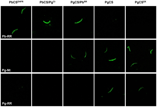
Figure 4. Sporozoite CSP expression.
Confocal immunofluorescence microphotographs of transgenic midgut sporozoites incubated at room temperature and developed with either a monoclonal antibody directed against the PbCSP repeat region (Pb-RR), a serum directed against the PgCSP N-terminal region (Pg-Nt), or a serum directed against the PgCSP repeat region (Pg-RR). Antibodies against the PbCSP and PgCSP repeat regions showed surface expression of the CSPs, whereas antibody against the PgCSP N-terminal region revealed mainly an intracellular pattern of expression.
Sporozoites from each of the transgenic clones, except PgCSSX and PgCS replacement parasites, showed a clear and uniform surface staining when incubated with the antibody against the PbCSP repeat region (Figure 4), similar to the staining observed for Pbwt sporozoites [33]. Although midgut sporozoites display limited gliding motility compared to salivary gland sporozoites, after incubation at 37°C with the PbCSP repeat region antibody, trails could be observed with Pbwt, PbCSDHFR, PbCS/PgCT and PgCS/PbRR parasites at a similar frequency, between 5 and 10% (Figure S3). However, the trails were extremely variable in shape and length and only very rarely were circular trails (<1% of total trails), similar to those seen for salivary gland sporozoites, observed (Figure S4). These results demonstrated that the presence of lengthy heterologous PgCS sequences in the chimeric CS genes does not significantly modify the surface expression of the proteins or the parasites' gliding motility. Therefore, the inability of the PgCS/PbRR parasites to infect the mosquito salivary glands or the vertebrate host does not appear to be due to incorrect CSP surface expression or motility.
Interestingly, analysis of the PgCS/PbRR, PgCSSX and PgCS replacement parasites with the PgCSP N-terminal region antiserum revealed the localization of the CSP to be mainly intracellular, throughout the cytosol (Figure 4). In contrast, PgCS/PbRR parasites, when analysed with the antibody against the PbCSP repeat region, displayed clear surface CSP expression (Figure 4) [33]. Similarly, PgCSSX and PgCS replacement sporozoites, incubated with a serum against the PgCSP repeat region, revealed normal surface staining (Figure 4), and also the presence of clumps of immunoreactive material either outside the parasite body or in proximity to its surface, following incubation at 37°C (Figure S3) [29], indicating that the introduction of the two restriction endonuclease sites in the PgCS sequence had no obvious effect on protein expression and release.
These IFA results are in agreement with recent data that report the absence of the N-terminal CSP sequence on the surface of P. berghei oocyst sporozoites [34]. The two antibodies are therefore reacting with different processed forms of CSP: the repeat region antibodies with a lower molecular weight (MW) form, lacking the N-terminal region and associated with the surface of the sporozoite, and the PgCSP N-terminal region serum with a higher MW form present in the cytoplasm.
Salivary gland sporozoites from Pbwt, PbCSDHFR and PbCS/PgCT infected mosquitoes, incubated with the PbCSP repeat region antibody, displayed a regular CSP surface expression and the characteristic circular gliding motility (Figure S4), with no differences observed either in the frequency of motile sporozoites or the percentage forming trails of at least one complete loop.
CSP processing in transgenic parasite lines
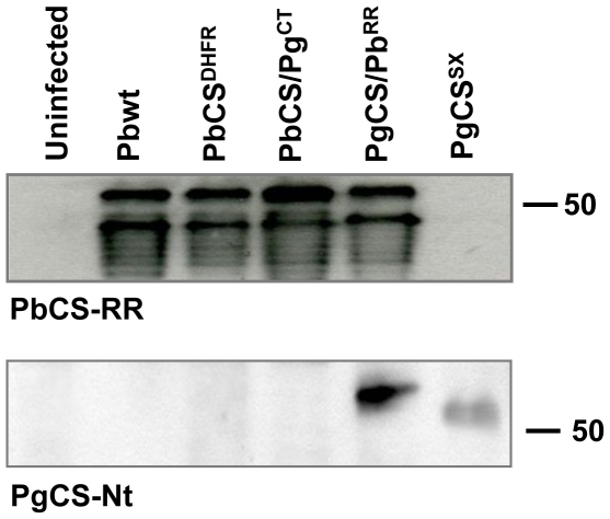
We investigated the expression and processing of CSP in the transgenic parasites by Western blot analysis of midgut sporozoite lysates collected 18 days p.i.. The PbCSP repeat region antibody revealed both the high and the low MW CSP forms in PbCSDHFR, PbCS/PgCT and PgCS/PbRR transgenic parasites (Figure 5) [11], [35]. A ladder of bands, weaker in intensity and ranging in size between ∼30 and 46 kDa, was also present and has previously been described [36], [37]. These results demonstrate that the CSPs in the transgenic parasite lines are expressed and processed similarly to CSP in Pbwt parasites.
Figure 5. Western blot analysis of CSP expression and processing in transgenic midgut sporozoites 18 days post-infection.
CSP expression was revealed by incubation with either a monoclonal antibody directed against the PbCSP repeat region (PbCS-RR) or a serum directed against the PgCSP N-terminal region (PgCS-Nt). As a control, midgut lysates from uninfected mosquitoes were also analysed. Antibody against the PbCSP repeat region reveals a higher molecular weight precursor polypeptide and a lower molecular weight processed polypeptide. A ladder of degradation products is also visible. Antibody against the PgCSP N-terminal region reveals only the higher molecular weight protein.
Incubation with the PgCSP N-terminal region antiserum revealed only the high MW form, corresponding to the CS precursor, in the PgCS/PbRR and PgCSSX parasite lines, in agreement with N-terminal region cleavage to yield the mature, processed form of CSP, containing the repeat region and C-terminal region. The band present in the PgCS/PbRR parasites appears to be slightly bigger than the expected ∼54 kDa band (revealed after incubation with the PbCSP repeat region antibody). This difference in size is likely due to the fact that the PbCSP repeat region is extremely proline rich and this has been shown to be responsible for anomalous migration patterns of proteins in SDS-PAGE [38].
Discussion
The aim of this work was to investigate the role of the CSP repeat region and the amino terminal region in the recognition of species-specific vector and host receptors. In previous studies, transgenic P. berghei parasite lines, in which either the entire endogenous CS gene or the repeat region was replaced with the P. falciparum orthologous sequence, both showed a ten fold reduction in A. stephensi salivary gland invasion [27], [29], suggesting a functional role of the repeat sequence in the recognition of species-specific host receptors. Our results indicate that the repeat region, on its own, is unlikely to represent the sporozoite species-specific salivary gland or hepatocyte ligand, because the replacement of the PgCS repeat region with the PbCS repeat region in the PgCS/PbRR transgenic parasites doesn't rescue sporozoite infectivity of either A. stephensi salivary glands or vertebrate hepatocytes. However, it is still possible that sequences in the N-terminal region and repeat region cooperate in, and are both required for, sporozoite infectivity.
The P. berghei transgenic parasites bearing a chimeric CSP displaying the P. falciparum CS repeat region, also had the RI motif and 14 residues upstream (RI-plus) exchanged with those of P. falciparum [27]. The change in either the repeat region or RI-plus could have been responsible for the 10-fold decrease in salivary gland invasion observed with these parasites. Although the RI core is the same between the two species (KLKQP), RI-plus differs between P. berghei and P. falciparum. In both P. falciparum and P. berghei nine of the fourteen residues are charged, indicating that the regions might be involved in interactions with host receptors. Nevertheless, it is possible that the repeat region has a function in infectivity acting in concert with other CSP regions. The CSP repeat region may fulfill a structural role, with the repeats from neighboring CSP molecules interlocking to form a rigid sheath surrounding the parasite [39]. Proline-rich tandem repeats generally form extended structures and flexible regions in proteins, and have been shown to function both as structural elements and in binding processes [40].
PbCS/PgCT parasites, containing both the PbCS N-terminal and repeat regions, were not impaired in salivary gland or vertebrate infectivity, pointing to a role for the N-terminal region in infection. Indeed, sera against the P. falciparum and P. berghei CSP N-terminal regions have been shown to inhibit sporozoite invasion of hepatocytes in vitro and HepG2 cells [41]. In previous investigations, a role in salivary gland infectivity was implied based on binding inhibition experiments, in which specific peptides were analysed for their ability to block recombinant CSP and sporozoite binding to the glands [13], [22]. However, the results of peptide inhibition studies can be due to steric hindrance. Therefore, gene targeting provides a more reliable system for determining the function of protein domains.
IFA analysis with a serum directed against the PgCSP N-terminal region appeared to localise the PgCS/PbRR and PgCSSX proteins mainly to the intracellular compartment, in contrast to antibodies against the PbCSP and PgCSP repeat regions, which revealed fluorescence at the surface of transgenic sporozoites (Figure 4). The results indicate that two forms of CSP are present in our transgenic lines: a full length protein localised intracellularly and a processed form, lacking the N-terminal region, and expressed uniformly on the sporozoite surface. This suggests possibly an intracellular site of CSP processing or alternatively, the protein may be cleaved either during its release from the apical organelles, before translocation onto the sporozoite surface, or after contact with target cells. Contradictory results support both intracellular [35], [42], [43] and surface [44], [45] sites of CSP cleavage. In Toxoplasma gondii, the N-terminal region of the adhesive protein, MIC2, is trimmed prior to secretion [46]. Another T. gondii protein, MIC3, which is re-localised to the surface of the parasite during invasion, is synthesised with an N-terminal propeptide that is cleaved during trafficking to the microneme. The processing was shown to be required for the protein to function as an adhesin [47] and it is possible that a similar mechanism operates in Plasmodium CSP.
Western blot analysis demonstrated that at least a large part of the N-terminal region is cleaved from the precursor polypeptide in each of the transgenic lines. Furthermore, data suggests that the protease that mediates CSP cleavage is parasite derived rather than host derived [45], which is compatible with an intracellular site of processing. The protease may therefore differ between P. berghei and P. gallinaceum parasites, which could in turn affect the correct processing of the transgenic CS proteins. However, the results indicate that the P. berghei protease is able to similarly cleave PbCSP and the chimeric CS proteins.
Our results demonstrate that the N-terminal region of CSP is important for infectivity, which suggests that at least part of this sequence is likely to be present on the sporozoite surface, possibly acting as a ligand for host receptors. This observation is partially in agreement with the recent results from Coppi et al. [34], in which they identify a higher MW CSP on the surface of the sporozoites. We have tried to determine the precise cleavage site in our transgenic lines by sequencing the low MW form of CSP, however we were unable to obtain enough material.
Recent results indicate that RI plays a critical role in CSP processing either because it is required for efficient cleavage or because it contains the cleavage site [34]. RI in P. gallinaceum (NLNQP) differs from mammalian Plasmodium spp. RI (KLKQP) and yet the PgCS/PbRR protein is still processed, probably by the same proteolytic enzyme. Both the high and low MW CSP bands of the PgCS/PbRR parasites run slightly higher compared to the equivalent bands of the PbCS/PgCT parasites in Western blot analysis (Figure 5). This is in part due to the fact that the chimeric proteins have different sizes: PgCS/PbRR and PbCS/PgCT contain 382 and 347 amino acids respectively. If the cleavage site is located at RI, one would expect the low MW bands from these two parasite lines to be the same size, since the processed CSPs in both these lines would contain the PbCS repeat region and the PgCS C-terminal region. Only the high MW bands, representing the precursor polypeptides, would differ in size. However, in our Western blot analysis, both the high and low MW bands from the PgCS/PbRR parasites appear to have larger MWs compared to the equivalent bands in the PbCS/PgCT parasites. This could be explained if the cleavage site is located upstream of RI, in the N-terminal region: an extra 25 amino acids are present just upstream of RI in the PgCSP sequence compared to the PbCSP sequence (Figure 1A), and this could explain the greater MW of the processed protein in PgCS/PbRR parasites compared to that of PbCS/PgCT parasites.
The sequence just upstream of RI contains a stretch of charged residues that, if included in the processed form of the protein, may act as a ligand. A peptide from PfCSP, spanning a region just upstream of RI (AA 93 to 113), was found to serve as a ligand for both hepatocytes [21] and salivary glands [13], [22]. Importantly, recognition of the short N-terminal peptide (PfCSP65–110) by sera from children living in a malaria-endemic region was associated with protection from disease [41]. The sequence of both RI and RI-plus varies considerably between P. berghei and P. gallinaceum (Figure 1A). In PbCSP, RI and the first 25 residues upstream of it contain 14 charged residues compared to the corresponding PgCSP sequence that contains only seven charged residues and this may explain the difference in infectivity observed between the PgCS/PbRR and the PbCS/PgCT parasites.
The numerous investigations into the functions of CSP's conserved motifs, RI and RII, have by no means led to a broad understanding of CSP's interactions during host infection. More recent work on CSP has focused on the N-terminal region of the protein [41] and the stretches of positively charged residues found throughout this region [19], [22]. The present investigation highlights, in vivo, a role for N-terminal residues in sporozoite infectivity in both the vertebrate and invertebrate hosts. These parasites could also provide a valuable rodent model to investigate the emerging role of the CSP N-terminal region in the induction of protective immune responses.
Materials and Methods
Ethics statement
Animal work conducted at Imperial College was performed according to UK Home Office Regulations and approved under Home Office License PPL 70/6453. Studies carried out at Perugia University have been performed according to the D.L 27 January 1992, n. 116, Italian legislation, and approved under Ethics Committee Licence n. PR 0161.
DNA constructs
The targeting constructs pPgCSSX, pPgCS/PbRR and pPbCSDHFR generated in this study contained the following structural elements: (i) the first 1,130 nucleotides of the PbCS 5′ UTR sequence immediately upstream of the start codon; (ii) PgCS, PbCS or a chimeric form of the CS coding sequence and (iii) the first 1,150 nucleotides of the PbCS 3′ UTR sequence immediately downstream of its stop codon, in which the T. gondii dihydrofolate reductase/thymidylate synthase (TgDHFR/TS) drug selectable marker cassette (5,150 bp) was inserted at its HindIII site.
All three constructs were generated from a targeting construct, pPgCS, already available in our laboratory which contains the full PgCS coding sequence [29]. The SpeI and XhoI sites were introduced on either side of the PgCS repeat region by PCR based site-directed mutagenesis. Primers used to introduce SpeI were: For1 5′-GAGAAAATGTTGTGAATCTTAATCAACCAACTAGTGTTGGAGGAAATGGTGGTGTTCAACCTGCTG-3′ (SpeI is underlined) and Rev1 complementary to the forward primer. Primers used to introduce XhoI were: For2 5′-CTGAAGAAGAAAAGGAGGATGAACCAATACCAGATCTCGAGCCAACTCAAGAAGAAATAGATAAATATTTAAAAAG-3′ (XhoI is underlined) and Rev2 complementary to the forward primer.
The 1.02 kb PbCS gene was amplified from P. berghei genomic DNA (Anka strain, clone 2.34) and sequenced. Forward and reverse primers were designed to amplify the PbCS repeat region from this sequence, introducing the SpeI and XhoI sites at either end of the repeat region (Spefor 5′-TAATAATAAATTGAAACAACCAACTAGTCCACCACCACCAAACCCAAATG-3′ (SpeI is underlined) and XhoRev 5′-CCGCTCGAGGATATAAGAATCGTCATTATTATTATTTTTGTTATTG-3′ (XhoI is underlined)). The PgCS repeat region was then replaced with the PbCS repeat region, via SpeI and XhoI digestions. Sequencing analysis and alignment with the wild type P. berghei (GenBank: M14145) or P. gallinaceum (GenBank: 65959) sequences confirmed the correct CS genes inserted in each construct.
Parasite transformation
Plasmids pPgCSSX and pPgCS/PbRR were digested with ApaI to release the ∼8.6 kb targeting insert from the plasmid backbone and the insert was purified by gel electrophoresis. Purified schizonts of the P. berghei ANKA strain (clone 2.34) were transformed with 5 µg of targeting DNA using an Amaxa Gene Pulser set at program U33. Mutant parasites were obtained by the standard method of drug (pyrimethamine) selection in mice [48]. Pyrimethamine resistant parasites were subsequently cloned by limiting dilution.
Southern blot analysis
Genomic DNA was isolated from parasites as previously described [33]. 1–3 µg of genomic DNA was digested with EcoRV alone or in combination with either SpeI or XhoI, separated on an agarose gel and blotted onto a nylon Hybond-N+ membrane (Amersham). The following DNA fragments were used as probes: (a) the 1.15 kb fragment of the PbCS 3′ UTR from the targeting constructs, (b) the entire N-terminal sequence of the PbCS coding region, (c) the entire N-terminal sequence of the PgCS coding region and (d) a 520 bp fragment amplified from the TgDHFR/TS coding sequence with the primers TgFor123 (5′-AGAGGGGCATCGGCATCAAC-3′) and TgRev124 (5′-TTGAAAGAATGTCATCTCCG-3′). All hybridization experiments were carried out as previously described [33], [49].
Sequencing of genomic DNA
The primers PbCS5′UTR3′F (5′-CCCTCACATAAGACAATCC-3′) and PbCS3′UTR5′R (5′-GTTTACACACGTCATATGTATG-3′), which anneal to the 5′ and 3′ UTR sequences of the CS locus, were used to amplify the full length CS gene from blood stage transgenic parasite genomic DNA, isolated from sporozoite-infected mice where possible. The PCR products were then sequenced directly (MWG-Biotech).
Parasite development in the mosquito
Female mosquitoes (4–6 days old) of A. stephensi (strain sd500), were fed for 30 minutes on either wild type (wt) or transgenic parasite infected BALB/c mice (six- to eight-week-old female Balb/c from Harlan Sprague) with a parasitaemia ranging from 5% to 10%. Mosquitoes were dissected at days 14 and 21 post infection (p.i.) to determine the presence of oocysts and the number of parasites in the midguts and salivary glands.
Parasite infectivity in mice
Midgut and salivary gland sporozoites were diluted with RPMI and intravenously (i.v.) injected into C57BL/6 mice (six- to eight-week-old female C57BL/6 from Charles River) in order to assess vertebrate infectivity. After sporozoite injection, blood samples were withdrawn from the tails at regular intervals up to 15 days p.i. and a minimum of 10,000 erythrocytes were examined on Giemsa stained blood smears. The pre-patent period was determined as the number of days between sporozoite injection and when parasites were first detected in the blood.
Western blot analysis
Total proteins were extracted from infected (day 18 p.i.) and uninfected A. stephensi mosquito midguts using reducing sample buffer and separated on a 12% gel by SDS-PAGE. Blots were incubated for 1 hour at RT with the appropriate primary antibody: either a 1∶200 dilution of the monoclonal antibody 3D11 raised in mice against the PbCSP repeat region [11] or a 1∶200 dilution of a mouse serum raised against the PgCSP N-terminal region (a gift from Dr. Dharmendar Rathore), then developed with the enhanced chemiluminescence detection system, according to the manufacturer's instructions (Amersham Pharmacia).
Immunofluorescence analysis
Freshly dissected sporozoites collected at day 21 p.i. from mosquito midguts and salivary glands were diluted in RPMI 1640 and spotted onto multiwell microscope slides. The slides were either incubated at 37°C, to induce sporozoite gliding [32], or at RT for 30 minutes. Parasites were fixed with 1% formaldehyde in 1× PBS and blocked with 1% BSA in 1× PBS for 30 minutes at RT. The slides were then incubated with one of three primary antibodies: the monoclonal antibody against the PbCSP repeat region [11], the serum against the PgCSP N-terminal region or a 1∶1000 dilution of a rabbit serum raised against the PgCSP repeat region. Antibody bound to the parasites or their trails was revealed by incubation with fluorescein isothiocyanate (FITC)-conjugated secondary antibody (Becton Dickinson). Analysis of expression was carried out using a Leica TCS SP 2 confocal microscope.
Supporting Information
Generation and southern blot analysis of transgenic PgCSSX parasite lines. (A) Schematic representation of (i) the PgCSSX targeting construct, (ii) the wt PbCS locus and (iii) the targeted locus after recombination between the PbCS 5′UTR and 3′UTR sequences. The vertically dashed box indicates the 1.13 kb 5′UTR sequence used in the construct and the horizontally dashed boxes indicate the 0.3 kb and 0.85 kb 3′UTR sequences between which the TgDHFR/TS selectable marker cassette (light grey) was inserted in the construct. The PgCSSX gene contains the full PgCS coding sequence (dark grey) into which the SpeI (S) and XhoI (X) sites were inserted on either side of the repeat region. Thick black lines indicate the probes used in southern blots. E = EcoRV. (B) Southern blot of EcoRV digested genomic DNA from Pbwt parasites and PgCS-replacement parasites (clone 14, [29]), that acted as negative and positive controls respectively, and transgenic PgCSSX parasites (clones 1 and 2). The blot was first hybridised with the PbCS 3′UTR probe, containing the full 1.15 kb fragment of the PbCS 3′UTR sequence present in the targeting construct. The 3′UTR probe hybridised with bands of 4.0 and 1.5 kb in digested Pbwt DNA and with bands of 4.2 and 2.0 kb in the transgenic parasite DNA. The size shift from 4.0 to 4.2 kb is due to the longer PgCS coding sequence. The 2.0 kb band indicates the presence of the TgDHFR/TS cassette in the PbCS locus. The membrane was also hybridized with probes encompassing the TgDHFR/TS gene (C), the PbCS gene (D) and the PgCS gene (E). The TgDHFR/TS probe revealed a band of 4.6 kb only in transgenic DNA (C). The PbCS probe revealed the 4.0 kb band only in the Pbwt DNA (D) while the PgCS probe bound to the correct 4.2 kb band only in the transgenic parasite DNA (E).
(TIF)
Generation and southern blot analysis of transgenic PbCSDHFR parasite lines. (A) Schematic representation of (i) the PgCS/PbRR targeting construct, (ii) the wt PbCS locus and (iii) the targeted locus after recombination between the two PbCS 3′ UTR sequences (horizontally dashed boxes) flanking the TgDHFR-TS selectable marker cassette (light grey box). The targeted locus therefore carried the endogenous wt PbCS gene (white box) and, inserted in the 3′UTR, the TgDHFR-TS selectable marker cassette. Thick black lines indicate the probes used in southern blots. E: EcoRV site, S: SpeI site and X: XhoI site. (B) Southern blot of EcoRV/SpeI digested genomic DNA from Pbwt parasites and transgenic PbCSDHFR parasites (clones 9 and 10), hybridised with the PbCS 3′ UTR probe. (C) Southern blot of EcoRV/XhoI digested genomic DNA hybridised with the PbCS N-terminal probe. Neither clone contained the SpeI or XhoI sites, as demonstrated by the presence of the 4.0 kb band in all lanes, indicating recombination had occurred between the short 0.3 kb PbCS 3′ UTR sequence upstream of the marker cassette. A second band in the transgenic parasite DNA of 2.0 kb, compared to the 1.5 kb band in the Pbwt DNA, indicated the insertion of the TgDHFR/TS cassette in the PbCS locus.
(TIF)
Sporozoite CSP expression and motility. Confocal immunofluorescence microphotographs of P. berghei wt and transgenic midgut sporozoites incubated at 37°C to induce motility and developed with either a monoclonal antibody directed against the PbCSP repeat region (Pb-RR), a serum directed against the PgCSP N-terminal region (Pg-Nt), or a serum directed against the PgCSP repeat region (Pg-RR). Sporozoites shed trails of material recognised by antibody against the PbCSP repeat region. Antibody against the PgCSP N-terminal region (Pg-Nt) revealed mainly an intracellular pattern of expression, and no trails, for the PgCS/PbRR, PgCSSX and PgCS replacement parasites. The antibody against the Pg-RR revealed the presence of clumps of immunoreactive material either outside the parasite body or in proximity to its surface.
(TIF)
Salivary gland sporozoite CSP expression and motility. Confocal immunofluorescence microphotographs of P. berghei wt and transgenic salivary gland sporozoites incubated at either room temperature (RT) or 37°C and developed with an antibody directed against the PbCSP repeat region (Pb-RR). The antibody revealed surface expression in PbCSDHFR and PbCS/PgCT salivary gland sporozoites, similar to P. berghei wt salivary gland sporozoites. Sporozoites incubated at 37°C shed trails of material recognised by the anti-PbCSP repeat region antibody.
(TIF)
Footnotes
Competing Interests: The authors have declared that no competing interests exist.
Funding: This work was supported by grants from the Italian Ministry of Research PRIN (Research Programmes of National Interest) n. 2005065913. The funders had no role in study design, data collection and analysis, decision to publish, or preparation of the manuscript.
References
- 1.Nardin E, Zavala F, Nussenzweig V, Nussenzweig RS. Pre-erythrocytic malaria vaccine: mechanisms of protective immunity and human vaccine trials. Parassitologia. 1999;41:397–402. [PubMed] [Google Scholar]
- 2.Birkett A, Lyons K, Schmidt A, Boyd D, Oliveira GA, et al. A modified hepatitis B virus core particle containing multiple epitopes of the Plasmodium falciparum circumsporozoite protein provides a highly immunogenic malaria vaccine in preclinical analyses in rodent and primate hosts. Infect Immun. 2002;70:6860–6870. doi: 10.1128/IAI.70.12.6860-6870.2002. [DOI] [PMC free article] [PubMed] [Google Scholar]
- 3.Ballou WR, Hoffman SL, Sherwood JA, Hollingdale MR, Neva FA, et al. Safety and efficacy of a recombinant DNA Plasmodium falciparum sporozoite vaccine. Lancet. 1987;1:1277–1281. doi: 10.1016/s0140-6736(87)90540-x. [DOI] [PubMed] [Google Scholar]
- 4.Herrington DA, Clyde DF, Losonsky G, Cortesia M, Murphy JR, et al. Safety and immunogenicity in man of a synthetic peptide malaria vaccine against Plasmodium falciparum sporozoites. Nature. 1987;328:257–259. doi: 10.1038/328257a0. [DOI] [PubMed] [Google Scholar]
- 5.Ballou WR. The development of the RTS,S malaria vaccine candidate: challenges and lessons. Parasite Immunol. 2009;31:492–500. doi: 10.1111/j.1365-3024.2009.01143.x. [DOI] [PubMed] [Google Scholar]
- 6.Nagasawa H, Procell PM, Atkinson CT, Campbell GH, Collins WE, et al. Localization of circumsporozoite protein of Plasmodium ovale in midgut oocysts. Infect Immun. 1987;55:2928–2932. doi: 10.1128/iai.55.12.2928-2932.1987. [DOI] [PMC free article] [PubMed] [Google Scholar]
- 7.Posthuma G, Meis JF, Verhave JP, Hollingdale MR, Ponnudurai T, et al. Immunogold localization of circumsporozoite protein of the malaria parasite Plasmodium falciparum during sporogony in Anopheles stephensi midguts. Eur J Cell Biol. 1988;46:18–24. [PubMed] [Google Scholar]
- 8.Stewart MJ, Nawrot RJ, Schulman S, Vanderberg JP. Plasmodium berghei sporozoite invasion is blocked in vitro by sporozoite-immobilizing antibodies. Infect Immun. 1986;51:859–864. doi: 10.1128/iai.51.3.859-864.1986. [DOI] [PMC free article] [PubMed] [Google Scholar]
- 9.Stewart MJ, Vanderberg JP. Malaria sporozoites leave behind trails of circumsporozoite protein during gliding motility. J Protozool. 1988;35:389–393. doi: 10.1111/j.1550-7408.1988.tb04115.x. [DOI] [PubMed] [Google Scholar]
- 10.Potocnjak P, Yoshida N, Nussenzweig RS, Nussenzweig V. Monovalent fragments (Fab) of monoclonal antibodies to a sporozoite surface antigen (Pb44) protect mice against malarial infection. J Exp Med. 1980;151:1504–1513. doi: 10.1084/jem.151.6.1504. [DOI] [PMC free article] [PubMed] [Google Scholar]
- 11.Yoshida N, Nussenzweig RS, Potocnjak P, Nussenzweig V, Aikawa M. Hybridoma produces protective antibodies directed against the sporozoite stage of malaria parasite. Science. 1980;207:71–73. doi: 10.1126/science.6985745. [DOI] [PubMed] [Google Scholar]
- 12.Warburg A, Touray M, Krettli AU, Miller LH. Plasmodium gallinaceum: antibodies to circumsporozoite protein prevent sporozoites from invading the salivary glands of Aedes aegypti. Exp Parasitol. 1992;75:303–307. doi: 10.1016/0014-4894(92)90215-v. [DOI] [PubMed] [Google Scholar]
- 13.Sidjanski SP, Vanderberg JP, Sinnis P. Anopheles stephensi salivary glands bear receptors for region I of the circumsporozoite protein of Plasmodium falciparum. Mol Biochem Parasitol. 1997;90:33–41. doi: 10.1016/s0166-6851(97)00124-2. [DOI] [PMC free article] [PubMed] [Google Scholar]
- 14.de Lara Capurro M, Coleman J, Beerntsen BT, Myles KM, Olson KE, et al. Virus-expressed, recombinant single-chain antibody blocks sporozoite infection of salivary glands in Plasmodium gallinaceum-infected Aedes aegypti. Am J Trop Med Hyg. 2000;62:427–433. doi: 10.4269/ajtmh.2000.62.427. [DOI] [PubMed] [Google Scholar]
- 15.Cerami C, Frevert U, Sinnis P, Takacs B, Clavijo P, et al. The basolateral domain of the hepatocyte plasma membrane bears receptors for the circumsporozoite protein of Plasmodium falciparum sporozoites. Cell. 1992;70:1021–1033. doi: 10.1016/0092-8674(92)90251-7. [DOI] [PubMed] [Google Scholar]
- 16.Cerami C, Frevert U, Sinnis P, Takacs B, Nussenzweig V. Rapid clearance of malaria circumsporozoite protein (CS) by hepatocytes. J Exp Med. 1994;179:695–701. doi: 10.1084/jem.179.2.695. [DOI] [PMC free article] [PubMed] [Google Scholar]
- 17.Pinzon-Ortiz C, Friedman J, Esko J, Sinnis P. The binding of the circumsporozoite protein to cell surface heparan sulfate proteoglycans is required for plasmodium sporozoite attachment to target cells. J Biol Chem. 2001;276:26784–26791. doi: 10.1074/jbc.M104038200. [DOI] [PMC free article] [PubMed] [Google Scholar]
- 18.Rathore D, McCutchan TF. Heparin can regulate the binding of Plasmodium falciparum circumsporozoite protein. Mol Biochem Parasitol. 2000;108:253–256. doi: 10.1016/s0166-6851(00)00214-0. [DOI] [PubMed] [Google Scholar]
- 19.Rathore D, Sacci JB, de la Vega P, McCutchan TF. Binding and invasion of liver cells by Plasmodium falciparum sporozoites. Essential involvement of the amino terminus of circumsporozoite protein. J Biol Chem. 2002;277:7092–7098. doi: 10.1074/jbc.M106862200. [DOI] [PubMed] [Google Scholar]
- 20.Ancsin JB, Kisilevsky R. A binding site for highly sulfated heparan sulfate is identified in the N terminus of the circumsporozoite protein: significance for malarial sporozoite attachment to hepatocytes. J Biol Chem. 2004;279:21824–21832. doi: 10.1074/jbc.M401979200. [DOI] [PubMed] [Google Scholar]
- 21.Rathore D, Nagarkatti R, Jani D, Chattopadhyay R, de la Vega P, et al. An immunologically cryptic epitope of Plasmodium falciparum circumsporozoite protein facilitates liver cell recognition and induces protective antibodies that block liver cell invasion. J Biol Chem. 2005;280:20524–20529. doi: 10.1074/jbc.M414254200. [DOI] [PubMed] [Google Scholar]
- 22.Myung JM, Marshall P, Sinnis P. The Plasmodium circumsporozoite protein is involved in mosquito salivary gland invasion by sporozoites. Mol Biochem Parasitol. 2004;133:53–59. doi: 10.1016/j.molbiopara.2003.09.002. [DOI] [PubMed] [Google Scholar]
- 23.Ying P, Shakibaei M, Patankar MS, Clavijo P, Beavis RC, et al. The malaria circumsporozoite protein: interaction of the conserved regions I and II-plus with heparin-like oligosaccharides in heparan sulfate. Exp Parasitol. 1997;85:168–182. doi: 10.1006/expr.1996.4134. [DOI] [PubMed] [Google Scholar]
- 24.Frevert U, Sinnis P, Cerami C, Shreffler W, Takacs B, et al. Malaria circumsporozoite protein binds to heparan sulfate proteoglycans associated with the surface membrane of hepatocytes. J Exp Med. 1993;177:1287–1298. doi: 10.1084/jem.177.5.1287. [DOI] [PMC free article] [PubMed] [Google Scholar]
- 25.Sinnis P, Clavijo P, Fenyo D, Chait BT, Cerami C, et al. Structural and functional properties of region II-plus of the malaria circumsporozoite protein. J Exp Med. 1994;180:297–306. doi: 10.1084/jem.180.1.297. [DOI] [PMC free article] [PubMed] [Google Scholar]
- 26.Nussenzweig V, Nussenzweig RS. Rationale for the development of an engineered sporozoite malaria vaccine. Adv Immunol. 1989;45:283–334. doi: 10.1016/s0065-2776(08)60695-1. [DOI] [PubMed] [Google Scholar]
- 27.Persson C, Oliveira GA, Sultan AA, Bhanot P, Nussenzweig V, et al. Cutting edge: a new tool to evaluate human pre-erythrocytic malaria vaccines: rodent parasites bearing a hybrid Plasmodium falciparum circumsporozoite protein. J Immunol. 2002;169:6681–6685. doi: 10.4049/jimmunol.169.12.6681. [DOI] [PubMed] [Google Scholar]
- 28.Rathore D, McCutchan TF, Garboczi DN, Toida T, Hernaiz MJ, et al. Direct measurement of the interactions of glycosaminoglycans and a heparin decasaccharide with the malaria circumsporozoite protein. Biochemistry. 2001;40:11518–11524. doi: 10.1021/bi0105476. [DOI] [PubMed] [Google Scholar]
- 29.Tewari R, Rathore D, Crisanti A. Motility and infectivity of Plasmodium berghei sporozoites expressing avian Plasmodium gallinaceum circumsporozoite protein. Cell Microbiol. 2005;7:699–707. doi: 10.1111/j.1462-5822.2005.00503.x. [DOI] [PubMed] [Google Scholar]
- 30.Gantt SM, Myung JM, Briones MR, Li WD, Corey EJ, et al. Proteasome inhibitors block development of Plasmodium spp. Antimicrob Agents Chemother. 1998;42:2731–2738. doi: 10.1128/aac.42.10.2731. [DOI] [PMC free article] [PubMed] [Google Scholar]
- 31.Vanderberg JP. Development of infectivity by the Plasmodium berghei sporozoite. J Parasitol. 1975;61:43–50. [PubMed] [Google Scholar]
- 32.Spaccapelo R, Naitza S, Robson KJ, Crisanti A. Thrombospondin-related adhesive protein (TRAP) of Plasmodium berghei and parasite motility. Lancet. 1997;350:335. doi: 10.1016/S0140-6736(97)24031-6. [DOI] [PubMed] [Google Scholar]
- 33.Tewari R, Spaccapelo R, Bistoni F, Holder AA, Crisanti A. Function of region I and II adhesive motifs of Plasmodium falciparum circumsporozoite protein in sporozoite motility and infectivity. J Biol Chem. 2002;277:47613–47618. doi: 10.1074/jbc.M208453200. [DOI] [PubMed] [Google Scholar]
- 34.Coppi A, Natarajan R, Pradel G, Bennett BL, James ER, et al. The malaria circumsporozoite protein has two functional domains, each with distinct roles as sporozoites journey from mosquito to mammalian host. J. 2011;208:341–356. doi: 10.1084/jem.20101488. [DOI] [PMC free article] [PubMed] [Google Scholar]
- 35.Yoshida N, Potocnjak P, Nussenzweig V, Nussenzweig RS. Biosynthesis of Pb44, the protective antigen of sporozoites of Plasmodium berghei. J Exp Med. 1981;154:1225–1236. doi: 10.1084/jem.154.4.1225. [DOI] [PMC free article] [PubMed] [Google Scholar]
- 36.Simonetti AB, Billingsley PF, Winger LA, Sinden RE. Kinetics of expression of two major Plasmodium berghei antigens in the mosquito vector, Anopheles stephensi. J Eukaryot Microbiol. 1993;40:569–576. doi: 10.1111/j.1550-7408.1993.tb06109.x. [DOI] [PubMed] [Google Scholar]
- 37.Boulanger N, Charoenvit Y, Krettli A, Betschart B. Developmental changes in the circumsporozoite proteins of Plasmodium berghei and P. gallinaceum in their mosquito vectors. Parasitol Res. 1995;81:58–65. doi: 10.1007/BF00932418. [DOI] [PubMed] [Google Scholar]
- 38.Robson KJ, Naitza S, Barker G, Sinden RE, Crisanti A. Cloning and expression of the thrombospondin related adhesive protein gene of Plasmodium berghei. Mol Biochem Parasitol. 1997;84:1–12. doi: 10.1016/s0166-6851(96)02774-0. [DOI] [PubMed] [Google Scholar]
- 39.Godson GN, Ellis J, Svec P, Schlesinger DH, Nussenzweig V. Identification and chemical synthesis of a tandemly repeated immunogenic region of Plasmodium knowlesi circumsporozoite protein. Nature. 1983;305:29–33. doi: 10.1038/305029a0. [DOI] [PubMed] [Google Scholar]
- 40.Williamson MP. The structure and function of proline-rich regions in proteins. Biochem J. 1994;297(Pt 2):249–260. doi: 10.1042/bj2970249. [DOI] [PMC free article] [PubMed] [Google Scholar]
- 41.Bongfen SE, Ntsama PM, Offner S, Smith T, Felger I, et al. The N-terminal domain of Plasmodium falciparum circumsporozoite protein represents a target of protective immunity. Vaccine. 2009;27:328–335. doi: 10.1016/j.vaccine.2008.09.097. [DOI] [PubMed] [Google Scholar]
- 42.Cochrane AH, Santoro F, Nussenzweig V, Gwadz RW, Nussenzweig RS. Monoclonal antibodies identify the protective antigens of sporozoites of Plasmodium knowlesi. Proc Natl Acad Sci U S A. 1982;79:5651–5655. doi: 10.1073/pnas.79.18.5651. [DOI] [PMC free article] [PubMed] [Google Scholar]
- 43.Santoro F, Cochrane AH, Nussenzweig V, Nardin EH, Nussenzweig RS, et al. Structural similarities among the protective antigens of sporozoites from different species of malaria parasites. J Biol Chem. 1983;258:3341–3345. [PubMed] [Google Scholar]
- 44.Gonzalez-Ceron L, Rodriguez MH, Wirtz RA, Sina BJ, Palomeque OL, et al. Plasmodium vivax: a monoclonal antibody recognizes a circumsporozoite protein precursor on the sporozoite surface. Exp Parasitol. 1998;90:203–211. doi: 10.1006/expr.1998.4334. [DOI] [PubMed] [Google Scholar]
- 45.Coppi A, Pinzon-Ortiz C, Hutter C, Sinnis P. The Plasmodium circumsporozoite protein is proteolytically processed during cell invasion. J Exp Med. 2005;201:27–33. doi: 10.1084/jem.20040989. [DOI] [PMC free article] [PubMed] [Google Scholar]
- 46.Carruthers VB, Sherman GD, Sibley LD. The Toxoplasma adhesive protein MIC2 is proteolytically processed at multiple sites by two parasite-derived proteases. J Biol Chem. 2000;275:14346–14353. doi: 10.1074/jbc.275.19.14346. [DOI] [PubMed] [Google Scholar]
- 47.Cerede O, Dubremetz JF, Bout D, Lebrun M. The Toxoplasma gondii protein MIC3 requires pro-peptide cleavage and dimerization to function as adhesin. Embo J. 2002;21:2526–2536. doi: 10.1093/emboj/21.11.2526. [DOI] [PMC free article] [PubMed] [Google Scholar]
- 48.Janse CJ, Ramesar J, Waters AP. High-efficiency transfection and drug selection of genetically transformed blood stages of the rodent malaria parasite Plasmodium berghei. Nat Protoc. 2006;1:346–356. doi: 10.1038/nprot.2006.53. [DOI] [PubMed] [Google Scholar]
- 49.Wengelnik K, Spaccapelo R, Naitza S, Robson KJ, Janse CJ, et al. The A-domain and the thrombospondin-related motif of Plasmodium falciparum TRAP are implicated in the invasion process of mosquito salivary glands. Embo J. 1999;18:5195–5204. doi: 10.1093/emboj/18.19.5195. [DOI] [PMC free article] [PubMed] [Google Scholar]
Associated Data
This section collects any data citations, data availability statements, or supplementary materials included in this article.
Supplementary Materials
Generation and southern blot analysis of transgenic PgCSSX parasite lines. (A) Schematic representation of (i) the PgCSSX targeting construct, (ii) the wt PbCS locus and (iii) the targeted locus after recombination between the PbCS 5′UTR and 3′UTR sequences. The vertically dashed box indicates the 1.13 kb 5′UTR sequence used in the construct and the horizontally dashed boxes indicate the 0.3 kb and 0.85 kb 3′UTR sequences between which the TgDHFR/TS selectable marker cassette (light grey) was inserted in the construct. The PgCSSX gene contains the full PgCS coding sequence (dark grey) into which the SpeI (S) and XhoI (X) sites were inserted on either side of the repeat region. Thick black lines indicate the probes used in southern blots. E = EcoRV. (B) Southern blot of EcoRV digested genomic DNA from Pbwt parasites and PgCS-replacement parasites (clone 14, [29]), that acted as negative and positive controls respectively, and transgenic PgCSSX parasites (clones 1 and 2). The blot was first hybridised with the PbCS 3′UTR probe, containing the full 1.15 kb fragment of the PbCS 3′UTR sequence present in the targeting construct. The 3′UTR probe hybridised with bands of 4.0 and 1.5 kb in digested Pbwt DNA and with bands of 4.2 and 2.0 kb in the transgenic parasite DNA. The size shift from 4.0 to 4.2 kb is due to the longer PgCS coding sequence. The 2.0 kb band indicates the presence of the TgDHFR/TS cassette in the PbCS locus. The membrane was also hybridized with probes encompassing the TgDHFR/TS gene (C), the PbCS gene (D) and the PgCS gene (E). The TgDHFR/TS probe revealed a band of 4.6 kb only in transgenic DNA (C). The PbCS probe revealed the 4.0 kb band only in the Pbwt DNA (D) while the PgCS probe bound to the correct 4.2 kb band only in the transgenic parasite DNA (E).
(TIF)
Generation and southern blot analysis of transgenic PbCSDHFR parasite lines. (A) Schematic representation of (i) the PgCS/PbRR targeting construct, (ii) the wt PbCS locus and (iii) the targeted locus after recombination between the two PbCS 3′ UTR sequences (horizontally dashed boxes) flanking the TgDHFR-TS selectable marker cassette (light grey box). The targeted locus therefore carried the endogenous wt PbCS gene (white box) and, inserted in the 3′UTR, the TgDHFR-TS selectable marker cassette. Thick black lines indicate the probes used in southern blots. E: EcoRV site, S: SpeI site and X: XhoI site. (B) Southern blot of EcoRV/SpeI digested genomic DNA from Pbwt parasites and transgenic PbCSDHFR parasites (clones 9 and 10), hybridised with the PbCS 3′ UTR probe. (C) Southern blot of EcoRV/XhoI digested genomic DNA hybridised with the PbCS N-terminal probe. Neither clone contained the SpeI or XhoI sites, as demonstrated by the presence of the 4.0 kb band in all lanes, indicating recombination had occurred between the short 0.3 kb PbCS 3′ UTR sequence upstream of the marker cassette. A second band in the transgenic parasite DNA of 2.0 kb, compared to the 1.5 kb band in the Pbwt DNA, indicated the insertion of the TgDHFR/TS cassette in the PbCS locus.
(TIF)
Sporozoite CSP expression and motility. Confocal immunofluorescence microphotographs of P. berghei wt and transgenic midgut sporozoites incubated at 37°C to induce motility and developed with either a monoclonal antibody directed against the PbCSP repeat region (Pb-RR), a serum directed against the PgCSP N-terminal region (Pg-Nt), or a serum directed against the PgCSP repeat region (Pg-RR). Sporozoites shed trails of material recognised by antibody against the PbCSP repeat region. Antibody against the PgCSP N-terminal region (Pg-Nt) revealed mainly an intracellular pattern of expression, and no trails, for the PgCS/PbRR, PgCSSX and PgCS replacement parasites. The antibody against the Pg-RR revealed the presence of clumps of immunoreactive material either outside the parasite body or in proximity to its surface.
(TIF)
Salivary gland sporozoite CSP expression and motility. Confocal immunofluorescence microphotographs of P. berghei wt and transgenic salivary gland sporozoites incubated at either room temperature (RT) or 37°C and developed with an antibody directed against the PbCSP repeat region (Pb-RR). The antibody revealed surface expression in PbCSDHFR and PbCS/PgCT salivary gland sporozoites, similar to P. berghei wt salivary gland sporozoites. Sporozoites incubated at 37°C shed trails of material recognised by the anti-PbCSP repeat region antibody.
(TIF)


