The outcome of a giant retroperitoneal liposarcoma is mainly dependent on its pathological features.
Case report
A 71-year-old man presented with a history of progressive abdominal distention during the previous two years, accompanied by increasing difficulty in moving his lower extremities and a recurrent right scrotal mass on standing in the past two months.
Physical examination revealed that the patient had prominent abdominal distention and a soft mass in the right scrotum (Figure 1).
Figure 1.
Physical examination revealing the patient had prominent abdominal distention and right scrotum soft mass
Computed tomography (CT) scan of the abdomen and pelvis revealed a large, well-encapsulated mass that filled the entire abdominal cavity (Figure 2), which caused left anterior compression of the right kidney (arrowhead) and compression of the bowels (arrow). The scan also showed that a part of the mass herniated into the right scrotum through the inguinal canal.
Figure 2.
Computed tomography scan of the abdomen and pelvis demonstrating a large, well-encapsulated mass that filled the entire abdominal cavity, left anterior compression of the right kidney (arrowhead) and of the bowel (arrow)
Exploratory operation revealed a lobulated fatty tumour covered by a layer of transparent membrane (Figure 3). Ninety percent of the tumour which weighed about 13 kg, was resected; the unresectable part was bound tightly to the mesenteric vessels.
Figure 3.
Exploratory operation revealing the mass to be a lobulated fatty tissue covered by a layer of transparent membrane
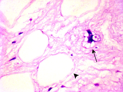
Pathological examination revealed a well-differentiated liposarcoma (lipoma-like subtype), composed of fat cells of various sizes, univacuolated (signet-ring cells, arrowhead) and multivacuolated (arrow) lipoblasts (Figure 4) and stromal cells with heteromorphous hyperchromatic nuclei (arrow) (Figure 5).
Figure 4.
Pathological examination revealing the mass to be a well-differentiated liposarcoma (lipoma-like subtype), characterized by fat cells with different sizes, univacuolated (signet-ring cells, arrowhead) and multivacuolated (arrow) lipoblast
Figure 5.
Pathological examination also revealing stromal cells with heteromorphous hyperchromatic nucleus (arrow)
The patient recovered and was discharged within a week after surgery. At a follow-up visit 5 months later, he remained free of symptoms.
Discussion
Liposarcoma is the most common type of retroperitoneal soft tissue sarcoma.1 Its diagnosis is often much delayed due to a lack of specific signs and symptoms at the early stage.2 Liposarcomas are classified under five basic histologic categories: well-differentiated, myxoid, round-cell, pleomorphic, and dedifferentiated liposarcomas, of which the well-differentiated liposarcoma is the common subtype.2 Complete resection is the fundamental treatment for retroperitoneal liposarcoma, whereas the efficacy of chemotherapy and radiotherapy as adjuvant treatment is still controversial.1 In 81% of the cases complete resection can be achieved,3 and in 57–77% of the cases resection of adjacent tissues is also necessary.1,3–5 About 5% of the patients with retroperitoneal liposarcoma may have distant metastases; however, the majority of the patients die from local invasion of adjacent critical organs by the tumor.3,4 The prognosis for liposarcoma depends on several factors, the most important of which are the histologic type and the location of the tumor. Usually, myxoid and well-differentiated/atypical lipomatous tumors have a better survival rate; retroperitoneal liposarcomas, probably because of the difficulty in achieving complete resection, have a poorer survival, type for type, than liposarcomas in the extremities.1
DECLARATIONS
Competing interests
None declared
Funding
None
Ethical approval
Written informed consent to publication was obtained from the patient or next of kin
Guarantor
YG
Contributorship
YG and JHW performed the operation; TQL read the radiographs; JZ did the pathological work; CQG conceptualized the report, drafted the manuscript, revised it critically and made final approval of the version to be published
Acknowledgements
None
Reviewer
John Yeboah
References
- 1.Perez EA, Gutierrez JC, Moffat FL Jr, et al. Retroperitoneal and truncal sarcomas: prognosis depends upon type not location. Ann Surg Oncol 2007;14:1114–22 [DOI] [PubMed] [Google Scholar]
- 2.Salemis NS, Tsiambas E, Karameris A, et al. Giant retroperitoneal liposarcoma with mixed histological pattern: a rare presentation and literature review. J Gastrointest Cancer 2009;40:138–41 [DOI] [PubMed] [Google Scholar]
- 3.Singer S, Antonescu CR, Riedel E, et al. Histologic subtype and margin of resection predict pattern of recurrence and survival for retroperitoneal liposarcoma. Ann Surg 2003;238:358–70, discussion 370–1 [DOI] [PMC free article] [PubMed] [Google Scholar]
- 4.Linehan DC, Lewis JJ, Leung D, et al. Influence of biologic factors and anatomic site in completely resected liposarcoma. J Clin Oncol 2000;18:1637–43 [DOI] [PubMed] [Google Scholar]
- 5.Lewis JJ, Leung D, Woodruff JM, et al. Retroperitoneal soft-tissue sarcoma: analysis of 500 patients treated and followed at a single institution. Ann Surg 1998;228:355–65 [DOI] [PMC free article] [PubMed] [Google Scholar]


