Abstract
Purpose
The relationship between glenoid version angle and rotator cuff pathology has been described. However, the effect of glenoid version angle on rotator cuff pathology is still unknown. The aim of this study was to investigate whether there is an impact of glenoid version angle on rotator cuff pathology.
Methods
All shoulder MRI examinations performed in the study centres between August 2008 and August 2009 were evaluated retrospectively. Shoulder MRI examinations having rotator cuff pathology such as trauma, degeneration, and acromion type 2-3-4 reported in previous studies were excluded from the study. Sixty-two shoulder MRIs with rotator cuff pathology having type 1 acromion morphology and 60 shoulder exams without rotator cuff pathology were included in the study. Glenoid version angle was calculated in axial images. Rotator cuff was evaluated in fat-suppressed T2-weighted and proton density-weighted images.
Result
The mean values for glenoid version angle were 2.41° and 0.61° in the control and the study groups, respectively. No statistically significant difference was found between the two groups (p > 0.05). In addition, 26.6% and 33.8% of the glenoids were retroverted and 73.4% and 66.2% were anteverted in the control and the study groups, respectively (all p > 0.05).
Conclusion
This study demonstrated no significant relationship between glenoid version angle and rotator cuff pathology. Therefore, the pathologies that can be related to the cuff itself should be investigated if the pathology cannot be explained by an extrinsic cause in subjects with rotator cuff pathology.
Introduction
Rotator cuff (RC) pathology is the most common cause of shoulder pain. Factors for RC disorders include repetitive overhead arm activities, trauma, degenerative diseases, and morphological features of the acromion and the coracoid [1–5]. Neer [6] has shown that most RC tears result from impingement of the RC under the anterior acromion as it passes beneath the coracoacromial arch. Bigliani et al. [7] found that acromion types are important and there was an increased incidence of full-thickness RC tears associated with a ‘hooked’ morphology of the acromion but not with ‘flat’ acromion. Other studies have also described glenoid version as a risk factor that influences the distribution of forces such that additional stress is focused on RC and causes injuries [1, 8, 9]. However, in the literature, none of the studies have indicated whether there were any other risk factors with glenoid version which cause rotator cuff pathology.
In this study, we tested the hypothesis that there is a relationship between the rotator cuff pathology and primary glenoid version angle excluding the other risk factors for RC injury described above.
Materials and methods
Study population
All shoulder MRI examinations performed in the study centres between August 2008 and August 2009 were evaluated retrospectively. Shoulder MRI examinations having rotator cuff pathology such as trauma, degeneration, and acromion type 2-3-4 reported in previous studies were not included in the evaluation. Sixty-two shoulder MRIs of 62 patients with rotator cuff pathology having type 1 acromion morphology and 60 shoulder exams of 59 patients without rotator cuff pathology (control group) were included in the study. Forty-one female and 21 male patients with a mean age of 48.7 years (age range, 25–73 years) were in the study group and 32 female and 27 male patients with a mean age of 37.3 years (age range, 14–61 years) were in the control group. The Institutional Ethics Committee approved the study protocol and all patients gave informed consent to participate in the study.
Anatomical measurements on MRI
The MRI examinations were performed on 1.5 Tesla systems (Philips, Achieva, Netherlands and Philips, Intera Nova, Netherlands). All patients were examined in the supine position with the arms close to the body, the palm facing up and the hand under the hip to keep shoulder immobile. The MRI study included imaging of the shoulder in the axial, sagittal and oblique coronal planes. Coronal oblique images were in a plane parallel to supraspinatus tendon. The following MRI pulse sequences were obtained: spin-echo T1-weighted sagittal oblique (TR range/TE range 450-640/12-24 ms), fast spin-echo T2-weighted axial (TR range/TE range 2520-3000/60-80 ms), fat-saturated T2-weighted coronal oblique (TR range/TE range 2600-3000/50-80 ms), spin-echo T1-weighted coronal oblique (TR range/TE range 540-720/14-26 ms) and fat-saturated proton density coronal oblique (PD) (TR range/TE range 2600-3000/20-30 ms). The matrix was 256 x 182, field of view (FOV) was 18–20 cm and slice thickness/interslice gap was 4/0.3 mm.
Angles were measured on a workstation with electronic calipers using the Extreme PACS program (Extreme PACS, Ankara). The measurements of all subjects were performed by an experienced musculoskeletal radiologist blinded to the study. In order to assess intra-observer variation, all measurements were repeated three times by the same radiologist and then means were calculated. The rotator cuff pathology was evaluated by two experienced musculoskeletal radiologists blinded to the study.
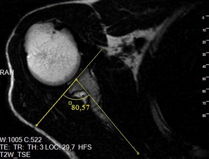
Rotator cuff was evaluated in fat-saturated T2-weighted and proton density-weighted images. Glenoid version angle (GVA) was measured on axial MR images (Fig. 1) as described by Tetrault et al. [8]. In the first axial image in which the posterior border of the glenoid neck was clearly visible, a line was drawn through the axis of the glenoid surface. The second line was drawn by joining the posterior glenoid neck and the junction of the scapular spine to the scapular body medially (Fig. 1), whereby ‘a’ was the angle in the posterior medial quadrant of the intersection of these two lines. GVA was calculated by subtracting 90° from the ‘a’ angle (GVA = a − 90°). Anteversion of the glenoid was indicated as positive (+) values for this angle, while retroversion was indicated as negative (−) values.
Fig. 1.
Measurement of glenoid version angle (GVA) on axial MR image (yellow lines)
Statistical analysis
Data were analysed with the SPSS software version 15.0 for Windows (SPSS Inc., Chicago, Illinois, USA). Continuous variables were presented as mean ± SD and categorical variables as frequency and percentage. The Kolmogorov–Smirnov test was applied to assess the distribution of continuous variables. The Student t test was used to compare continuous variables with normal distribution. The Mann–Whitney U test was used to compare continuous variables without normal distribution. The χ2 test was used to compare categorical variables. A two-tailed p-value of <0.05 was considered statistically significant.
Results
Table 1 summarises the characteristics of the study population. The study group consisted of 62 patients with a mean age of 48.7 years (range 25–73 years) and the control group consisted of 59 patients with a mean age of 37.3 years (range 14–61 years). A significant difference was found between the patients and controls for age (p < 0.001). There were no statistically significant differences between males and females, and right and left shoulders for all measurements in the patient and control groups (all p > 0.05).
Table 1.
Characteristics of the study population
| Characteristics | Study group (n = 62) | Control group (n = 60) | p value |
|---|---|---|---|
| Age (range) years | 48.7 ± 10.5 (25–73) | 37.3 ± 12.5 (14–61) | <0.001 |
| Examination number | 62 | 60 | --- |
| Bilateral cases | 0 | 1 | --- |
| Gender (male/female) | 21 (%34) / 41 (%66) | 27 (%47) / 32 (%53) | 0.149 |
| Side (right/left) | 43 (%69) / 19 (%31) | 36 (%60) / 24 (%40) | 0.280 |
In the control group, 26.6 % of the glenoids were retroverted from −12.88 to −0.55° and 73.4% were anteverted from 0.03 to 10.99° (Table 2). The mean value for GVA was 2.41°. In the study group, 33.8% of the glenoids were retroverted from −16.82 to −0.9° and 66.2% were anteverted from 0.03° to 16.28°. The mean value for GVA was 0.61°. There was no statistically significant difference for GVA between the control and patient groups.
Table 2.
Measurements of the patient and control groups
| Measurement | Patient group (n = 62) | Control group (n = 60) | p value |
|---|---|---|---|
| GVA | 0.61° ± 7.36 | 2.41° ± 4.75 | 0.112 |
| Anteversion (n); range | 41 | 44 | 0.434 |
| 0.03–16.28° | 0.03–10.99° | ||
| Retroversion (n); range | 21 | 16 | |
| −16.82° to −0.9° | −12.88° to −0.55° |
GVA glenoid version angle
In the control group, none of the shoulders had any rotator cuff injury. In the study group, 20% of patients had supraspinatus tendinosis, 16% had partial rupture of supraspinatus, 5% had complete rupture, 6% had both tendinosis and partial rupture of supraspinatus, 2% had complete rupture of supraspinatus and partial rupture of subscapularis tendon, 1% had supraspinatus tendinosis and partial rupture of subscapularis tendon, 1% had supraspinatus tendinosis and both partial rupture and tendinosis of subscapularis tendon.
Discussion
The glenohumeral joint is responsible for most movements of the shoulder joint, especially during internal rotation [10]. The main cause of shoulder pain in young people and athletes is glenohumeral instability, although it is rotator cuff pathology in older people. Many different risk factors play an important role in the aetiology of the RC diseases which have been described. The most studied mechanism is extrinsic mechanical compression. Attrition of the aponeurosis against the undersurface of the acromion, which was first described by Meyer in 1931, is the main cause of damage to supraspinatus tendon [11]. Neer has stated that chronic impingement beneath the coracoacromial arch causes 95% of rotator cuff ruptures [12]. Bigliani et al. have described the relationship between the variation in the shape of the acromion and RC tears [7]. The ‘hooked’ shape of the acromion was found to have the highest correlation with RC ruptures [13, 14].
In the literature, GVA was measured and compared in patients with RC tears and in controls. The authors found that GVA was more retroverted in the patient group, and the difference was statistically significant [8, 9, 15–19].
Glenoid version has been examined in several studies. Das et al. [15] reported that the mean GVA in ten normal subjects as determined by plain radiography was −4.9°. In 1983, Cyprien et al. [16] reported the mean GVA as −7.6° in 50 normal men (100 shoulders) using measurements from plain radiographs. The other studies with computed tomography (CT) and MR images have reported similar values of GVA [16, 17, 18]. Tokgoz et al. [9] have reported the mean GVA as −7.1° in patients with supraspinatus tendon tears and −4.8° in the control group. Tetrault et al. [8] found a correlation between the area of RC tears and the glenoid version. They reported that retroversion (mean value, −5° ± 4) was associated with a relatively high probability of supraspinatus tendon injury. However, a control group was not included in their study.
In contrast to previous studies that have demonstrated normal GVA to be negative, Friedman et al. [20] have determined the mean GVA as positive (2° ± 5). In our study, we found the GVA as 2.41°± 4.75 in the control group, similar to Friedman’s study and 0.61° ± 7.36 in the patient group in contrast to the literature with a significant difference. There was no statistically significant difference for GVA between the control and patient groups (p > 0.05).
This is the first study in the literature that defines the effect of primary GVA on RC pathology, because we excluded subjects having any other risk factors that might cause RC pathology such as hooked acromion, degenerative changes and trauma. In other words, in this radiological study the effect of the primary glenoid axis on RC tears was evaluated in a homogenous study population. In patients with anteverted flat-type acromions especially, the evaluation of RC pathologies itself seems to be more appropriate if there is no extrinsic pathology causing RC tear. However, this study has the level of evidence ‘B’. Therefore, for more robust results on this topic, studies having the level of evidence ‘A’ are needed [21].
Finally, some limitations were present in our study. The most serious limitation is the small sample size. During the one-year period, only 62 shoulders with rotator cuff pathology having type 1 acromion morphology were detected retrospectively. For a more healthy comparison between two groups, a similar sample size of 60 shoulders without rotator cuff pathology of 59 patients was included in the study. This limitation could be overcome by increasing the study period. Another limitation of the study was its retrospective nature. In fact, rotator cuff pathology should be confirmed arthroscopically. However, the patients were diagnosed only by MRI. To rule out misdiagnosis, images were reported independently at different times by two different radiologists.
In conclusion, glenoid version and acromion types are different morphological features of scapula; however, they may combine to provoke rotator cuff injury. Further studies with large sample sizes are needed to confirm this hypothesis.
Conflict of interest
The authors declare that they have no conflict of interest.
References
- 1.Hughes RE, Bryant CR, Hall JM, Wening J, Huston LJ, Kuhn JE, Carpenter JE, Blasier RB. Glenoid inclination is associated with full-thickness rotator cuff tears. Clin Orthop Relat Res. 2003;407:86–91. doi: 10.1097/00003086-200302000-00016. [DOI] [PubMed] [Google Scholar]
- 2.Banas MP, Miller RJ, Totterman S. Relationship between lateral acromion angle and rotator cuff disease. J Shoulder Elbow Surg. 1995;4:454–461. doi: 10.1016/S1058-2746(05)80038-2. [DOI] [PubMed] [Google Scholar]
- 3.Epstein RE, Schweitzer ME, Frieman BG, Fenlin JM, Jr, Mitchell DG. Hooked acromion: prevalence on MR images of painful shoulders. Radiology. 1993;187:479–481. doi: 10.1148/radiology.187.2.8475294. [DOI] [PubMed] [Google Scholar]
- 4.Gerber C, Ganz R, Terrier F. The role of coracoid process in the chronic impingement syndrome. J Bone Joint Surg Br. 1985;67:703–708. doi: 10.1302/0301-620X.67B5.4055864. [DOI] [PubMed] [Google Scholar]
- 5.Prato N, Peloso D, Francioneri A, Tegaldo G, Ravera GB, Silvestri E, Derchi LE. The anterior tilt of the acromion: radiographic evaluation and correlation with shoulder diseases. Eur Radiol. 1998;8:1639–1646. doi: 10.1007/s003300050602. [DOI] [PubMed] [Google Scholar]
- 6.Neer CS., 2nd Anterior acromioplasty for the chronic impingement syndrome in the shoulder: a preliminary report. J Bone Joint Surg Am. 1972;54:41–50. [PubMed] [Google Scholar]
- 7.Bigliani LU, Morrison DS, April EW. The morphology of the acromion and its relationship to rotator cuff tears. Orthop Trans. 1986;10:228. [Google Scholar]
- 8.Tetreault P, Krueger A, Zurakowski D, Gerber C. Glenoid version and rotator cuff tears. J Orthop Res. 2004;22:202–207. doi: 10.1016/S0736-0266(03)00116-5. [DOI] [PubMed] [Google Scholar]
- 9.Tokgoz N, Kanatli U, Voyvoda NK, Gultekin S, Bolukbasi S, Tali ET. The relationship of glenoid and humeral version with supraspinatus tendon tears. Skeletal Radiol. 2007;36:509–514. doi: 10.1007/s00256-007-0290-x. [DOI] [PubMed] [Google Scholar]
- 10.Koishi H, Goto A, Tanaka M, Omori Y, Futai K, Yoshikawa H, Sugamoto K (2011) In vivo three-dimensional motion analysis of the shoulder joint during internal and external rotation. Int Orthop. doi:10.1007/s00264-011-1219-5, Epub ahead of print [DOI] [PMC free article] [PubMed]
- 11.Meyer AW. The minuter anatomy of attrition lesions. J Bone Joint Surg. 1931;13:41–60. [Google Scholar]
- 12.Neer CS., 2nd Impingement lesions. Clin Orthop. 1983;173:70–77. [PubMed] [Google Scholar]
- 13.Nicholson GP, Goodman DA, Flatow EL, Bigliani LU. The acromion: morphologic condition and age-related changes. A study of 420 scapulas. J Shoulder Elbow Surg. 1996;5:1–11. doi: 10.1016/S1058-2746(96)80024-3. [DOI] [PubMed] [Google Scholar]
- 14.Seeger LL, Gold RH, Bassett LW, Elman H. Shoulder impingement syndrome: MR findings in 53 shoulders. AJR Am J Roentgenol. 1988;150:343–347. doi: 10.2214/ajr.150.2.343. [DOI] [PubMed] [Google Scholar]
- 15.Das SP, Ray GS, Saha AK. Observations on the tilt of the glenoid cavity of scapula. J Anat Soc India. 1996;15:114–118. [Google Scholar]
- 16.Cyprien JM, Vasey HM, Burdet A, Bonvin JC, Kritsikis N, Vuagnat P. Humeral retrotorsion and glenohumeral relationship in the normal shoulder and in recurrent anterior dislocation (scapulometry) Clin Orthop Relat Res. 1983;175:8–17. [PubMed] [Google Scholar]
- 17.Laumann U, Kramps HA. Computer tomography on recurrent shoulder dislocation. In: Bateman YE, Welsh RP, editors. Surgery of the shoulder. Philadelphia: Decker; 1984. p. 84. [Google Scholar]
- 18.Randelli M, Gambrioli PL. Glenohumeral osteometry by computed tomography in normal and unstable shoulders. Clin Orthop. 1986;208:151–156. [PubMed] [Google Scholar]
- 19.Brewer BJ, Wubben RC, Carrera GF. Excessive retroversion of the glenoid cavity. J Bone Joint Surg. 1986;68:724–731. [PubMed] [Google Scholar]
- 20.Friedman RJ, Hawthorne KB, Genez BM. The use of computerized tomography in the measurement of glenoid version. J Bone Joint Surg Am. 1992;74:1032–1037. [PubMed] [Google Scholar]
- 21.Hadorn DC, Baker D, Hodges JS, Hicks N. Rating the quality of evidence for clinical practice guidelines. J Clin Epidemiol. 1996;49:749–754. doi: 10.1016/0895-4356(96)00019-4. [DOI] [PubMed] [Google Scholar]


