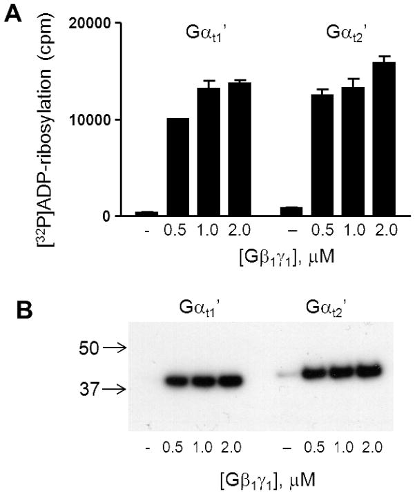
Figure 4. The Gβ1γ1-dependent ADP-ribosylation of Gαt1′ and Gαt2′.
Pertussis toxin-catalyzed ADP-ribosylation of Gαt1′ and Gαt2′ (0.5 μM each) was carried out in the presence of increasing concentrations of Gβ1γ1. (A) [32P]ADP-ribosylation of Gαt1′ and Gαt2′ is analyzed by liquid scintillation counting (mean±SE, n=3). (B) Aliquots from the ADP-ribosylation reaction mixtures were analyzed by SDS-PAGE followed by autoradiography.

