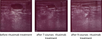
Figure 2. Sonographic recording of the popliteal left metastasis during rituximab therapy.
The metastasis substantially shrinks during therapy from approximately 722 mm2 to 135 mm2, i.e., a reduction by 80%; note that the blood vessel immediately under the metastasis showed nearly the same diameter in these projections.

