Abstract
INTRODUCTION
Difficulty may be encountered with retrograde access for rigid and flexible ureterorenoscopy (URS) due to anatomic abnormalities, a narrow ureteric lumen, tortuous ureteric path or previous instrumentation. Ureteric dilatation using a balloon or tapered dilator can occasionally fail and will usually lead to the placement of a ureteric stent. We present our experience and incidence of pre-stenting after failed standard access and dilatation techniques, the aim being to quote a figure for the patient at the time of consent.
PATIENTS AND METHODS
Data were collected prospectively from a single surgeon at a regional tertiary referral stone unit. The outcomes of those patients pre-stented, for failed access, were recorded.
RESULTS
Between December 2007 and December 2008, a total of 119 patients underwent flexible and rigid URS. Mean patient age was 49 years (range, 19–86 years). Of these, 107 cases were undertaken for urolithiasis and 12 cases for diagnosis of upper tract malignancy. 12% (13/107) of cases were for pain and non-diagnostic imaging and 8.4% (9/107) of patients were pre-stented because of failed access, without complication, and subsequently had successful interval treatment. Of the remaining successful cases of confirmed urolithiasis, 33% (28/85) and 67% (56/85) were undertaken for ureteric and renal calculi, respectively. Stone clearance rates were 83% (19/23) and 75% (3/4) for lower pole renal calculi 5–10 mm and > 10 mm in size, respectively. The overall clearance rate for lower pole calculi was 81% (22/27). The ureteric stone clearance rate was 86% (24/28) rising to 92% (24/26) in those solitary stones less than 10 mm in size.
CONCLUSIONS
The incidence of ureteric pre-stenting in a tertiary referral unit was 8% and should be considered and indeed discussed with patients when obtaining pre-operative consent, especially for purely elective, non-urgent, upper tract cases. The alternative for these difficult, tight ureters is extensive balloon dilatation, with the risk of trauma and the potential for long-term stricture formation.
Keywords: Ureterorenoscopy, Ureter, Pre-stenting
Approximately 10% of Caucasian men will expect to suffer from renal stone disease by the age of 70 years. It is a growing problem in the UK, with a cross-sectional prevalence of approximately 1.2%. These statistics mean there are approximately 720,000 individuals with a history of kidney stones in the UK.1
The increased prevalence of kidney stones parallels the well-publicised increase in the nation’s prevalence of obesity and its well-documented relationship to urolithiasis.2
In 1912, the first visualisation of the upper urinary tract was performed by Hampton Young. He achieved this by passing a cystoscope into a mega-ureter of a paediatric patient. Subsequent developments in optics have revolutionised endourology and established the ureteroscopic treatment of ureteric and renal calculi.
Although a comparison between extracorporeal shockwave lithotripsy (ESWL) and ureterorenoscopy (URS) removal of stones from the lower calyx of the kidney has failed to show a significantly better result with URS,3 the updated 2007 American Urological Association/European Association of Urology (AUA/EAU) guidelines, and recent Cochrane meta-analysis, suggest that stone-free rates are superior with URS for all stone sizes and for all positions in the ureter, apart from stones in the upper third less than 10 mm in size.4
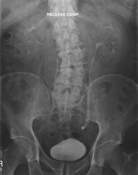
Standard access to the ureter for endoscopic management of stone disease may be difficult due to anatomic abnormalities, a narrow ureteric lumen, tortuous ureteric path or previous instrumentation (Fig. 1). Failure of access will usually lead to the placement of a ureteric stent. The alternative for these difficult, tight ureters is extensive balloon dilatation, with the risk of trauma and the potential for long-term stricture formation.
Figure 1.

Intravenous urogram highlighting a tortuous narrow distal left ureter.
Patients and Methods
Data were collected prospectively from a single surgeon at a regional tertiary referral stone unit. Between December 2007 and December 2008, a total of 119 patients underwent flexible or rigid URS. At our institution we use a 7.5/6.0-Fr rigid ureteroscope and an 8.4/7.5-Fr Flex-X2™ flexible ureteroscope. Standard graduated ureteric dilatation was undertaken with a Nottingham™ 12-Fr dilator and 11/13-Fr Navigator™ ureteric access sheath.
The outcomes of those patients who were pre-stented, for failed access, were recorded.
In addition, the stone-free rates for all URS were documented with regards to site and size of the stone. A patient’s stone-free status was determined under direct vision and fluoroscopy at the time of the operation and/or follow-up plain X-ray imaging.
Results
Of the 119 patients, 107 cases were undertaken for urolithiasis and 12 cases for the diagnosis of upper tract malignancy. Mean patient age was 49 years (range, 19–86 years). Female to male ratio was 1:1.4. Only 12% (13/107) of the cases of urolithiasis were for on-going pain and non-diagnostic imaging, but were found to be stone-free on URS.
Standard dilatation was undertaken with a Nottingham™ in 11% (10/107) of cases, and with a Navigator™ access sheath in 17% (18/107).
Of the patients with stones, 8.4% (9/107) were pre-stented because of failed access, without complication, and subsequently had successful interval treatment. Of these, three were female patients and six were male patients, representing 6.9% of those female patients undergoing URS for presumed urolithiasis and 9.3% of male patients.
There were 85 cases of confirmed urolithiasis which were accessed successfully. Of these, 33% (28/85) and 67% (56/85) were undertaken for ureteric and renal calculi, respectively; 35/85 were evaluated for stone clearance at the time of the procedure, by direct vision and fluoroscopy. The remaining patients were also evaluated with a plain KUB X-ray at a mean follow-up of 4.7 months. Stone clearance rates were 100% (13/13) and 75% (3/4) for solitary lower pole renal calculi 5–10 mm and > 10 mm in size, respectively. Total clearance rate for lower pole calculi was 81% (22/27), compared to 44% (8/18) of renal pelvic stones (Table 1).
Table 1.
Percentage stone clearance rates for patients with renal calculi treated with URS in regards to stone size and position
| Renal pelvis (%) | Lower pole (%) | Interpolar (%) | Upper pole (%) | Total | |
|---|---|---|---|---|---|
| 5–10 mm | 60 (6/10) | 83 (19/23) | 100 (4/4) | 67 (4/6) | 77 |
| > 10 mm | 25 (2/8) | 75 (3/4) | 0 (0/1) | 0 (0/1) | 36 |
| Total | 44 | 81 | 80 | 57 |
The total ureteric stone clearance rate was 86% (24/28), and 92% (22/24) in those solitary stones less than 10 mm in size (Table 2).
Table 2.
Percentage stone clearance rates for patients with ureteric calculi treated with URS in regards to stone size and position
| Distal third ureter (%) | Mid third ureter (%) | Proximal third ureter (%) | Total (%) | |
|---|---|---|---|---|
| 5–10mm | 92 (12/13) | 100 (3/3) | 90 (9/10) | 92 |
| > 10 mm | 0 (0/1) | n/a | 0 (0/1) | 0 |
| Total | 86 | 100 | 82 |
The ureteric perforation rate in this series was 0.9% (1/107). This one case occurred without dilatation and was managed successfully with ureteric stenting. Other complications were postoperative sepsis, with one case of perinephric abscess (3/107), acute retention of urine (1/107), re-admission for pain (5/107) and un-explained ileus (1/107).
Discussion
The General Medical Council set out guidance for consent in 2008. This expanded on Good Medical Practice (1998), which required doctors to be satisfied that they had consent from a patient, or other valid authority, before undertaking any examination, investigation or providing treatment. When seeking consent for a treatment, the guidelines state the clinician must outline all potential serious adverse outcomes, even if the likelihood is very small; and also tell patients about less serious side effects or complications if they occur frequently.5
In our study, the incidence of ureteric pre-stenting in a tertiary referral unit was 8%, and should be considered and indeed discussed with patients when obtaining pre-operative consent, especially for purely elective, non-urgent, upper tract cases. The alternative for these difficult, tight ureters is extensive balloon dilatation and the risk of long-term stricture formation. The mechanism of this is not fully understood but, in experimental models, balloon dilatation caused longitudinal incisions in the ureteric mucosa similar to those seen at endoureterotomy,6 and the resulting extravasation of irrigating fluid and urine is believed to cause fibrosis.7
In a study from Pardalidis and colleagues,8 98 consecutive patients with small (< 10 mm) impacted lower third ureteric stones were randomly managed with both a 12/14-Fr co-axial ureteric dilator/sheath and a 7.5-Fr flexible ureteroscope, or with balloon dilatation and the 7.5-Fr flexible ureteroscope. In the latter group, who had balloon dilatation, ureteric perforation rates were higher (8% versus 0%).8
Our study shows a higher failure rate for access in male patients, although the total number were small. Failed access occurred throughout the ureter, with no consistent failed point of access across the sexes that could be attributed to their specific anatomical differences.
Our data compare favourably with the URS data from the EAU/AUA meta-analysis, and confirm the conclusion that URS is an effective treatment for ureteric calculi with minimal complications (Table 3). In addition, our audit shows, contrary to the paper by Pearle and colleagues,3 that stone-free rates of 83% can be achieved for lower pole renal calculi < 10 mm in size with flexible URS, with the caveat that this was not a randomised trial and patient selection, as occurs in normal practice, was determined by the likelihood of a favourable outcome.
Table 3.
URS stone-free rates for patients with ureteric stones < 10 mm in size in regards to ureteric level: a comparison of the AUA/EAU meta-analysis and our study
| This study | AUA/EAU meta-analysis4 | |
|---|---|---|
| Distal ureter | 92% | 97% |
| Mid ureter | 100% | 91% |
| Proximal ureter | 90% | 80% |
The poor results of clearance seen for stones located in the renal pelvis, in our study, may reflect the displacement of these stones during fragmentation into a previously unappreciated, inaccessible lower pole.
Conclusions
The Endourological Society (CROES) international URS audit will publish next year. Before then, we recommend quoting an 8% pre-stenting rate to patients at the time of consent, prior to URS for urolithiasis.
References
- 1.Johri N, Cooper B, Robertson W, Choong S, Rickards D, Unwin R. An update and practical guide to renal stone management. Nephron Clin Pract. 2010;116:159–71. doi: 10.1159/000317196. [DOI] [PubMed] [Google Scholar]
- 2.Vujovic A, Keoghane S. Management of renal stone disease in obese patients. Nat Clin Pract Urol. 2007;4:671–6. doi: 10.1038/ncpuro0988. [DOI] [PubMed] [Google Scholar]
- 3.Pearle MS, Lingeman JE, Leveillee R, Kuo R, Preminger GM, et al. Prospective randomized trial comparing shock wave lithotripsy and ureteroscopy for lower pole caliceal calculi 1 cm or less. J Urol. 2005;173:2005–9. doi: 10.1097/01.ju.0000158458.51706.56. [DOI] [PubMed] [Google Scholar]
- 4.Preminger GM, Tiselius HG, Assimos DG, Alken P, Buck AC, et al. American Urological Association Education and Research, Inc.; European Association of Urology. 2007 Guideline for the management of ureteral calculi. Eur Urol. 2007;52:1610–31. doi: 10.1016/j.eururo.2007.09.039. [DOI] [PubMed] [Google Scholar]
- 5. General Medical Council. Consent: patients and doctors making decisions together. 2008. < http://www.gmc-uk.org/guidance/ethical_guidance/consent_guidance_index.asp> [accessed July 2010]
- 6.Nakada SY, Soble JJ, Gardner SM, Wolf JS, Jr, Figenshau RS, et al. Comparison of acucise endopyelotomy and endoballoon rupture for management of secondary proximal ureteral stricture in the porcine model. J Endourol. 1996;10:311–8. doi: 10.1089/end.1996.10.311. [DOI] [PubMed] [Google Scholar]
- 7.Roberts WW, Cadeddu JA, Micali S, Kavoussi LR, Moore RG. Ureteral stricture formation after removal of impacted calculi. J Urol. 1998;159:723–6. [PubMed] [Google Scholar]
- 8.Pardalidis NP, Papatsoris AG, Kapotis CG, Kosmaoglou EV. Treatment of impacted lower third ureteral stones with the use of the ureteral access sheath. Urol Res. 2006;34:211–4. doi: 10.1007/s00240-006-0044-6. [DOI] [PubMed] [Google Scholar]


