Abstract
Background
Observational studies examining the role of estrogen in the risk of kidney stone formation have shown conflicting results. However, randomized trial evidence on nephrolithiasis risk with estrogen therapy in postmenopausal women is lacking.
Methods
We reviewed the incidence of nephrolithiasis in the Women’s Health Initiative estrogen-alone and estrogen plus progestin trials conducted at 40 US clinical centers. A total of 10 739 postmenopausal women with hysterectomy were randomized to receive 0.625 mg/d of conjugated equine estrogens (CEE) or placebo, and 16 608 postmenopausal women without hysterectomy were randomized to receive placebo or estrogen plus progestin given as CEE plus medroxyprogesterone acetate (2.5 mg/d). The incidence of nephrolithiasis was determined for an average follow-up of 7.1 years for the CEE trial and 5.6 years for the estrogen plus progestin trial.
Results
Baseline demographic characteristics and risk factors for nephrolithiasis were similar in the placebo and treatment arms. Estrogen therapy was associated with a significant increase in nephrolithiasis risk from 34 to 39 cases per 10 000 person-years (hazard ratio, 1.21; 95% confidence interval, 1.03-1.44). Censoring data from women when they ceased to adhere to study medication increased the hazard ratio to 1.39 (95% confidence interval, 1.08-1.78). The increased nephrolithiasis risk was independent of progestin coadministration, and effects did not vary significantly according to prerandomization history of nephrolithiasis.
Conclusions
These data suggest that estrogen therapy increases the risk of nephrolithiasis in healthy postmenopausal women. These findings should be considered in decision making regarding postmenopausal estrogen use. The mechanisms underlying this higher susceptibility remain to be determined.
Nephrolithiasis is a common condition that affects 5% to 7% of postmenopausal women in the United States.1 In addition to the suffering caused by an acute kidney stone event, long-term complications can include renal insufficiency.2 Treatment of nephrolithiasis also incurs substantial costs, estimated at $2 billion yearly in the United States.3 Although kidney stones occur less commonly in women than in men younger than 50 years, this disparity becomes less prominent in the sixth decade of life in parallel with the on-set of menopause in women.4,5 The sex difference in the incidence of nephrolithiasis has been ascribed to a possible protective role of estrogen against kidney stone formation in premenopausal women.6
Observational studies examining the role of estrogen therapy on the risk of nephrolithiasis have shown conflicting results. Cross-sectional studies of postmenopausal kidney stone–forming women suggest that estrogen therapy may potentially be protective against nephrolithiasis based on 24-hour urinary parameters.6,7 On the other hand, analysis of data from the Nurses’ Health Study did not find an association between postmenopausal hormone therapy (HT) use and incident kidney stones.8
Because the process of kidney stone formation is influenced by a variety of lifestyle and other health-related factors, the true impact of estrogen therapy on the risk of kidney stone formation is difficult to infer from observational studies. To our knowledge, there are no previous randomized trials examining the outcome of kidney stone formation after estrogen therapy in postmenopausal women. The Women’s Health Initiative (WHI) postmenopausal HT trials included 2 separate studies that examined the impact of HT in women with and without a hysterectomy.9,10 Their results on the risk-benefit profile of postmenopausal estrogen use on a variety of outcomes have been reported previously.11,12 This report provides new evidence on the effect of estrogen therapy on the incidence of nephrolithiasis.
METHODS
PARTICIPANTS
A total of 27 347 postmenopausal women aged 50 to 79 years were enrolled in the WHI-HT trials at 40 US clinical centers between 1993 and 1998: 10 739 postmenopausal women with hysterectomy were enrolled in the estrogen-alone trial, while 16 608 postmenopausal women without hysterectomy were enrolled in the estrogen plus progestin (E+P) trial. The design of these 2 trials has previously been described in detail.9,10 The trials were approved by the National Institutes of Health and by the local institutional review boards of all participating institutions. All participants provided informed consent.
INTERVENTIONS
Women in the estrogen-alone trial were randomized to receive 0.625 mg/d of conjugated equine estrogens (CEE) (Premarin; Wyeth, Philadelphia, Pennsylvania) or matching placebo. Women in the E+P trial were given a single tablet of CEE plus 2.5 mg/d of medroxyprogesterone acetate (Prempro; Wyeth-Ayerst; St Davids, Pennsylvania) or matching placebo. The E+P trial was stopped early after a mean follow-up period of 5.6 years because the overall benefits of E+P were outweighed by the harms.11 The CEE trial was stopped early after a mean follow-up period of 7.1 years because of increased strokes with no benefit for coronary heart disease.12
DATA COLLECTION AND OUTCOMES
Baseline evaluation included detailed self-administered questionnaires that assessed demographic, clinical, social, behavioral, and dietary characteristics of study participants. Height and weight were measured by a wall-mounted stadiometer and a balance beam scale, respectively, and body mass index was calculated as weight in kilograms divided by height in meters squared. Participants in the HT trials also underwent baseline clinical and gynecological examinations.
Six weeks after randomization, study participants were contacted by telephone to assess symptoms and to reinforce adherence. Standardized information on specific symptoms, safety concerns, and major health events was collected at 6-month intervals afterward, with a mandatory clinic visit annually.
Kidney stone disease was self-reported by the participants in the baseline and semiannual medical history update questionnaires. The specific wording in the questionnaires was “Has a doctor told you that you have kidney or bladder stones (renal or urinary calculi)?” No further ascertainment of stone disease was made in the WHI.
Incident stone is used to designate a participant’s self-report of a urinary calculus after enrollment in the HT trial and is applied to all participants, including women with a known history of kidney stones at study entry.
STATISTICAL ANALYSIS
Selected risk factors were described for the study population by HT randomization assignment. Kaplan-Meier curves of reported kidney stone events were plotted by treatment assignment for all participants in the HT trials and for those who remained adherent throughout the trials. Nonadherence was prospectively defined for adherence monitoring purposes as consuming more than 80% of study pills (by pill weight), starting nonstudy prescribed HT during the most recent 6-month study interval, or reporting nonstudy HT use during the most recent medication inventory assessment, which occurred at baseline and years 1, 3, 6, and 9.
Cox proportional hazards models were used to analyze the effect of HT on the risk of kidney stones using an intention-to-treat approach. The main models were stratified on participation in the E+P and the CEE trials and randomization assignment for the WHI dietary modification trial. Adjustment for the randomization arm of the WHI calcium and vitamin D (CaD) clinical trial was done using a time-dependent covariate, since participants were randomized to the CaD trial 1 to 2 years after randomization to the HT trial. Also, models were stratified according to age group at screening (50-54, 55-59, 60-69, and 70-79 years), ethnicity, body mass index, and history of kidney stones at baseline.
A sensitivity analysis looking at the effect of nonadherence was performed by repeating the main analysis with additional censoring for events occurring after the estimated date of non-adherence. Subgroup analyses looking at the effect of HT on kidney stones within categories of major risk factors were modeled in the same fashion as the main model. The only time an adjustment variable from the main model was not included was when that variable was the risk factor being categorized. P values were obtained from a Wald test for the interaction term between treatment assignment and potential risk factor, whereby the risk factor was treated as a continuous variable. Under the null hypothesis, approximately 1 of the 11 interactions investigated would be expected to be nominally significant (P<.05) by chance alone.
RESULTS
The baseline characteristics of study participants are shown in Table 1. The control and treatment arms were balanced with respect to baseline demographic and risk factors for kidney stone formation including lifestyle and dietary elements. A history of nephrolithiasis was reported by about 4% of the study population at the time of the screening visit. Approximately 26% of women enrolled in the E+P trial reported prior or current use of HT at study entry (before washout), while 47% of women with hysterectomy had used HT. The only statistically significant difference (P<.05) between the CEE and the CEE-placebo groups at baseline was dietary oxalate intake (based on dietary recall), although the difference was small (289 mg/d vs 295 mg/d; P=.02). Similarly, a small but statistically significant difference was noted between the E+P and the placebo groups in terms of baseline intake of vitamin C (314 mg/d vs 332 mg/d; P=.02).
Table 1.
Baseline Characteristics of the Study Participants in the Women’s Health Initiative Hormone Therapy (HT) Trialsa
| CEE Trial |
E+P Trial |
|||
|---|---|---|---|---|
| Variable | CEE Placebo (n=5429) |
CEE (n=5310) |
E+P Placebo (n=8102) |
E+P (n=8506) |
| Age, mean (SD), y | 63.6 (7.3) | 63.6 (7.2) | 63.3 (7.1) | 63.2 (7.1) |
| 50-59 | 30.8 | 30.8 | 33.1 | 33.4 |
| 60-69 | 45.4 | 45.0 | 45.1 | 45.3 |
| 70-79 | 23.8 | 24.2 | 21.8 | 21.3 |
| Race/ethnicity | ||||
| White | 75.1 | 75.5 | 84.0 | 83.9 |
| Black | 15.4 | 14.7 | 7.1 | 6.5 |
| Hispanic | 5.1 | 6.1 | 5.1 | 5.6 |
| American Indian | 0.6 | 0.8 | 0.4 | 0.3 |
| Asian/Pacific Islander | 1.4 | 1.6 | 2.1 | 2.3 |
| Unknown | 1.4 | 1.4 | 1.3 | 1.5 |
| BMI, mean (SD) | 30.1 (6.2) | 30.1 (6.1) | 28.5 (5.9) | 28.5 (5.8) |
| <25.0 | 20.3 | 21.0 | 30.8 | 30.5 |
| 25.0-29.9 | 35.5 | 34.0 | 35.0 | 35.3 |
| ≥30.0 | 44.2 | 45.0 | 34.0 | 34.2 |
| HT use ever | 49.0 | 47.9 | 25.7 | 26.2 |
| History of kidney or bladder stones at randomization | 4.9 | 4.7 | 3.5 | 3.3 |
| Hypertensive ever | 40.1 | 40.5 | 30.0 | 29.9 |
| History of diabetes | 9.6 | 9.5 | 5.8 | 5.7 |
| Thiazide use | 5.5 | 5.5 | 4.0 | 4.0 |
| Regular physical activityb | 32.0 | 32.9 | 39.2 | 38.4 |
| Dietary animal protein, g/d | 49.0 (25.7) | 48.9 (24.9) | 49.1 (23.8) | 49.4 (24.1) |
| Dietary sodium, mg/d | 2771 (1266) | 2751 (1218) | 2783 (1195) | 2788 (1187) |
| Dietary calcium, mg/d | 780 (446) | 781 (450) | 827 (444) | 835 (456) |
| Dietary oxalic acid, mg/d | 295 (146) | 289 (138)c | 305 (143) | 305 (139) |
| Dietary potassium, mg/d | 2537 (1006) | 2507 (985) | 2640 (970) | 2653 (981) |
| Dietary magnesium, mg/d | 246 (99) | 243 (96) | 256 (96) | 256 (96) |
| Dietary sucrose, g/d | 35 (20) | 34 (20) | 35 (19) | 35 (20) |
| Calcium from supplements, mg/d | 260 (477) | 248 (472) | 312 (517) | 302 (485) |
| Supplemental vitamin D, μg/d | 3.8 (5.5) | 3.8 (5.5) | 4.3 (6.0) | 4.4 (6.0) |
| Total vitamin C, diet plus supplements, mg/d | 306 (532) | 312 (515) | 332 (510) | 314 (484)d |
Abbreviations: BMI, body mass index (calculated as weight in kilograms divided by height in meters squared); CEE, conjugated equine estrogens; E+P, estrogen plus progestin.
Conversion factor: To convert vitamin D from micrograms to international units, multiply by 40.
Values are expressed as percentage or mean (SD).
Regular physical activity was defined as 2 or more 20-minute episodes of moderate or strenuous activity per week.
P<.05 between the CEE and the CEE placebo groups.
P<.05 between the E+P and the E+P placebo groups.
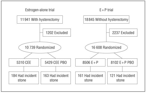
Of the 27 347 women randomized to the HT trials, 2727 had missing data regarding a history of kidney stones at study entry and were excluded from the Cox models, which included stratification based on baseline history of kidney stones. Therefore, a total of 24 620 women were included in the Cox models analysis: 9607 from the E+P trial and 15 103 from the CEE trial. A total of 335 incident cases of nephrolithiasis were reported in the active treatment group, while 284 cases occurred in the placebo group (Figure 1). The corresponding annualized incidence rate per 10 000 person-years was 39 in the treatment group and 34 in the placebo group (Table 2).These rates translate to a hazard ratio (HR) of 1.21 (95% confidence interval [CI], 1.03-1.44) with postmenopausal hormone use.
Figure 1.

Profile of the hormone therapy trials in the Women’s Health Initiative included in the current analysis. CEE indicates conjugated equine estrogens; E+P, estrogen plus progestin; and PBO, placebo.
Table 2.
Incidence (Annualized Percentage) and Hazard Ratio of Kidney Stones Reported During the Hormone Therapy (HT) Trials in the Women’s Health Initiative (WHI) According to Treatment Group and Baseline Characteristicsa
| No. of Participants With Kidney Stones During Follow-up per Year, No. (%) |
||||
|---|---|---|---|---|
| Variable | Placebo | HT | HR (95% CI) |
P Value for Interactionb |
| Overall | 284 (0.34) | 335 (0.39) | 1.21 (1.03-1.44) | |
| Overall, censored for nonadherence | 119 (0.23) | 157 (0.32) | 1.39 (1.08-1.78) | |
| History of kidney or bladder stones | .47 | |||
| No | 203 (0.29) | 254 (0.36) | 1.25 (1.04-1.50) | |
| Yes | 48 (1.65) | 48 (1.74) | 1.07 (0.71-1.61) | |
| HT trial participation | .79 | |||
| E+P | 121 (0.27) | 151 (0.31) | 1.24 (0.97-1.60) | |
| CEE | 163 (0.42) | 184 (0.49) | 1.19 (0.95-1.49) | |
| Age group at screening, y | .28 | |||
| 50-54 | 36 (0.32) | 44 (0.38) | 1.43 (0.88-2.34) | |
| 55-59 | 54 (0.32) | 55 (0.32) | 1.00 (0.67-1.52) | |
| 60-64 | 65 (0.35) | 95 (0.49) | 1.57 (1.12-2.19) | |
| 65-69 | 60 (0.32) | 73 (0.39) | 1.26 (0.89-1.80) | |
| 70-74 | 44 (0.35) | 45 (0.35) | 0.96 (0.63-1.48) | |
| 75-79 | 25 (0.46) | 23 (0.43) | 0.91 (0.50-1.65) | |
| Race/ethnicity | .22 | |||
| White | 210 (0.31) | 255 (0.37) | 1.22 (1.00-1.48) | |
| Black | 27 (0.30) | 41 (0.48) | 1.83 (1.07-3.12) | |
| Hispanic | 28 (0.61) | 22 (0.46) | 1.00 (0.55-1.82) | |
| Other | 19 (0.66) | 17 (0.54) | 1.04 (0.52-2.05) | |
| BMI | .51 | |||
| <25.0 | 46 (0.21) | 73 (0.32) | 1.49 (1.02-2.19) | |
| 25.0-29.9 | 110 (0.37) | 109 (0.37) | 1.00 (0.75-1.32) | |
| ≥30.0 | 127 (0.40) | 152 (0.46) | 1.27 (0.99-1.64) | |
| HT use status | .76 | |||
| Never used | 177 (0.34) | 219 (0.40) | 1.23 (1.00-1.52) | |
| Past user | 74 (0.32) | 85 (0.37) | 1.26 (0.90-1.75) | |
| Current user | 33 (0.42) | 31 (0.39) | 0.99 (0.58-1.70) | |
| Thiazide use | .25 | |||
| No | 267 (0.34) | 320 (0.39) | 1.24 (1.05-1.48) | |
| Yes | 17 (0.44) | 15 (0.39) | 0.94 (0.43-2.03) | |
| Cups/d of caffeinated coffee | .55 | |||
| 0 | 27 (0.29) | 32 (0.34) | 1.05 (0.59-1.88) | |
| 1-2 | 91 (0.32) | 108 (0.37) | 1.26 (0.94-1.70) | |
| >3 | 84 (0.34) | 91 (0.36) | 1.03 (0.75-1.40) | |
| DM trial participant | .46 | |||
| No | 193 (0.33) | 241 (0.41) | 1.26 (1.03-1.53) | |
| Yes | 91 (0.36) | 94 (0.36) | 1.11 (0.80-1.52) | |
| CaD trial arm | .11 | |||
| Placebo | 57 (0.28) | 50 (0.24) | 0.86 (0.58-1.27) | |
| Active | 53 (0.25) | 69 (0.33) | 1.40 (0.95-2.05) | |
| Time since menopause at study entry, y | .06 | |||
| ≤5 | 40 (0.34) | 42 (0.33) | 1.06 (0.67-1.68) | |
| 6-10 | 28 (0.23) | 49 (0.40) | 1.91 (1.17-3.12) | |
| 11-15 | 34 (0.25) | 48 (0.35) | 1.42 (0.90-2.23) | |
| 16-20 | 59 (0.46) | 56 (0.44) | 1.00 (0.69-1.46) | |
| ≥21 | 90 (0.39) | 90 (0.40) | 1.03 (0.76-1.40) | |
Abbreviations: BMI, body mass index (calculated as weight in kilograms divided by height in meters squared); CaD, calcium and vitamin D; CEE, conjugated equine estrogens; CI, confidence Interval; DM, dietary modification; E+P, estrogen plus progestin; HR, hazard ratio for incident nephrolithiasis.
The annualized rates are shown as the percentage of participants reporting kidney stones during follow-up. Cox models were stratified according to WHI-HT, DM, and calcium and vitamin D trial randomization; age group; and history of kidney stone unless used in a subgroup analysis. Cox models analyses are based on 24 620 women only, as 2727 participants with no data on history of kidney stones at study entry had to be excluded from the analyses.
P values were obtained from an interaction term between treatment assignment and potential risk factor.
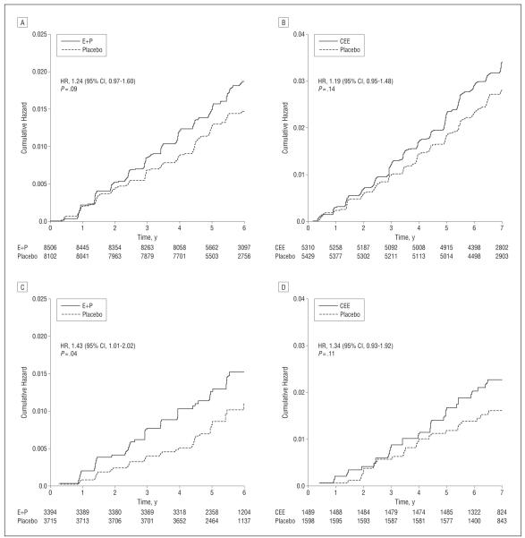
After data were censored from women when they ceased to adhere to the study medication, HT resulted in a 39% increase in incident nephrolithiasis (HR, 1.39; 95% CI, 1.08-1.78). Hormone therapy increased the risk of nephrolithiasis to a similar extent in both trials (HRs, 1.24 and 1.19 for the E+P and the CEE trials, respectively). Nephrolithiasis occurrence was 5 times more common in women with a history of kidney stones at study entry, although this higher incidence was not significantly altered by estrogen therapy (Table 2).
No significant interactions were found with age, ethnicity, body mass index, prior HT, or use of coffee or thiazide diuretics (all P values for interaction, ≥.20). There was a suggestion of a decreased HR in women with longer time since menopause at study entry (P=.06); however, this association was likely driven by the relatively higher HR in women who entered the trials 6 to 10 years after menopause.
The Kaplan-Meier estimates of cumulative hazards of nephrolithiasis for all participants and adherent participants only are shown in Figure 2. These curves showed a divergence starting in the second year after randomization. Because this was the time when some women were also enrolled in the WHI-CaD supplementation clinical trial, we examined the possibility of a potential interaction between HT and CaD use on the risk of kidney stones. Of the women in the WHI-HT trials, 16 089 were also randomized into the WHI-CaD trial (8117 in HT active arms; 7972 in HT placebo arms). There was a suggestion (P=.11) of an increased risk with combined CaD and HT use vs CaD and placebo (HR, 1.40; 95% CI, 0.95-2.05).
Figure 2.
Kaplan-Meier estimates of cumulative hazards for kidney stones reported during the Women’s Health Initiative hormone therapy trials. CEE indicates conjugated equine estrogens; CI, confidence interval; E+P, estrogen plus progestin; and HR, hazard ratio. A, E+P trial: all participants. B, CEE trial: all participants. C, E+P trial: adherent participants. D, CEE trial: adherent participants.
COMMENT
These WHI findings demonstrate, for the first time in a randomized, placebo-controlled trial, an increased risk of nephrolithiasis in postmenopausal women receiving estrogen therapy. The risk of incident kidney stones was increased with CEE alone and with CEE combined with progestin, with similar HRs. Before this report, the only other published study examining the association between postmenopausal estrogen therapy and the incidence of nephrolithiasis found no relationship between HT and incident kidney stones.8 In that report from the Nurses’ Health Study,8 estrogen use was not randomized. The disparity in the findings between the WHI and the Nurses’ Health Study may therefore be attributable to the differences between HT users and nonusers, which could not be controlled for in that nonrandomized study. Although the current study represents a post hoc analysis, the randomized design led to similar baseline characteristics with respect to nephrolithiasis risk in placebo- and actively treated participants (Table 1).
The pathogenesis of kidney stone formation is complex and influenced by genetic and environmental factors.13 Estrogens may affect several key steps in kidney stone formation, including the urinary excretion of kidney stone constituents and of urinary promoters and inhibitors of kidney stone formation. One potential mechanism for the higher incidence of stone disease with HT could be through enhanced urinary uric acid excretion with estrogen use.14,15 Greater uric acid excretion, in turn, could lead to heterogeneous nucleation of calcium oxalate.16,17 In healthy postmenopausal women, estrogen therapy enhances intestinal calcium absorption,18 reduces bone resorption,19 and increases renal tubular re-absorption of calcium.20 Compared with nontreated postmenopausal kidney stone–forming women, those treated with estrogen therapy had lower urinary calcium excretion in one previous study6 and a higher urinary calcium excretion in another report.7 Similarly, conflicting results have been reported on the effects of estrogen therapy on urinary citrate, a major inhibitor of calcium oxalate and calcium phosphate crystal growth and kidney stone formation.6,7 Finally, while oral estrogen use increases serum concentration of the prothrombin fragment 1,21 a molecule found both within calcium oxalate stones and more abundantly in the kidneys of kidney stone formers than in healthy individuals,22 its effects on urinary prothrombin fragment 1 have not been reported. The impact of estrogen therapy on urinary biochemical composition of stone components was not evaluated in the WHI study.
The incidence of nephrolithiasis in the WHI-HT trials was approximately 35 per 10 000 person-years. In comparison, kidney stone incidence in women older than 50 years in Rochester, Minnesota, was approximately 10 per 10 000 person-years.23 In the Nurses’ Health Study I, the kidney stone incidence was 10 in 10 000 person-years for the overall population, 20.5 per 10 000 person-years in postmenopausal women, and 26.2 per 10 000 person-years in women with surgical menopause.8 The higher kidney stone incidence in the WHI compared with the Nurses’ Health Study could in part be attributable to the higher body mass index in the WHI, as greater body size is associated with heightened nephrolithiasis risk.24 Furthermore, kidney stone incidence in the Nurses’ Health Study was based on data collected between 1980 and 1998, while WHI data were collected between 1993 and 2003. Therefore, the higher incidence of stone disease in the WHI-HT trials may in part reflect the rise in disease incidence23 and prevalence.1 Finally, in the WHI, kidney stone disease was self-reported by participants in the medical history update questionnaire without further confirmation. In comparison, self-reported stone disease was corroborated by an additional questionnaire in the Nurses’ Health Study, while kidney stone incidence in the report from Rochester was based on International Classification of Diseases, Ninth Revision, codes.23 Differences in ascertainment could contribute to higher rates of kidney stone disease in the WHI.
In the WHI, the incidence of nephrolithiasis was higher in the CEE trial (in women after hysterectomy) than the E+P trial (in women with intact uterus) (Table 2). This finding was observed irrespective of treatment arm, suggesting that progesterone coadministration is not responsible for this effect. These results are compatible with the Nurses’ Health Study results, which found a higher incidence of nephrolithiasis in women with surgical menopause.8 The mechanism underlying this observation is not clear. However, surgical menopause results in a more sudden loss of ovarian production of estrogens and androgens, which may enhance bone loss25 and urinary calcium excretion, potentially raising kidney stone risk.
Since the publications of the principal results from the WHI-HT trials,10,11 postmenopausal estrogen use has declined considerably in the United States.26,27 A concomitant decline in the incidence of breast cancer has been seen,28 which has been attributed in large part to reduced estrogen use.28,29 While the link between estrogen and nephrolithiasis is not as strong as that with breast cancer, it would be interesting to evaluate whether the incidence of nephrolithiasis among postmenopausal women changed over the past decade.
One limitation of the current study is that the incidence of nephrolithiasis was measured by self-report in questionnaires and was not confirmed by review of records. However, reporting bias is unlikely to have occurred to a different degree in placebo- vs actively treated participants. Furthermore, self-reported incident kidney stone disease could be ascertained by record review in more than 95% of participants in other studies, such as the Health Professionals Follow-up Study and the Nurses’ Health Study I.30,31 Another limitation is that only dosages of 0.625 mg/d of CEE or 0.625 mg/d of CEE plus 2.5 mg/d of medroxyprogesterone acetate were studied in the WHI-HT trials. Therefore, our ability to generalize these findings to women taking other HT formulations is limited. Further research is needed on the effects of different estrogen formulations on the risk of nephrolithiasis and changes in urine composition.
In conclusion, these results from a large, double-blind, placebo-controlled, randomized clinical trial indicate that estrogen therapy increases the risk of nephrolithiasis in healthy postmenopausal women. The mechanisms underlying this higher propensity remain to be determined. In view of the sizable prevalence of nephrolithiasis in this segment of the population, these findings need to be considered in the decision-making process regarding postmenopausal estrogen use.
WHI Investigators
Program Office National Heart, Lung, and Blood Institute, Bethesda, Maryland: Elizabeth Nabel, Jacques Rossouw, Shari Ludlam, Joan McGowan, Leslie Ford, and Nancy Geller.
Clinical Coordinating Center
Fred Hutchinson Cancer Research Center, Seattle, Washington: Ross Prentice, Garnet Anderson, Andrea LaCroix, Charles Kooperberg, Martin McIntosh, Ching-Yung Wang, Chu Chen, Deborah Bowen, Alan Kristal, Janet Stanford, Nicole Urban, Noel Weiss, and Emily White. Medical Research Laboratories, Highland Heights, Kentucky: Evan Stein. University of California at San Francisco: Steven R. Cummings.
Clinical Centers
Albert Einstein College of Medicine, Bronx, New York: Sylvia Wassertheil-Smoller. Baylor College of Medicine, Houston, Texas: J. Haleh Sangi-Haghpeykary. Brigham and Women’s Hospital, Harvard Medical School, Boston, Massachusetts: JoAnn E. Manson. Brown University, Providence, Rhode Island: Charles B. Eaton. Emory University, Atlanta, Georgia: Lawrence S. Phillips. Fred Hutchinson Cancer Research Center: Shirley Beresford. George Washington University Medical Center, Washington, DC: Lisa Martin. Los Angeles Biomedical Research Institute at Harbor–UCLA Medical Center, Torrance, Calif: Rowan Chlebowski. Kaiser Permanente Center for Health Research, Portland, Oregon: Erin LeBlanc. Kaiser Permanente Division of Research, Oakland, California: Bette Caan. Medical College of Wisconsin, Milwaukee: Jane Morley Kotchen. MedStar Research Institute/Howard University, Washington, DC: Barbara V. Howard. Northwestern University, Chicago/Evanston, Illinois: Linda Van Horn. Rush University Medical Center, Chicago: Henry Black. Stanford Prevention Research Center, Stanford, California: Marcia L. Stefanick. State University of New York at Stony Brook: Dorothy Lane. The Ohio State University, Columbus: Rebecca Jackson. University of Alabama at Birmingham: Cora E. Lewis. University of Arizona, Tucson/Phoenix: Cynthia A. Thomson. University of Buffalo, Buffalo, New York: Jean Wactawski-Wende. University of California at Davis, Sacramento: John Robbins. University of California at Irvine: F. Allan Hubbell. University of California at Los Angeles: Lauren Nathan. University of California at San Diego, LaJolla/Chula Vista: Robert D. Langer. University of Cincinnati, Cincinnati, Ohio: Margery Gass. University of Florida, Gainesville/Jacksonville: Marian Limacher. University of Hawaii, Honolulu: J. David Curb. University of Iowa, Iowa City/Davenport: Robert Wallace. University of Massachusetts/Fallon Clinic, Worcester: Judith Ockenen. University of Medicine and Dentistry of New Jersey, Newark: Norman Lasser. University of Miami, Miami, Florida: Mary Jo O’Sullivan. University of Minnesota, Minneapolis: Karen L. Margolis. University of Nevada, Reno: Robert Brunner. University of North Carolina, Chapel Hill: Gerardo Heiss. University of Pittsburgh, Pittsburgh, Pennsylvania: Lewis Kuller. University of Tennessee Health Science Center, Memphis: Karen C. Johnson, Suzanne Satterfield, Raymond W. Ke, Stephanie Connelly, and Fran Tylavsky. University of Texas Health Science Center, San Antonio: Robert Brzyski. University of Wisconsin, Madison: Gloria E. Sarto. Wake Forest University School of Medicine, Winston-Salem, North Carolina: Mara Vitolins. Wayne State University School of Medicine/Hutzel Hospital, Detroit, Michigan: Michael S. Simon.
Acknowledgments
Funding/Support: The WHI program is funded by the National Heart, Lung, and Blood Institute, National Institutes of Health, US Department of Health and Human Services, through contracts N01WH22110, 24152, 32100-2, 32105-6, 32108-9, 32111-13, 32115, 32118-32119, 32122, 42107-26, 42129-32, and 44221.
Footnotes
Author Contributions: Study concept and design: Maalouf, Welch, and Sakhaee. Acquisition of data: Howard and Robbins. Analysis and interpretation of data: Maalouf, Sato, Howard, Cochrane, and Sakhaee. Drafting of the manuscript: Maalouf, Sato, and Welch. Critical revision of the manuscript for important intellectual content: Howard, Cochrane, Sakhaee, and Robbins. Statistical analysis: Sato. Obtained funding: Robbins. Administrative, technical, and material support: Cochrane and Robbins. Study supervision: Howard and Sakhaee.
Financial Disclosure: None reported.
Additional Contributions: Mary Pettinger helped with the manuscript.
REFERENCES
- 1.Stamatelou KK, Francis ME, Jones CA, Nyberg LM, Curhan GC. Time trends in reported prevalence of kidney stones in the United States: 1976-1994. Kidney Int. 2003;63(5):1817–1823. doi: 10.1046/j.1523-1755.2003.00917.x. [DOI] [PubMed] [Google Scholar]
- 2.Rule AD, Bergstralh EJ, Melton LJ, III, Li X, Weaver AL, Lieske JC. Kidney stones and the risk for chronic kidney disease. Clin J Am Soc Nephrol. 2009;4(4):804–811. doi: 10.2215/CJN.05811108. [DOI] [PMC free article] [PubMed] [Google Scholar]
- 3.Pearle MS, Calhoun EA, Curhan GC. Urologic Diseases of America Project. Urologic diseases in America project: urolithiasis. J Urol. 2005;173(3):848–857. doi: 10.1097/01.ju.0000152082.14384.d7. [DOI] [PubMed] [Google Scholar]
- 4.Marshall V, White RH, De Saintonge MC, Tresidder GC, Blandy JP. The natural history of renal and ureteric calculi. Br J Urol. 1975;47(2):117–124. doi: 10.1111/j.1464-410x.1975.tb03930.x. [DOI] [PubMed] [Google Scholar]
- 5.Johnson CM, Wilson DM, O’Fallon WM, Malek RS, Kurland LT. Renal stone epidemiology: a 25-year study in Rochester, Minnesota. Kidney Int. 1979;16(5):624–631. doi: 10.1038/ki.1979.173. [DOI] [PubMed] [Google Scholar]
- 6.Heller HJ, Sakhaee K, Moe OW, Pak CY. Etiological role of estrogen status in renal stone formation. J Urol. 2002;168(5):1923–1927. doi: 10.1016/S0022-5347(05)64264-4. [DOI] [PubMed] [Google Scholar]
- 7.Dey J, Creighton A, Lindberg JS, et al. Estrogen replacement increased the citrate and calcium excretion rates in postmenopausal women with recurrent urolithiasis. J Urol. 2002;167(1):169–171. [PubMed] [Google Scholar]
- 8.Kramer HJ Mattix, Grodstein F, Stampfer MJ, Curhan GC. Menopause and postmenopausal hormone use and risk of incident kidney stones. J Am Soc Nephrol. 2003;14(5):1272–1277. doi: 10.1097/01.asn.0000060682.25472.c3. [DOI] [PubMed] [Google Scholar]
- 9.The Women’s Health Initiative Study Group Design of the Women’s Health Initiative clinical trial and observational study. Control Clin Trials. 1998;19(1):61–109. doi: 10.1016/s0197-2456(97)00078-0. [DOI] [PubMed] [Google Scholar]
- 10.Stefanick ML, Cochrane BB, Hsia J, Barad DH, Liu JH, Johnson SR. The Women’s Health Initiative postmenopausal hormone trials: overview and baseline characteristics of participants. Ann Epidemiol. 2003;13(9)(suppl):S78–S86. doi: 10.1016/s1047-2797(03)00045-0. [DOI] [PubMed] [Google Scholar]
- 11.Rossouw JE, Anderson GL, Prentice RL, et al. Writing Group for the Women’s Health Initiative Investigators Risks and benefits of estrogen plus progestin in healthy postmenopausal women: principal results from the Women’s Health Initiative randomized controlled trial. JAMA. 2002;288(3):321–333. doi: 10.1001/jama.288.3.321. [DOI] [PubMed] [Google Scholar]
- 12.Anderson GL, Limacher M, Assaf AR, et al. Women’s Health Initiative Steering Committee Effects of conjugated equine estrogen in postmenopausal women with hysterectomy: the Women’s Health Initiative randomized controlled trial. JAMA. 2004;291(14):1701–1712. doi: 10.1001/jama.291.14.1701. [DOI] [PubMed] [Google Scholar]
- 13.Moe OW. Kidney stones: pathophysiology and medical management. Lancet. 2006;367(9507):333–344. doi: 10.1016/S0140-6736(06)68071-9. [DOI] [PubMed] [Google Scholar]
- 14.Nicholls A, Snaith ML, Scott JT. Effect of oestrogen therapy on plasma and urinary levels of uric acid. Br Med J. 1973;1(5851):449–451. doi: 10.1136/bmj.1.5851.449. [DOI] [PMC free article] [PubMed] [Google Scholar]
- 15.Adamopoulos D, Vlassopoulos C, Seitanides B, Contoyiannis P, Vassilopoulos P. The relationship of sex steroids to uric acid levels in plasma and urine. Acta Endocrinol (Copenh) 1977;85(1):198–208. doi: 10.1530/acta.0.0850198. [DOI] [PubMed] [Google Scholar]
- 16.Pak CY, Arnold LH. Heterogeneous nucleation of calcium oxalate by seeds of mono-sodium urate. Proc Soc Exp Biol Med. 1975;149(4):930–932. doi: 10.3181/00379727-149-38929. [DOI] [PubMed] [Google Scholar]
- 17.Coe FL, Lawton RL, Goldstein RB, Tembe V. Sodium urate accelerates precipitation of calcium oxalate in vitro. Proc Soc Exp Biol Med. 1975;149(4):926–929. doi: 10.3181/00379727-149-38928. [DOI] [PubMed] [Google Scholar]
- 18.Gallagher JC, Riggs BL, DeLuca HF. Effect of estrogen on calcium absorption and serum vitamin D metabolites in postmenopausal osteoporosis. J Clin Endocrinol Metab. 1980;51(6):1359–1364. doi: 10.1210/jcem-51-6-1359. [DOI] [PubMed] [Google Scholar]
- 19.Hassager C, Colwell A, Assiri AM, Eastell R, Russell RG, Christiansen C. Effect of menopause and hormone replacement therapy on urinary excretion of pyridinium cross-links: a longitudinal and cross-sectional study. Clin Endocrinol (Oxf ) 1992;37(1):45–50. doi: 10.1111/j.1365-2265.1992.tb02282.x. [DOI] [PubMed] [Google Scholar]
- 20.McKane WR, Khosla S, Burritt MF, et al. Mechanism of renal calcium conservation with estrogen replacement therapy in women in early postmenopause—a clinical research center study. J Clin Endocrinol Metab. 1995;80(12):3458–3464. doi: 10.1210/jcem.80.12.8530583. [DOI] [PubMed] [Google Scholar]
- 21.Kluft C, Meijer P, LaGuardia KD, Fisher AC. Comparison of a transdermal contraceptive patch vs. oral contraceptives on hemostasis variables. Contraception. 2008;77(2):77–83. doi: 10.1016/j.contraception.2007.10.004. [DOI] [PubMed] [Google Scholar]
- 22.Stapleton AM, Seymour AE, Brennan JS, Doyle IR, Marshall VR, Ryall RL. Immunohistochemical distribution and quantification of crystal matrix protein. Kidney Int. 1993;44(4):817–824. doi: 10.1038/ki.1993.316. [DOI] [PubMed] [Google Scholar]
- 23.Lieske JC, de la Vega LS Peña, Slezak JM, et al. Renal stone epidemiology in Rochester, Minnesota: an update. Kidney Int. 2006;69(4):760–764. doi: 10.1038/sj.ki.5000150. [DOI] [PubMed] [Google Scholar]
- 24.Taylor EN, Stampfer MJ, Curhan GC. Obesity, weight gain, and the risk of kidney stones. JAMA. 2005;293(4):455–462. doi: 10.1001/jama.293.4.455. [DOI] [PubMed] [Google Scholar]
- 25.Hreshchyshyn MM, Hopkins A, Zylstra S, Anbar M. Effects of natural menopause, hysterectomy, and oophorectomy on lumbar spine and femoral neck bone densities. Obstet Gynecol. 1988;72(4):631–638. [PubMed] [Google Scholar]
- 26.Hersh AL, Stefanick ML, Stafford RS. National use of postmenopausal hormone therapy: annual trends and response to recent evidence. JAMA. 2004;291(1):47–53. doi: 10.1001/jama.291.1.47. [DOI] [PubMed] [Google Scholar]
- 27.Haas JS, Kaplan CP, Gerstenberger EP, Kerlikowske K. Changes in the use of postmenopausal hormone therapy after the publication of clinical trial results. Ann Intern Med. 2004;140(3):184–188. doi: 10.7326/0003-4819-140-3-200402030-00009. [DOI] [PubMed] [Google Scholar]
- 28.Ravdin PM, Cronin KA, Howlader N, et al. The decrease in breast-cancer incidence in 2003 in the United States. N Engl J Med. 2007;356(16):1670–1674. doi: 10.1056/NEJMsr070105. [DOI] [PubMed] [Google Scholar]
- 29.Chlebowski RT, Kuller LH, Prentice RL, et al. WHI Investigators Breast cancer after use of estrogen plus progestin in postmenopausal women. N Engl J Med. 2009;360(6):573–587. doi: 10.1056/NEJMoa0807684. [DOI] [PMC free article] [PubMed] [Google Scholar]
- 30.Curhan GC, Willett WC, Rimm EB, Stampfer MJ. A prospective study of dietary calcium and other nutrients and the risk of symptomatic kidney stones. N Engl J Med. 1993;328(12):833–838. doi: 10.1056/NEJM199303253281203. [DOI] [PubMed] [Google Scholar]
- 31.Curhan GC, Willett WC, Speizer FE, Spiegelman D, Stampfer MJ. Comparison of dietary calcium with supplemental calcium and other nutrients as factors affecting the risk for kidney stones in women. Ann Intern Med. 1997;126(7):497–504. doi: 10.7326/0003-4819-126-7-199704010-00001. [DOI] [PubMed] [Google Scholar]


