Abstract
Antigen detection has been reported to be a promising method for rapid diagnosis of coccidioidomycosis in humans. Coccidioides antigen detection has not been previously reported in dogs with coccidioidomycosis and was evaluated in 60 cases diagnosed based on detection of anti-Coccidioides antibodies at titers of 1:16 or more in serum. Controls included dogs with presumed histoplasmosis or blastomycosis, other fungal infections, or nonfungal diseases and healthy dogs. Urine and serum specimens were tested using an enzyme immunoassay for Coccidioides galactomannan antigen. Antibody testing was performed at commercial veterinary reference laboratories. Antigen was detected in urine or serum of 12 of 60 (20.0%), urine only in 2 of 57 (3.5%), and serum only in 11 of 58 (19.0%) dogs with coccidioidomycosis. Antigen was detected in the urine of 3 of 43 (7.0%) and serum of 1 of 37 (2.7%) dogs with histoplasmosis or blastomycosis but not in 13 dogs with other fungal infections (serum, 9; urine, 13), 41 dogs with nonfungal diseases (urine, 41; serum, 18), or healthy dogs (serum, 21; urine, 21). Detection of antigen was an insensitive method for diagnosis of coccidioidomycosis in dogs in which the diagnosis was based primarily upon detection of antibodies at titers of 1:16 or higher, and the highest sensitivity was in serum.
INTRODUCTION
The diagnosis of coccidioidomycosis in dogs is usually based upon the detection of immunoglobulin G (IgG) anti-Coccidioides antibodies by agar gel immunodiffusion (AGID) (8), which is positive in over 90% of affected dogs (7). Antibodies also may be detected in apparently healthy dogs, however (10). In a prospective, longitudinal study from Arizona, only 5 of 28 dogs (18%) with positive antibody tests for coccidioidomycosis were judged to have clinical disease (10). Furthermore, titers overlapped in dogs with clinical coccidioidomycosis and those without clinical disease, supporting the need for additional tests, such as cytology, histopathology, and culture, to establish the diagnosis of clinical coccidioidomycosis (10). Nevertheless, AGID titers of at least 1:16 are highly suggestive of clinically relevant coccidioidomycosis in ill dogs (6).
More recently, detection of antigen in urine (2) and serum (3) has been reported to complement the results of serologic testing for antibodies and histopathology in human patients with coccidioidomycosis. Coccidioides antigenuria was detected in 71% of patients with moderately severe or severe coccidioidomycosis (2). Furthermore, in milder cases, of which 50% exhibited antigenuria, an additional 21% were detected if serum was tested (3). Specificity was 99% in humans without fungal infection, but cross-reactions were noted in 10% of those with other endemic mycoses (2). Reproducibility was 100%, and interassay precision was good, with coefficients of variation of 7.3 to 12.7%. The objective of this study was to determine the sensitivity of Coccidioides antigen detection in dogs with coccidioidomycosis and specificity in dogs with other conditions and healthy subjects.
MATERIALS AND METHODS
Experimental design and animals.
Dogs with coccidioidomycosis were recruited from two veterinary internal medicine practices: Phoenix Veterinary Internal Medicine Services (R. T. Greene) and the Southern Arizona Veterinary Specialty and Emergency Center (A. Prahl). Sixty dogs were enrolled based on detection of IgG antibody titers of ≥1:16 determined by AGID at one or the other of two commercial laboratories (Antech, Phoenix, AZ; or Idexx, Phoenix, AZ). Histopathology or cytologic examination of body fluids or tissues was not performed for any of these dogs. Urine and/or serum samples were obtained with the informed consent of the dog owners and were stored at −20°C at the collaborating veterinarians' laboratories. The specimens were shipped to MiraVista Diagnostics on ice packs via overnight delivery, where they were stored at −20°C until they were tested together as a single batch.
Controls included dogs with proven blastomycosis based upon visualization of yeast by cytologic or histopathologic examination of tissues or fluids, which also had positive tests for Blastomyces antigen and dogs with presumed histoplasmosis based on positive antigen tests for Histoplasma antigen in urine and/or serum in the absence of cytologic or histopathologic examination of tissues or fluids. Additional controls included dogs from California with systemic mold infections, dogs from California or Arizona with nonfungal diseases, and healthy dogs from Arizona. IgG antibodies were measured on serum of the control dogs from California and Arizona by AGID using a commercial test according to the manufacturer's instructions (Meridian Bioscience, Cincinnati, OH).
Antigen assay.
The Coccidioides quantitative antigen assay was performed as previously reported (2), using microplates coated (VWR, Batavia, IL) with anti-Coccidioides antibodies selected for maximum sensitivity and specificity for detection of the galactomannan antigen. Serum specimens were first treated by adding 200 μl of 4% EDTA (Midwest Scientific, St. Louis, MO) at pH 4.6 to 600 μl of serum, vortexing the mixture, and placing it in a heat block (Fisher Scientific, Pittsburgh, PA) at 104°C for 6 min, after which the samples were centrifuged and the supernatant was collected (3). Following incubation of the test specimen in the precoated microplate, antigen that had attached to the capture antibody was quantified with biotinylated rabbit anti-Coccidioides detector antibody. The standards used for quantification were prepared from urine containing known concentrations of Coccidioides galactomannan, based upon comparison to purified galactomannan from Coccidioides mold culture supernatant (2). Results greater than or equal to the 0.07-ng/ml galactomannan calibrator were considered positive, and the concentration was determined by comparison to the calibration curve.
Statistical analysis.
The respective proportion of patients with positive results was compared using Fisher's exact test with MedCalc software (Mariakerke, Belgium). P values of ≤0.05 were considered significant.
RESULTS
Sensitivity.
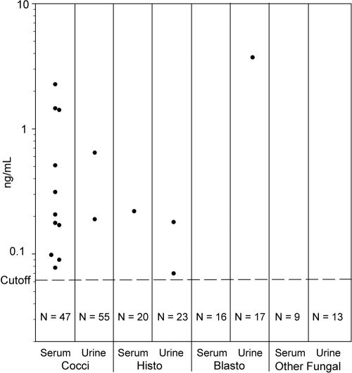
Antigen was detected in the serum of 11 (19.0%) of 58 dogs, urine of two (3.5%) of 57 dogs, and the urine or serum of 12 (20.0%) of 60 dogs. One dog with a positive urine antigen test result did not have a matching serum sample. The antigen concentration in serum ranged from 0.08 ng/ml to 2.3 ng/ml (median, 0.2 ng/ml; mean, 0.5 ng/ml) (Fig. 1).
Fig 1.
Detection of Coccidioides antigen in dogs with coccidioidomycosis, presumed histoplasmosis or blastomycosis, or other fungal infections. Cocci, coccidioidomycosis; Blasto, blastomycosis; Histo, histoplasmosis; Other Fungal, other fungal infections.
Specificity.
Among 151 control dogs, results were antigen positive in 4 (2.6%) dogs, including 3 of 45 presumed to have histoplasmosis (6.7%) based on detection of antigen in urine and/or serum and 1 of 31 (3.2%) dogs with cytologically or histopathologically proven blastomycosis (Table 1). Results for individual controls are shown in Fig. 1. Seventy-five controls were from areas where coccidioidomycosis is endemic, and these included 13 dogs with other fungal infections (Aspergillus [n = 9], Paecilomyces spp. [n = 2], and Penicillium spp. or Zygomyces spp. [1 dog each]), 41 dogs with conditions other than fungal infection (other infection, 12; malignancy, 6; immunological disease, 5; polyarthritis, 3; and other conditions, 14 [renal disease and diabetes mellitus, 2 each; and lymphoma, hypothyroidism, allergic bronchitis, polymyositis, multiple endocrine disorder, pancreatitis with diabetes mellitus, sterile inflammatory brain disease, undiagnosed pulmonary disease, diskospondylitis, and vomiting, 1 each]), and 21 healthy dogs. Coccidioides antigen was not detected in any of these controls (Table 1). Furthermore, in a subset of controls from areas of endemicity with sufficient serum to test for anti-Coccidioides antibodies, antibodies were detected by AGID in 2 of 48 (4.2%) dogs.
Table 1.
Detection of Coccidioides antigen in dogs with coccidioidomycosis, other fungal infections, or no fungal infection
| Groupa | No. of dogs with antigen detected/total (%) in: |
||
|---|---|---|---|
| Serum | Urine | Serum or urine | |
| Coccidioidomycosis | 11/58 (19.0) | 2/57 (3.5) | 12/60 (20.0)b |
| Histoplasmosis | 1/21 (4.8) | 2/25 (8.0) | 3/45 (6.7)c |
| Blastomycosis | 0/16 | 1/18 | 1/31 (3.2)d |
| Other mycosis | 0/9 | 0/13 | 0/13 |
| Nonfungal | 0/18 | 0/41 | 0/41 |
| Healthy | 0/21 | 0/21 | 0/21 |
“Other mycosis” represents dogs from California with other fungal infections. “Nonfungal” represents dogs from California or Arizona with diseases other than fungal infection. “Healthy” represents dogs from California or Arizona with no underlying conditions.
Serum only was available for two dogs and was positive for one.
Both serum and urine were available for one dog, for which only the urine was positive.
Both serum and urine were available for 10 dogs, for which only the urine was positive for one.
DISCUSSION
Antigen was detected in the serum in only 19% of dogs with coccidioidomycosis. The sensitivity for detection of antigenuria was much lower, about 4%. In humans with acute coccidioidomycosis, antigenemia occurred during the first month of infection, before antibodies could be detected, and cleared after antibodies appeared (4). Whether antigen detection could be useful for diagnosis earlier in the course of the illness, before high levels of antibodies develop, remains to be determined in dogs.
The sensitivity was higher in humans: 70.8% in one study (2) and 71.4% in another (3). A possible explanation for the difference in sensitivity between human patients and dogs is that the organism burden may have been higher in the human patients (2, 3), as most of the human cases had severe coccidioidomycosis and were immunocompromised. Information was not collected about the severity of coccidioidomycosis, the presence of immunocompromising conditions, species or strain characteristics of the infecting strain of Coccidioides spp., or prior antifungal therapy, precluding assessment of the role of these factors in antigen production in dogs.
Positive test results for Coccidioides antigen occurred in the urine of 7.0% and serum of 2.7% of dogs with presumed histoplasmosis or blastomycosis and positive tests for Histoplasma or Blastomyces antigen, probably reflecting cross-reactivity. Antigenuria and/or antigenemia has been detected in 17 of 18 (94.4%) cats with histoplasmosis (1), 40 of 43 (93.0%) dogs with blastomycosis (11), and 17 of 17 (100%) dogs with disseminated aspergillosis (5). Cross-reactivity also has been observed in about 10% of urine samples from humans with histoplasmosis (2), paracoccidioidomycosis (2), or blastomycosis (3). Cross-reactivity was not observed in 13 dogs with other fungal infections (mostly aspergillosis), 41 dogs with nonfungal diseases, or 21 healthy dogs from California or Arizona, while anti-Coccidioides antibodies were detected in 2 of 48 (4.2%) dogs with sufficient serum for AGID testing. In humans without systemic fungal infections, the specificities were 99% (95% confidence interval [CI], 96.6% to 99.9%) in urine (2) and 100% (95% CI, 96.4% to 100%) in serum (3).
The study has several limitations. First, identification of the site of tissue involvement in the coccidioidomycosis cases was largely based on history and physical examination findings, as imaging studies and histopathology were not performed in most cases. Second, specificity is uncertain as only 75 controls were from areas where coccidioidomycosis is endemic. Specificity was 99% in urine and serum from healthy humans from areas of endemicity, however (2, 3). Third, clinical information was not available for the control dogs with histoplasmosis. However, specificity was 97% in specimens from dogs with proven blastomycosis. Also, the studies in humans have established cross-reactivity in the Coccidioides antigen assay in urine and serum from 5 to 10% of patients with histoplasmosis or blastomycosis (2, 3, 9).
In summary, detection of antigen in dog urine and/or serum was an insensitive method for diagnosis of coccidioidomycosis in this study.
ACKNOWLEDGMENTS
We thank the veterinarians who collaborated in the study and the dog owners who allowed the participation of their pets in the study.
E.J.K., M.M.D., and L.J.W. are employees of MiraVista Diagnostics, a company that performs fungal antigen testing commercially.
The study was funded by MiraVista Diagnostics, Indianapolis, IN.
Footnotes
Published ahead of print 25 January 2012
REFERENCES
- 1. Cook AC, Cunningham LY, Cowell AK, Wheat LJ. Clinical evaluation of urine Histoplasma capsulatum antigen measurement in cats with suspected disseminated histoplasmosis. J. Feline Med. Surg., in press. [DOI] [PMC free article] [PubMed] [Google Scholar]
- 2. Durkin M, et al. 2008. Diagnosis of coccidioidomycosis with use of the Coccidioides antigen enzyme immunoassay. Clin. Infect. Dis. 47:e69–e73 [DOI] [PubMed] [Google Scholar]
- 3. Durkin M, et al. 2009. Detection of Coccidioides antigenemia following dissociation of immune complexes. Clin. Vaccine Immunol. 16:1453–1456 [DOI] [PMC free article] [PubMed] [Google Scholar]
- 4. Galgiani JN, Grace GM, Lundergan LL. 1991. New serologic tests for early detection of coccidioidomycosis. J. Infect. Dis. 163:671–674 [DOI] [PubMed] [Google Scholar]
- 5. Garcia RS, Kirsch E, Wheat LJ, Cook AC, Sykes JE. 2011. Sensitivity and specificity of a blood and urine galactomannan antigen assay for diagnosis of systemic aspergillosis in dogs. J. Vet. Intern. Med., in press [DOI] [PubMed] [Google Scholar]
- 6. Graupmann-Kuzma A, et al. 2008. Coccidioidomycosis in dogs and cats: a review. J. Am. Anim. Hosp. Assoc. 44:226–235 [DOI] [PubMed] [Google Scholar]
- 7. Johnson LR, Herrgesell EJ, Davidson AP, Pappagianis D. 2003. Clinical, clinicopathologic, and radiographic findings in dogs with coccidioidomycosis: 24 cases (1995–2000). J. Am. Vet. Med. Assoc. 222:461–466 [DOI] [PubMed] [Google Scholar]
- 8. Kerl ME. 2003. Update on canine and feline fungal diseases. Vet. Clin. North Am. Small Anim. Pract. 33:721–747 [DOI] [PubMed] [Google Scholar]
- 9. Kuberski T, et al. 2007. Diagnosis of coccidioidomycosis by antigen detection using cross-reaction with a Histoplasma antigen. Clin. Infect. Dis. 44:e50–e54 [DOI] [PubMed] [Google Scholar]
- 10. Shubitz LE, Butkiewicz CD, Dial SM, Lindan CP. 2005. Incidence of coccidioides infection among dogs residing in a region in which the organism is endemic. J. Am. Vet. Med. Assoc. 226:1846–1850 [DOI] [PubMed] [Google Scholar]
- 11. Spector D, et al. 2008. Antigen and antibody testing for the diagnosis of blastomycosis in dogs. J. Vet. Intern. Med. 22:839–843 [DOI] [PubMed] [Google Scholar]


