Abstract
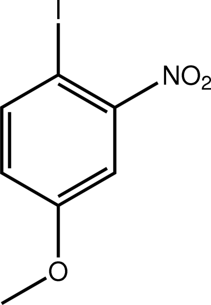
In the title compound, C7H6INO3, the 12 non-H atoms are planar, with an r.m.s. deviation of 0.016 Å. A close intramolecular I⋯O interaction [3.0295 (13) Å] is present. Intermolecular I⋯O interactions [3.3448 (13) Å] lead to the formation of zigzag chains along the b axis. These are assembled into layers by weak π–π interactions [centroid–centroid distance = 3.8416 (9) Å], and the layers stack along the a axis, being connected by C—H⋯O contacts.
Related literature
For general background to halogen bonding, see: Metrangolo et al. (2008 ▶); Pennington et al. (2008 ▶). For previous structural studies probing iodo–nitro interactions, see: Glidewell et al. (2002 ▶, 2004 ▶); Garden et al. (2002 ▶). For van der Waals radii, see: Bondi (1964 ▶).
Experimental
Crystal data
C7H6INO3
M r = 279.03
Orthorhombic,

a = 18.6370 (13) Å
b = 11.6257 (5) Å
c = 7.4740 (3) Å
V = 1619.38 (15) Å3
Z = 8
Mo Kα radiation
μ = 3.92 mm−1
T = 100 K
0.13 × 0.09 × 0.01 mm
Data collection
Rigaku Saturn724+ (2 × 2 bin mode) diffractometer
Absorption correction: multi-scan (CrystalClear-SM Expert; Rigaku, 2011 ▶) T min = 0.755, T max = 1.000
8053 measured reflections
1841 independent reflections
1615 reflections with I > 2σ(I)
R int = 0.017
Refinement
R[F 2 > 2σ(F 2)] = 0.014
wR(F 2) = 0.039
S = 1.04
1841 reflections
110 parameters
H-atom parameters constrained
Δρmax = 0.42 e Å−3
Δρmin = −0.25 e Å−3
Data collection: CrystalClear-SM Expert (Rigaku, 2011 ▶); cell refinement: CrystalClear-SM Expert; data reduction: CrystalClear-SM Expert; program(s) used to solve structure: SHELXS97 (Sheldrick, 2008 ▶); program(s) used to refine structure: SHELXL97 (Sheldrick, 2008 ▶); molecular graphics: ORTEP-3 (Farrugia, 1997 ▶) and DIAMOND (Brandenburg, 2006 ▶); software used to prepare material for publication: publCIF (Westrip, 2010 ▶).
Supplementary Material
Crystal structure: contains datablock(s) global, I. DOI: 10.1107/S1600536812006484/hg5176sup1.cif
Structure factors: contains datablock(s) I. DOI: 10.1107/S1600536812006484/hg5176Isup2.hkl
Supplementary material file. DOI: 10.1107/S1600536812006484/hg5176Isup3.cml
Additional supplementary materials: crystallographic information; 3D view; checkCIF report
Table 1. Hydrogen-bond geometry (Å, °).
| D—H⋯A | D—H | H⋯A | D⋯A | D—H⋯A |
|---|---|---|---|---|
| C7—H7B⋯O1i | 0.98 | 2.58 | 3.4503 (19) | 148 |
Symmetry code: (i)  .
.
Acknowledgments
The use of the EPSRC X-ray crystallographic service at the University of Southampton, England, and the valuable assistance of the staff there is gratefully acknowledged. JLW acknowledges support from CAPES (Brazil). We also thank the Ministry of Higher Education (Malaysia) for funding structural studies through the High-Impact Research scheme (UM·C/HIR/MOHE/SC/12).
supplementary crystallographic information
Comment
In connection with previous structural studies of iodo···nitro interactions (Glidewell et al., 2002; Garden et al., 2002; Glidewell et al., 2004), the crystal and molecular structure of the title compound (I) was undertaken in order to probe the structure for possible I···O halogen bonding (Metrangolo et al., 2008; Pennington et al., 2008).
The 12 non-hydrogen atoms comprising (I), Fig. 1, are co-planar with a rm.s. deviation = 0.016 Å; the maximum deviations of ±0.026 Å being found for the nitro-O atoms. A close intramolecular I1···O1 interaction of 3.0295 (13) Å is noted that is significantly less than the sum of the van der Waals radii for these atoms, i.e. 3.50 Å (Bondi, 1964). There are also notable intermolecular I1···O interactions, the shortest of 3.3448 (13) Å occurs with the O1i atom [symmetry operation i: 3/2 - x, 1/2 + y, z]. A longer interaction [3.4530 (13) Å] is formed with the O2 atom of the same nitro group. The I1···O1i interactions lead to the formation of zigzag chains along the b axis, Fig. 2. The supramolecular chains are assembled into layers in the bc plane by weak π–π interactions [ring centroid···centroid distance = 3.8416 (9) Å for symmetry operation x, 3/2 - y, -1/2 + z]. Layers stack along the a axis and are connected by C—H···O contacts, Fig. 3 and Table 1.
Experimental
The commercial compound (Aldrich) was recrystallized from EtOH; M.pt: 334–335 K.
Refinement
The C-bound H atoms were geometrically placed (C—H = 0.95–0.98 Å) and refined as riding with Uiso(H) = 1.2–1.5Ueq(C).
Figures
Fig. 1.
The molecular structure of (I) showing the atom-labelling scheme and displacement ellipsoids at the 50% probability level.
Fig. 2.
A view of the linear supramolecular chain along the b axis in (I). The I···O interactions are shown as blue dashed lines.
Fig. 3.
A view in projection down the c axis of the packing of supramolecular chains in (I). The I···O, C—H···O and π–π interactions are shown as blue, orange and purple dashed lines, respectively.
Crystal data
| C7H6INO3 | F(000) = 1056 |
| Mr = 279.03 | Dx = 2.289 Mg m−3 |
| Orthorhombic, Pbca | Mo Kα radiation, λ = 0.71073 Å |
| Hall symbol: -P 2ac 2ab | Cell parameters from 6840 reflections |
| a = 18.6370 (13) Å | θ = 3.4–27.4° |
| b = 11.6257 (5) Å | µ = 3.92 mm−1 |
| c = 7.4740 (3) Å | T = 100 K |
| V = 1619.38 (15) Å3 | Plate, dark-orange |
| Z = 8 | 0.13 × 0.09 × 0.01 mm |
Data collection
| Rigaku Saturn724+ (2x2 bin mode) diffractometer | 1841 independent reflections |
| Radiation source: Rotating Anode | 1615 reflections with I > 2σ(I) |
| Confocal monochromator | Rint = 0.017 |
| Detector resolution: 28.5714 pixels mm-1 | θmax = 27.5°, θmin = 3.4° |
| profile data from ω–scans | h = −15→24 |
| Absorption correction: multi-scan (CrystalClear-SM Expert; Rigaku, 2011) | k = −14→14 |
| Tmin = 0.755, Tmax = 1.000 | l = −9→8 |
| 8053 measured reflections |
Refinement
| Refinement on F2 | Primary atom site location: structure-invariant direct methods |
| Least-squares matrix: full | Secondary atom site location: difference Fourier map |
| R[F2 > 2σ(F2)] = 0.014 | Hydrogen site location: inferred from neighbouring sites |
| wR(F2) = 0.039 | H-atom parameters constrained |
| S = 1.04 | w = 1/[σ2(Fo2) + (0.0188P)2 + 0.9351P] where P = (Fo2 + 2Fc2)/3 |
| 1841 reflections | (Δ/σ)max = 0.002 |
| 110 parameters | Δρmax = 0.42 e Å−3 |
| 0 restraints | Δρmin = −0.25 e Å−3 |
Special details
| Geometry. All s.u.'s (except the s.u. in the dihedral angle between two l.s. planes) are estimated using the full covariance matrix. The cell s.u.'s are taken into account individually in the estimation of s.u.'s in distances, angles and torsion angles; correlations between s.u.'s in cell parameters are only used when they are defined by crystal symmetry. An approximate (isotropic) treatment of cell s.u.'s is used for estimating s.u.'s involving l.s. planes. |
| Refinement. Refinement of F2 against ALL reflections. The weighted R-factor wR and goodness of fit S are based on F2, conventional R-factors R are based on F, with F set to zero for negative F2. The threshold expression of F2 > 2σ(F2) is used only for calculating R-factors(gt) etc. and is not relevant to the choice of reflections for refinement. R-factors based on F2 are statistically about twice as large as those based on F, and R- factors based on ALL data will be even larger. |
Fractional atomic coordinates and isotropic or equivalent isotropic displacement parameters (Å2)
| x | y | z | Uiso*/Ueq | ||
| I1 | 0.697282 (5) | 0.925416 (9) | −0.003362 (13) | 0.01546 (5) | |
| O1 | 0.70514 (6) | 0.66598 (11) | −0.03589 (18) | 0.0231 (3) | |
| O2 | 0.62939 (7) | 0.52986 (10) | 0.01236 (15) | 0.0214 (3) | |
| O3 | 0.41118 (6) | 0.68759 (8) | 0.23820 (15) | 0.0148 (2) | |
| N1 | 0.64607 (7) | 0.63180 (12) | 0.01188 (16) | 0.0133 (3) | |
| C1 | 0.60441 (8) | 0.83551 (12) | 0.0734 (2) | 0.0119 (3) | |
| C2 | 0.59202 (8) | 0.71660 (12) | 0.0719 (2) | 0.0116 (3) | |
| C3 | 0.52698 (8) | 0.67047 (12) | 0.12652 (19) | 0.0122 (3) | |
| H3 | 0.5199 | 0.5896 | 0.1236 | 0.015* | |
| C4 | 0.47214 (8) | 0.74173 (12) | 0.1854 (2) | 0.0115 (3) | |
| C5 | 0.48273 (8) | 0.86045 (12) | 0.1870 (2) | 0.0128 (3) | |
| H5 | 0.4455 | 0.9104 | 0.2254 | 0.015* | |
| C6 | 0.54812 (8) | 0.90498 (12) | 0.1319 (2) | 0.0131 (3) | |
| H6 | 0.5548 | 0.9860 | 0.1342 | 0.016* | |
| C7 | 0.35196 (8) | 0.75849 (13) | 0.2943 (2) | 0.0157 (3) | |
| H7A | 0.3658 | 0.8025 | 0.4007 | 0.024* | |
| H7B | 0.3107 | 0.7096 | 0.3228 | 0.024* | |
| H7C | 0.3391 | 0.8116 | 0.1977 | 0.024* |
Atomic displacement parameters (Å2)
| U11 | U22 | U33 | U12 | U13 | U23 | |
| I1 | 0.01244 (8) | 0.01476 (8) | 0.01918 (8) | −0.00280 (4) | 0.00198 (3) | 0.00187 (4) |
| O1 | 0.0129 (6) | 0.0209 (6) | 0.0354 (7) | 0.0015 (5) | 0.0054 (5) | −0.0037 (5) |
| O2 | 0.0223 (7) | 0.0104 (5) | 0.0315 (7) | 0.0033 (5) | 0.0035 (5) | −0.0004 (4) |
| O3 | 0.0109 (5) | 0.0121 (5) | 0.0214 (6) | −0.0013 (4) | 0.0029 (4) | −0.0001 (4) |
| N1 | 0.0128 (6) | 0.0149 (6) | 0.0124 (6) | 0.0025 (5) | −0.0020 (4) | −0.0002 (5) |
| C1 | 0.0108 (7) | 0.0137 (7) | 0.0113 (7) | −0.0019 (6) | −0.0011 (5) | 0.0018 (5) |
| C2 | 0.0122 (7) | 0.0115 (6) | 0.0111 (7) | 0.0038 (6) | −0.0016 (5) | −0.0007 (5) |
| C3 | 0.0146 (7) | 0.0091 (6) | 0.0130 (7) | 0.0003 (5) | −0.0024 (5) | 0.0004 (5) |
| C4 | 0.0101 (6) | 0.0135 (7) | 0.0109 (7) | −0.0013 (5) | −0.0014 (5) | 0.0007 (5) |
| C5 | 0.0116 (7) | 0.0121 (6) | 0.0148 (7) | 0.0027 (6) | −0.0009 (5) | −0.0010 (5) |
| C6 | 0.0154 (7) | 0.0097 (6) | 0.0143 (7) | −0.0006 (6) | −0.0014 (6) | 0.0003 (6) |
| C7 | 0.0110 (7) | 0.0170 (7) | 0.0191 (8) | 0.0009 (6) | 0.0019 (6) | 0.0001 (6) |
Geometric parameters (Å, º)
| I1—C1 | 2.1017 (14) | C3—C4 | 1.387 (2) |
| O1—N1 | 1.2238 (18) | C3—H3 | 0.9500 |
| O2—N1 | 1.225 (2) | C4—C5 | 1.394 (2) |
| O3—C4 | 1.3574 (18) | C5—C6 | 1.386 (2) |
| O3—C7 | 1.4398 (18) | C5—H5 | 0.9500 |
| N1—C2 | 1.4791 (19) | C6—H6 | 0.9500 |
| C1—C6 | 1.395 (2) | C7—H7A | 0.9800 |
| C1—C2 | 1.4017 (19) | C7—H7B | 0.9800 |
| C2—C3 | 1.387 (2) | C7—H7C | 0.9800 |
| C4—O3—C7 | 117.44 (11) | O3—C4—C5 | 125.10 (13) |
| O1—N1—O2 | 122.90 (14) | C3—C4—C5 | 119.30 (13) |
| O1—N1—C2 | 119.00 (13) | C6—C5—C4 | 119.44 (13) |
| O2—N1—C2 | 118.10 (13) | C6—C5—H5 | 120.3 |
| C6—C1—C2 | 116.72 (13) | C4—C5—H5 | 120.3 |
| C6—C1—I1 | 114.66 (10) | C5—C6—C1 | 122.56 (13) |
| C2—C1—I1 | 128.62 (11) | C5—C6—H6 | 118.7 |
| C3—C2—C1 | 121.54 (13) | C1—C6—H6 | 118.7 |
| C3—C2—N1 | 115.27 (12) | O3—C7—H7A | 109.5 |
| C1—C2—N1 | 123.19 (13) | O3—C7—H7B | 109.5 |
| C2—C3—C4 | 120.42 (13) | H7A—C7—H7B | 109.5 |
| C2—C3—H3 | 119.8 | O3—C7—H7C | 109.5 |
| C4—C3—H3 | 119.8 | H7A—C7—H7C | 109.5 |
| O3—C4—C3 | 115.59 (12) | H7B—C7—H7C | 109.5 |
| C6—C1—C2—C3 | −0.2 (2) | C7—O3—C4—C3 | −177.84 (13) |
| I1—C1—C2—C3 | −179.68 (11) | C7—O3—C4—C5 | 2.3 (2) |
| C6—C1—C2—N1 | 179.41 (13) | C2—C3—C4—O3 | −179.07 (13) |
| I1—C1—C2—N1 | −0.1 (2) | C2—C3—C4—C5 | 0.8 (2) |
| O1—N1—C2—C3 | −178.97 (13) | O3—C4—C5—C6 | 179.05 (14) |
| O2—N1—C2—C3 | 1.04 (19) | C3—C4—C5—C6 | −0.9 (2) |
| O1—N1—C2—C1 | 1.5 (2) | C4—C5—C6—C1 | 0.4 (2) |
| O2—N1—C2—C1 | −178.54 (14) | C2—C1—C6—C5 | 0.1 (2) |
| C1—C2—C3—C4 | −0.3 (2) | I1—C1—C6—C5 | 179.73 (12) |
| N1—C2—C3—C4 | −179.93 (13) |
Hydrogen-bond geometry (Å, º)
| D—H···A | D—H | H···A | D···A | D—H···A |
| C7—H7B···O1i | 0.98 | 2.58 | 3.4503 (19) | 148 |
Symmetry code: (i) x−1/2, y, −z+1/2.
Footnotes
Supplementary data and figures for this paper are available from the IUCr electronic archives (Reference: HG5176).
References
- Bondi, A. (1964). J. Phys. Chem. 68, 441–452.
- Brandenburg, K. (2006). DIAMOND Crystal Impact GbR, Bonn, Germany.
- Farrugia, L. J. (1997). J. Appl. Cryst. 30, 565.
- Garden, S. J., Fontes, S. P., Wardell, J. L., Skakle, J. M. S., Low, J. N. & Glidewell, C. (2002). Acta Cryst. B58, 701–709. [DOI] [PubMed]
- Glidewell, C., Howie, R. A., Low, J. N., Skakle, J. M. S., Wardell, S. M. S. V. & Wardell, J. L. (2002). Acta Cryst. B58, 864–876. [DOI] [PubMed]
- Glidewell, C., Low, J. N., Skakle, J. M. S., Wardell, S. M. S. V. & Wardell, J. L. (2004). Acta Cryst. B60, 472–480. [DOI] [PubMed]
- Metrangolo, P., Resnati, G., Pilati, T. & Biella, S. (2008). Struct. Bond. 126, 105–136.
- Pennington, W. T., Hanks, T. W. & Arman, H. D. (2008). Struct. Bond. 126, 65–104.
- Rigaku (2011). CrystalClear-SM Expert Rigaku Corporation, Tokyo, Japan.
- Sheldrick, G. M. (2008). Acta Cryst. A64, 112–122. [DOI] [PubMed]
- Westrip, S. P. (2010). J. Appl. Cryst. 43, 920–925.
Associated Data
This section collects any data citations, data availability statements, or supplementary materials included in this article.
Supplementary Materials
Crystal structure: contains datablock(s) global, I. DOI: 10.1107/S1600536812006484/hg5176sup1.cif
Structure factors: contains datablock(s) I. DOI: 10.1107/S1600536812006484/hg5176Isup2.hkl
Supplementary material file. DOI: 10.1107/S1600536812006484/hg5176Isup3.cml
Additional supplementary materials: crystallographic information; 3D view; checkCIF report


