Abstract
Introduction
Abnormal uterine bleeding (AUB) is the commonest presenting symptom in gynaecology out-patient department. Endometrial sampling could be effectively used as the first diagnostic step in AUB, although at times, its interpretation could be quite challenging to the practicing pathologists. This study was done to evaluate histopathology of endometrium for identifying the endometrial causes of AUB. We also tried to observe the incidence of various pathology in different age groups presenting with abnormal uterine bleeding.
Material and Methods
This was a study done at Sri Ramachandra Medical College and Research Institute, Chennai, India on 620 patients who presented with AUB from June 2005–June 2006. Out of which 409 cases of isolated endometrial lesions diagnosed on histopathology were selected for the final analyses. A statistical analysis between age of presentation and specific endometrial causes was done using χ2 test.
Results
The most common age group presenting with AUB was 41–50 years (33.5%). The commonest pattern in these patients was normal cycling endometrium (28.4%). The commonest pathology irrespective of the age group was disordered proliferative pattern (20.5%). Other causes identified were complications of pregnancy (22.7%), benign endometrial polyp (11.2%), endometrial hyperplasias (6.1%), carcinomas (4.4%) and chronic endometritis (4.2%). Endometrial causes of AUB and age pattern was statistically significant with P value <0.05.
Conclusion
There is an age specific association of endometrial lesions. In perimenopausal women AUB is most commonly dysfunctional in origin and in reproductive age group, one should first rule out complications of pregnancy. The incidence of disordered proliferative pattern was significantly high in this study, suggesting an early presentation of these patients.
Keywords: Abnormal uterine bleeding, Endometrium
Introduction
Abnormal uterine bleeding (AUB) is a common reason for women of all ages to consult their gynaecologist. It includes both organic and non organic causes of uterine bleeding. Endometrial biopsy or curettage could be a safe and effective diagnostic step in evaluation of abnormal uterine bleeding after ruling out medical causes. This study was done to evaluate the endometrial causes of AUB and to determine the specific pathology in different age groups.
Material and Methods
This was a prospective study done on patients presenting with AUB from June 2005 to June 2006 in the department of Pathology in collaboration with the Department of Obstetrics & Gynaecology of Sri Ramachandra Medical College and Research Institute, Chennai (India). Patients were selected based on clinical details. The study material included a total number of 620 specimens consisting of 408 endometrial samples (endometrial curettage and biopsy) and 212 hysterectomy specimens.
Patients with isolated endometrial causes of abnormal uterine bleeding were included for the study and those with leiomyoma, cervical, vaginal pathology and hemostatic disorders were excluded. All specimens were transported in 10% formalin to the pathology laboratory. The gross morphology was recorded with total submission of endometrial samples and representative bits were taken from the hysterectomy specimens. The tissue bits were processed in LIECA automatic tissue processor and paraffin blocks were prepared. Tissue sections (4–6 μ) were cut and stained with hematoxylin and eosin stain (H&E). Microscopic examination was done by two pathologists, individually to reduce observer bias. The data collected for the study was statistically analysed using χ2 test.
Results
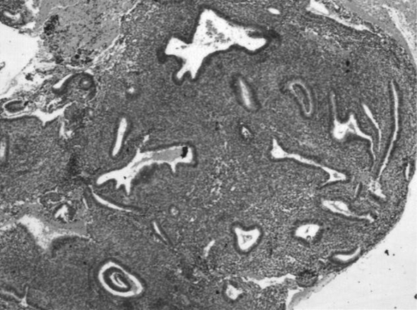
Isolated endometrial pathology as a cause of AUB was observed in 409 patients (Table 1). The remaining 211 patients who had leiomyoma, adenomyosis and cervical pathology with or without endometrial lesions were excluded from our final analysis. The age of 409 patients studied, were categorised into seven groups (Table 2). Age of patients with AUB ranged from 17 to 79 years in our study. Abnormal uterine bleeding was commonly seen in the 41 to 50 years age group and the predominant pattern noted was normal cycling endometrium closely followed by disordered proliferative pattern (Fig. 1). Except in the 71–80 years age group, a significant statistical association was seen between causes of AUB and age group with P value <0.001.
Table 1.
Details of cases presenting with AUB from June 2005 to June 2006
| Total no of cases | 620 |
| Isolated endometrial pathology | 409 |
| Leiomyoma ± endometrial pathology | 185 |
| Cervical pathology | 26 |
| Endometrial samples | 408 |
| Hysterectomy specimens | 212 |
Table 2.
Distribution of cases of AUB with isolated endometrial lesions according to age group
| Age groups | <20 years | 21–30 years | 31–40 years | 41–50 years | 51–60 years | 61–70 years | 71–80 years | Total |
|---|---|---|---|---|---|---|---|---|
| Normal cyclical patterns | 2 1.7% |
9 7.8% |
42 36.2% |
48 41.4% |
12 10.3% |
3 2.6% |
116 100% |
|
| Disordered proliferative pattern | 5 6% |
28 33.3% |
40 47.6% |
10 11.9% |
1 1.2% |
84 100% |
||
| Hyperplasia | 2 8% |
17 68% |
6 24% |
25 100% |
||||
| Atrophic pattern | 1 10% |
3 30% |
4 40% |
2 20% |
10 100% |
|||
| Benign endometrial polyp | 5 10.9% |
13 28.3% |
18 39.1% |
6 13% |
4 8.7% |
46 100% |
||
| Chronic endometritis | 2 11.8% |
5 29.4% |
6 35.3% |
3 17.6% |
1 5.9% |
17 100% |
||
| Endometrial carcinoma | 5 27.8% |
4 22.2% |
7 38.9% |
2 11.1% |
18 100% |
|||
| Complications of pregnancy | 4 4.3% |
63 67.7% |
26 28% |
93 100% |
||||
| Total | 6 1.5% |
85 20.8% |
116 28.4% |
137 33.5% |
45 11% |
18 4.4% |
2 0.4% |
409 100% |
Fig. 1.

Endometrial glands in irregular shapes with focal crowding of glands and presence of dense compact stroma (Hematoxylin and eosin ×20)
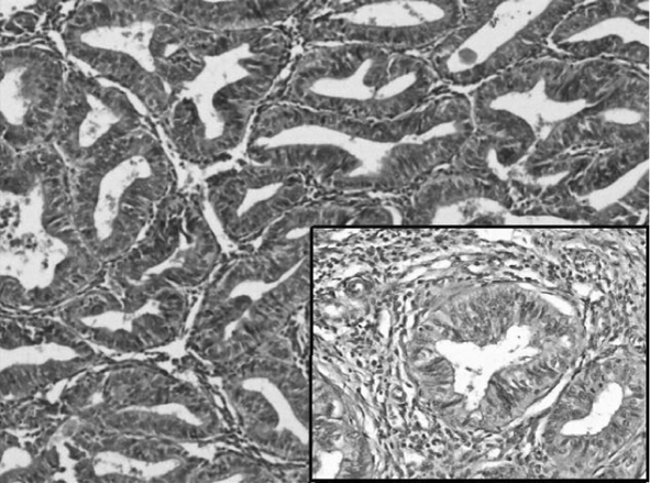
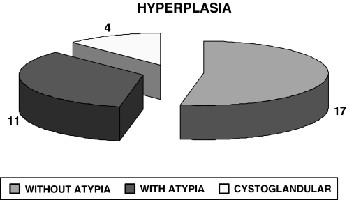
Histopathologic examination showed various pattern in AUB consisting of normal cyclical pattern showing proliferative, secretory and shedding phases in 116 patients of the total 409 cases (Table 2). Hyperplasia was observed in 25 patients (Graph 1) of which 8 patients presented with atypia (Fig. 2). Chronic endometritis was seen in 17 patients, including one case of tuberculous endometritis (Table 2). Complications of pregnancy were seen in 93 cases (Table 2) with abortion being the predominant cause (80 cases). Other causes were ectopic gestation (8 cases), partial mole (3 cases) and complete mole (2 cases). A total of 84/409 cases showed disordered proliferative pattern which were most commonly seen between 41 and 50 years of age.
Graph 1.
Distribution of cases of hyperplasia
Fig. 2.

Closely packed endometrial glands with sparse intervening stroma (Inset shows stratification of lining epithelium and epithelial cells show cytological atypia with high nucleocytoplasmic ratio and irregular clumping of nuclear chromatin on higher magnification)
Discussion
The term abnormal uterine bleeding has been used to describe any bleeding not fulfilling the criteria of normal menstrual bleeding. The causes of abnormal uterine bleeding include a wide spectrum of diseases of the reproductive system and non-gynecologic causes as well. Organic cause of abnormal uterine bleeding maybe subdivided into reproductive tract disease, iatrogenic causes and systemic disease. When an organic cause of AUB cannot be found, then by exclusion, a diagnosis of dysfunctional uterine bleeding (DUB) is assumed. In about 25% of the patients, the abnormal uterine bleeding is the result of a well defined organic abnormality [1].
The routine non invasive investigations for abnormal uterine bleeding include complete blood count, platelet count, prothrombin time (PT), Activated partial thromboplastin time (APTT) and liver function test to rule out any coagulation and bleeding disorders. In women of reproductive age group, serum and urine human chorionic gonadotropin (HCG) levels are evaluated to rule out pregnancy. To rule out an endocrine etiology, thyroid function test, follicle stimulating hormone (FSH), lutenizing hormone (LH), prolactin levels are assessed. On ruling out these causes, gynaecologists turn to imaging studies such as pelvic ultrasound (USG), and transvaginal USG and tissue sampling. Dilation and curettage can be a diagnostic as well as therapeutic procedure [2]. The sensitivity of endometrial biopsy for the detection of endometrial abnormalities has been reported to be as high as 96% [2, 3].
The most likely etiology of AUB relates to the patient’s age as to whether the patient is premenopausal, perimenopausal or postmenopausal [4]. The youngest patient in our study was a 17 year old girl and the oldest was a 79 year old lady. Newborn girls may have spotting within first few days of life because of withdrawal form high levels of maternal estrogen, which had stimulated the endometrium in utero. Beyond the neonatal period, causes such as precocious puberty and functional ovarian tumor have to be considered. In this age group the attending pediatrician should also do a careful search for urinary or cervical cause of bleeding [5].
The adolescent age group (<20 years) accounted for 1.5% of cases and their endometrium showed normal cyclical pattern. This may not be representative of true incidence because invasive procedures are avoided in this age group. Although most cases of abnormal uterine bleeding do not cause acute medical complications, bleeding can be traumatic for young patients and their families. The prevalence of a primary coagulation disorder in adolescent requiring hospitalization ranges from 3 to 20%, hence all adolescents with menorrhagia should undergo evaluation for coagulopathy [6].
Complications of pregnancy was common in the age group 21–30 years. This can be explained by the fact that most women conceive at this age, hence pregnancy should be considered a complication of pregnancy until proven otherwise. Patient’s presenting in this age group with abnormal uterine bleeding should be investigated and evaluated for pregnancy by doing a urine gravindex test [7].
Our study significantly revealed that the occurrence of menstrual disorders increases with advancing age. The commonest age group presenting with excessive bleeding in our study was 41–50 years. A similar incidence was reported by Yusuf et al. and Muzaffar et al. in their study of endometrium [8, 9]. Our study like several others showed that proliferative lesions like disordered proliferative pattern, hyperplasia, and benign endometrial polyp occur more commonly in the age group 41–50 years [9]. The reason for increased incidence of abnormal uterine bleeding in this age group (41–50 years) may be due to the fact that these patients are in their climacteric period. As women approach menopause, cycles shorten and often become intermittently anovulatory due to a decline in the number of ovarian follicles and the estradiol level.
The incidence of AUB between 51 and 70 years was lower as compared to those between 41 and 50 years. The reason for this finding may be due to the fact that the patients were evaluated much earlier and treated appropriately thereby decreasing the incidence in later age group. We had only 2 patients with AUB in the age group of 71 to 80 years and both of them had endometrial carcinomas.
Predominant number of cases in this study showed normal physiologic phases such as proliferative, secretory and atrophic menstrual pattern. The bleeding in the proliferative phase may be due to anovulatory cycles and bleeding in the secretory phase is due to ovulatory dysfunctional uterine bleeding.
A significant number of cases showed disordered proliferative pattern in this study. Disordered proliferative pattern lies at one end of the spectrum of proliferative lesions of the endometrium that includes carcinoma at the other end with intervening stages of hyperplasias. The term “disordered proliferative endometrium” has been used in a number of ways and is somewhat difficult to define. It denotes an endometrial appearance that is hyperplastic but without an increase in endometrial volume [10]. It also refers to a proliferative phase endometrium that does not seem appropriate for any one time in the menstrual cycle, but is not abnormal enough to be considered hyperplastic. Disordered proliferative pattern resembles a simple hyperplasia, but the process is focal rather than diffuse. A higher incidence of disordered proliferative pattern was found in our study as compared to Cho Nam-Hoon et al. [11]. An earlier stage of presentation due to increase health awareness could explain the high incidence in our study. Diagnosing the patients at the earliest stage of this spectrum will be of definitive help to the practicing gynaecologists to prevent the disease progression. But pathologists should have clear cut criteria for diagnosing disordered proliferative pattern and this should become a waste paper basket diagnosis.
Atrophic endometrium was seen predominantly in the 51–60 years age group. The incidence is slightly lower when compared with results shown by Gredmark et al. [12]. The exact cause of bleeding from the atrophic endometrium is not known. It is postulated to be due to anatomic vascular variations or local abnormal hemostatic mechanisms. Thin walled veins, superficial to the expanding cystic glands make the vessel vulnerable to injury.
The incidence of endometrial hyperplasias in this study was less as compared to others [12]. The possible explanation could be that most of patients here belong to lower socioeconomic status and the occurrence of risk factors like obesity, diabetes, increased intake of animal fat and sedentary life style is low. Another reason could be that most of these patients are being identified at a much earlier stage that is in the disordered proliferative phase. Identification of endometrial hyperplasia is important because they are thought to be precursors of endometrial carcinoma.
The incidence of benign endometrial polyps in this study was high in 41–50 years age group. Lower incidence of the endometrial polyps in the younger age group may be attributed to a possible spontaneous regression mechanism, which is characteristic of the cycling endometrium in reproductive age group. There is significant difference between the endometrial polyp and normal endometrium in receptor expression, cell proliferation and apoptosis regulation. These differences combined with non-random chromosomal aberrations and monoclonality suggests that polyp may provide a suitable microenvironment for the development of malignancy [13].
In the present study incidence of carcinoma endometrium was more common in the 51–60 years age group. The result of this study was almost similar to data mentioned by Yusuf et al. and Escoffery et al. in their study [8, 14]. A study done by Dangal et al. in Nepal documented a lower incidence of endometrial cancer in Nepalese woman attributing it to the practice of early childbearing and multiparty [15]. Possibly, the same factors contributed to a lower incidence of carcinoma in our patients.
One of the patient in this study who presented with AUB had malignant mixed mullerian tumour (MMMT) with a clinical stage I. Patients with MMMT are postmenopauasal women who present commonly with AUB but can also present with complaints of mass descending per vagina as well.
Abnormal uterine bleeding due to abortion formed a significant group in younger women. Hence, in reproductive age group complications of pregnancy should be ruled out in any patient with AUB. Chronic endometritis was observed in few patients. One case showed epithelioid granulomas suggestive of tuberculosis. Patient with chronic endometritis can present with AUB, pelvic pain and infertility. This condition needs to be diagnosed because with specific treatment, endometrium starts functioning normally.
Conclusion
Endometrial cause of AUB is age related pathology. Histopathological examination of endometrial biopsy is a major diagnostic tool in evaluation of AUB and a specific diagnosis could help the physician to plan therapy for successful management of AUB.
References
- 1.Brenner PF. Differential diagnosis of AUB. Am J Obstet Gynecol. 1996;175:766–769. doi: 10.1016/S0002-9378(96)80082-2. [DOI] [PubMed] [Google Scholar]
- 2.Albers JR, Hull SK, Wesley RM. Abnormal uterine bleeding. Am Fam Phys. 2004;69:1915–1926. [PubMed] [Google Scholar]
- 3.Litta P, Merlin F, Saccardi C, et al. Role of hysteroscopy with endometrial biopsy to rule out endometrial cancer in post menopausal women with abnormal uterine bleeding. Maturitas. 2005;50:117–123. doi: 10.1016/j.maturitas.2004.05.003. [DOI] [PubMed] [Google Scholar]
- 4.Dahlenbach-Hellweg G. Histopathology of endometrium. 4. New York: Springer-Verlag; 1993. [Google Scholar]
- 5.Emans SJ, Woods ER, Flagg NT, et al. Genital findings in sexually abused, symptomatic and asymptomatic girls. Paediatrics. 1987;79:778–785. [PubMed] [Google Scholar]
- 6.Shankar M, Lee CA, Sabin CA, et al. Von Willebrand disease in women with menorrhagia: a systematic review. BJOG. 2004;111:734–740. doi: 10.1111/j.1471-0528.2004.00176.x. [DOI] [PubMed] [Google Scholar]
- 7.Kilbourn CL, Richards CS. Abnormal uterine bleeding, diagnostic considerations, management options. Postgrad Med. 2001;109:137–150. doi: 10.3810/pgm.2001.01.832. [DOI] [PubMed] [Google Scholar]
- 8.Yusuf NW, Nadeem R, Yusuf AW, et al. Dysfunctional uterine bleeding. A retrospective clinicopathological study over 2 years. Pak J Obstet Gynaecol. 1996;9:27–30. [Google Scholar]
- 9.Muzaffar M, Akhtar KAK, Yasmin S, et al. Menstrual irregularities with excessive blood loss: a clinico-pathologic correlation. J Pak Med Assoc. 2005;55:486–489. [PubMed] [Google Scholar]
- 10.Steven SG. Problems in the differential diagnosis of endometrial hyperplasia and carcinoma. Mod Pathol. 2000;13:309–327. doi: 10.1038/modpathol.3880053. [DOI] [PubMed] [Google Scholar]
- 11.Nam-Hoon C, Chan-II P, In-Joon C. Clinicopathologic study of the endometrium. Dysfunctional uterine bleeding. Korean J Pathol. 1989;23:65–74. [Google Scholar]
- 12.Gredmark T, Kvint S, Havel G, et al. Histopathological findings in women with postmenopausal bleeding. B J Obstet Gynaecol. 1998;102:133–136. doi: 10.1111/j.1471-0528.1995.tb09066.x. [DOI] [PubMed] [Google Scholar]
- 13.Hileeto D, Fadare O, Martel M, et al. Age dependent association of endometrial polyps with increased risk of cancer involvement. World J Surg Oncol. 2005;3:8. doi: 10.1186/1477-7819-3-8. [DOI] [PMC free article] [PubMed] [Google Scholar]
- 14.Escoffery CT, Blake GO, Sargenat LA. Histopathological findings in women with postmenopausal bleeding in Jamaica. West Indian Med J. 2002;51:232–235. [PubMed] [Google Scholar]
- 15.Dangal G. A study of endometrium of patients with abnormal uterine bleeding at chitwan valley. Kathmandu Univ Med J. 2003;1:110–112. [PubMed] [Google Scholar]


