Abstract
Objective:
The aim of the study was to investigate differences in the palatal rugae patterns in males and females of a cross-sectional hospital-based coastal Andhra population and application of discriminant function analysis in sex identification.
Materials and Methods:
One hundred pre-orthodontic plaster casts, equally distributed between males and females belonging to an age range of 15-30 years, were examined for different rugae patterns. Thomas classification was adopted for analysis. Association between rugae patterns and sexual dimorphism were tested using Unpaired t test, Chi square test and discriminant function analysis developed using SAS package.
Results:
Difference in unification pattern among males and females was found to be statistically significant. The total number of the rugae was not statistically significant between the sexes. Association between rugae length and shape with sex determination was computed using discriminant analysis which enabled sex differentiation in this population with an accuracy of 78%.
Conclusion:
Palatal rugae revealed a specific pattern in unification among males and females of the coastal Andhra population. Discriminant function analysis enabled sex determination of individuals. However, these interpretations were precluded by the small sample size and further research work on larger samples and use of different classification systems is required to validate its use in forensic science.
Keywords: Coastal Andhra Pradesh, discriminant function analysis, palatal rugae, rugoscopy, sex determination
Introduction
The identification of sex is of significance in case of major disasters where bodies are often damaged beyond recognition. On predicting the sex it builds the biological profile of the unidentified human remains thereby excluding about half the population in search operations.
Palatal rugae (rugae palatinae or plicae palatinae transversae) refer to a series of transverse ridges on the anterior part of the palatal mucosa on each side of the median palatal raphe and behind the incisive papillae. According to a histological study of the development in mice, palatal rugae develop in the third month of intrauterine life as localized regions of epithelial proliferation and thickening even before the elevation of the palatal shelves.[1] Later, fibroblasts and collagen fibers accumulate in the connective tissue beneath the thickened epithelium and attain a distinctive orientation.[2] Physiologically the palatal rugae aid in oral swallowing, taste perception, participate in speech, suction in children and in the medico-legal identification process.[3]
Palatoscopy or palatal rugoscopy is the study of palatal rugae in order to establish a person's identity. This palatal rugoscopy was first proposed in 1932 by a Spanish investigator, Trobo Hermosa.[4] Palatal rugae is selected in forensics as it is relevant for human identification due to its internal position, stability, perennity[5] i.e., it persists throughout life. Also, its design and structure are invariably unchanged and not altered by routine chemicals like nicotine, ethanol, acetyl salicylate etc., consumed heat, disease or trauma. There are reports published that the number of rugae remains unchanged throughout life but the size and pattern of arrangement changes with palatal development.[6] In addition, the rugae pattern appears to be specific to racial groups facilitating population identification.[7]
The basic theory of discriminant analysis was developed by Fisher.[8–10] This technique is designed to generate rules for classifying individuals into a defined group on the basis of a set of measurements of the individual.[11] In our study this technique was used for sex determination.
The objective of the present study is to investigate the palatal rugae patterns in males and females of a cross-sectional hospital-based coastal Andhra population and apply discriminant analysis in sex identification.
Materials and Methods
The study sample consisted of 100 pre- orthodontic dental casts which included 50 males and 50 females in the age group of 15-30 years from the Department of Orthodontics, Vishnu Dental College, Bhimavaram, West Godavari District, Andhra Pradesh. All individuals of the study belonged to the same geographical population and were healthy, free of any diagnosed congenital abnormalities, inflammation, trauma or orthodontic treatment. All selected casts from the individuals were free of air bubbles or voids, especially at the anterior third of the palate. Initially rugae patterns on the study models were delineated using 0.1 HB graphite pencil under adequate light and magnification using hand lens. Delineation enhanced the visualization of the palatal rugae on these casts. Measurements were made directly from the cast using digital slide calipers with an accuracy of 0.05 mm from the origin near the mid- palatine raphe to the terminal end transversely.
The method of identification of the palatal rugae pattern was based on the classification of Thomas et al.,[11] (1983). This classification includes number, length, shape, and unification patterns of rugae.
Having determined the length of all rugae, three categories were formed:
-
1)
Primary rugae (5-10 mm)
-
2)
Secondary rugae (3-5 mm)
-
3)
Fragmentary rugae (less than 3 mm)
All rugae were considered for the study irrespective of their length.
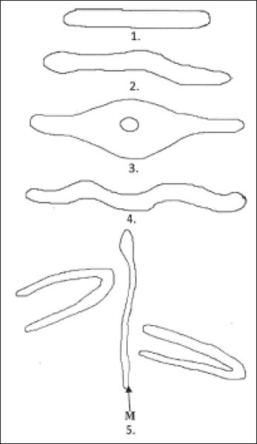
Considering that the rugae originate from the mid- palatine raphe and terminates Transversly, the shapes of individual rugae were classified into four major types [Figure 1]: 1. STRAIGHT- runs directly from the origin to termination 2. CURVY- a simple crescent shape which was curved gently 3. CIRCULAR- a definite continuous ring formation 4. WAVY- serpentine form. In case of circular shape the diameter from the origin to termination was considered. Additionally, a specific pattern called unification occurs when rugae have two arms which are joined either at their origin or termination. The unification pattern is further subdivided into diverging and converging type. A diverging pattern occurs when two rugae begin from the same origin but immediately diverge transversely. Similarly, a converging pattern occurs when two rugae arise with different origins and converge transversely.
Figure 1.

Patterns of palatal rugae based on shape and unification
Every identification and measurement was done by one examiner and the readings were repeated three times for each cast.
Statistical analysis
Unpaired t test and Chi square test were used in assessing sex differences in the number and unification pattern of rugae respectively. Association of palatal rugae length and shape in sex determination were tested using univariate discriminatory analysis developed using the SAS (Statistical Analysis Software) 9.0 version.
Results
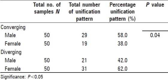
The average number of rugae in females was slightly more when compared to males, but it was statistically insignificant [Table 1]. [Table 2] showed gender-wise distribution of mode of unification of the rugae showing a P value of 0.04 which shows that the difference is statistically significant. [Table 3] shows that variables of length and shape of the palatal rugae are statistically significant and on further discriminant analysis these variables can classify the sex of an individual. From the details obtained from [Table 4] an equation was constructed which can be executed for sex determination when both length and shape of the palatal rugae are considered.
Table 1.
Unpaired t test analysis for assessing the difference in the total number of rugae in males and females

Table 2.
Chi square test analysis for assessing the difference in the unification pattern of rugae in males and females

Table 3.
Univariate discriminant analysis of the variables involved in the length and shape of rugae

Table 4.
Discriminant function coefficients for sex determination considering both length and shape of the rugae

GENDER = –0.2620 (PR)–0.5133 (SR) – 0.6614 (FR) + 0.3366 (STRAIGHT) + 0.4582 (WAVY) + 0.4353 (CURVED) + 0.5096 (CIRCULAR)
After executing the above equation with the new data, sex determination could be made with the help of the adjusted canonical centroids of –0.3088 to 0.3088 i.e. if the product obtained is close to –0.3088 then the proposed gender of the individual is female and if the other centroid is close to 0.3088 then the proposed gender of the individual is male. This model when tested with the present data as well as new data it derived a ‘F’ likelihood ratio test with model accuracy of 73.08%. From the details obtained from Table 5 an equation was constructed which can determine sex when only the length of the palatal rugae is considered.
Table 5.
Discriminant function co-efficients for sex determination considering only length of the rugae

GENDER = – 0.9878 (PR) – 0.2713 (SR)–0.2933 (FR)
After executing the above equation with the new data, sex determination could be made with the help of the adjusted canonical centroids of –0.2144 to 0.2144 i.e. if the product obtained is close to –0.2144 then the classified sex of the individual is male and if the obtained product is close to 0.2144 then the classified sex of the individual is female. This model when tested with the present data as well as new data it derived a ‘F’ likelihood ratio test with model accuracy of 78%.
Discussion
Determination of sex is the key analysis that forensic investigators perform in order to construct the biological profile of human remains.[12] In this context, the assessment of sex can substantially narrow the biological profile for unidentified remains. Indeed, in most cases, clarification of the events and later judicial decisions will depend on the precision and the reliability of the identification procedures.[13] Routinely, teeth and bones are used for sex determination in forensics but in cases of their absence in identification, mucosal tissues are important since these structures provide interesting data for identification. Application of palatal rugae patterns to personal identification was first suggested by Allen in 1889.[14] Since then various studies were done in association of palatal rugae and its use in forensic identification. The key factors which make palatal rugae one of the investigative tools in forensics are its internal position, stability, perennity etc.[5]
In our study dental casts were used as an antemortem record as these present the advantages of simple analysis, reduced cost and easy fabrication when compared to digital photographs[5] which are highly viable and require the development of a specific software for image superimposition and location of landmarks to allow identification. Also, photography is a skill which needs a specific angulation and position to attain a good photograph.
Researchers had found difficulty in the task of classification of the rugae patterns due to the subjective nature of observation and interpretation within and between observers. Since the study of Lysell[15] (1955) specific anatomical investigations on palatal rugae patterns have been reported by many researchers. Numerous classifications have been devised by several authors to record the palatal rugae patterns; among all, the Silva,[16] Carrea,[17] Lysell,[18] Thomas and Kotze[16–18] classifications are often used in recording the patterns. Thomas and Kotze in their literature highlighted the difficulties in observing, classifying and interpreting the limitless and minute variations in palatal rugae and emphasized the necessity for standardizing the procedures in recording. After a thorough review on all classifications from the literature, the method of identification used in this study (Thomas et al., 1983) was the most practical and easy to apply compared with other methods.
Various studies reported the regional variation in the palatal rugae patterns by comparing the patterns in males and females. Dohke and Osato[19] reported that among the Japanese, the females had fewer rugae than males. In a comparative study between Indians and the Tibetan population by Shetty et al.,[20] it was reported that Indian males had more primary rugae on the left side as compared to females and vice versa for the Tibetan population. Also, Indian males had more number of curved rugae on both the right and left sides than Tibetan males, and Tibetan females had more wavy rugae on the right and left sides than Indian females. Due to this variation of patterns in males and females, we hypothesized and conducted this study to know the degree of sexual dimorphism of the palatal rugae and its role in sex determination. Results showed that there exists a significant difference in the total number and unification pattern of rugae among males and females. On discriminant analysis of the length and shape of the rugae, an equation for sex discrimination was obtained which was 73.08% accurate, this could be due to the increased involvement of variables in the analysis. When an equation was made by discriminant analysis considering only the length of the rugae, the accuracy was 78%. Reduced accuracy may be due to the reduced sample number. Further research is indicated with a larger sample size in order to validate our findings and application of advanced statistical methods in attaining better accuracy levels for the use of palatal rugae patterns in sex determination.
Conclusion
Palatal rugae revealed a specific pattern in unification among males and females of coastal Andhra population. Discriminant function analysis enabled sex determination of individuals. However, these interpretations are precluded by the small sample size and further research work on larger samples and application of advanced statistical methods is required to validate its use in forensic application.
Acknowledgments
We wish to thank Mr. Venugopal Sharma, SAS Analyst, V Infosystems Pvt Ltd, Bangalore, for his valuable contribution.
Footnotes
Source of Support: Nil
Conflict of Interest: None declared
References
- 1.Amasaki H, Ogawa M, Nagasao J, Mutoh K, Ichihara N, Asari M, et al. Distributional changes of BrdU, PCNA, E2F1 and PAL31 molecules in developing murine palatal rugae. Ann Anat. 2003;185:517–23. doi: 10.1016/s0940-9602(03)80116-4. [DOI] [PubMed] [Google Scholar]
- 2.Hauser G, Daponte A, Roberts MJ. Palatal rugae. J Anat. 1989;133:41–4. [PMC free article] [PubMed] [Google Scholar]
- 3.Venegas VH, Valenzuela JS, Lopez MC, Suazo IC. Palatal rugae: Systematic analysis of its shape and dimensions for use in human Identification. Int J Morphol. 2009;27:819–25. [Google Scholar]
- 4.Pueyo VM, Garrido BR, Sánchez JS. Odontología legaly forense, Masson, Barcelona. 1994;23:277–92. [Google Scholar]
- 5.Filho EM, Helena SP, Arsenio SP, Suzana MC. Palatal rugae patterns as bioindicator of identification in forensic dentistry. RFO. 2009;14:227–33. [Google Scholar]
- 6.Silva L. Ficha rugoscopica palatine. Brasil Odonto. 1938;14:307–16. [Google Scholar]
- 7.Carrea JU. Gaumenfalten-fotostenogramme, ein neues identifizierungsverfahren. Dtsch Zahnarztl Z. 1955;10:11–7. [PubMed] [Google Scholar]
- 8.Fisher RA. The use of multiple measurements in taxonomic problems. Ann Eugen. 1936;7:179–188. [Google Scholar]
- 9.Fisher RA. The statistical utilization of multiple measurements. Ann Eugen. 1936;8:376–86. [Google Scholar]
- 10.Fisher RA. The precision of discriminant function. Ann Eugen. 1940;10:422–9. [Google Scholar]
- 11.Huberty CJ. Applied discriminant analysis. New York: Wiley; 1994. [Google Scholar]
- 12.Williams BA, Rogers T. Evaluating the accuracy and precision of cranial morphological traits for sex determination. J Forensic Sci. 2006;51:729–35. doi: 10.1111/j.1556-4029.2006.00177.x. [DOI] [PubMed] [Google Scholar]
- 13.Barrio PA, Trancho GJ, Sanchez JA. Metacarpal sexual determination in a Spanish population. J Forensic Sci. 2006;51:990–5. doi: 10.1111/j.1556-4029.2006.00237.x. [DOI] [PubMed] [Google Scholar]
- 14.Allen H. The palatal rugae in man. Dent Cosmos. 1889;31:66–80. [Google Scholar]
- 15.Lysell L. Plicae palatinae transversae and papilla incisive in man: A morphologic and genetic study. Acta Odontol Scand. 1955;13(suppl 18):1–137. [PubMed] [Google Scholar]
- 16.Thomas CJ, Kotze TJ. The palatal rugae pattern in six Southern African human populations, Part I: A description of the population and a method for its investigation. J Dent Assoc S Afr. 1983;38:547–53. [PubMed] [Google Scholar]
- 17.Thomas CJ, Kotze TJ. The palatal ruga pattern in six Southern African human populations, Part II: Inter-racial differences. J Dent Assoc S Afr. 1983;38:166–72. [PubMed] [Google Scholar]
- 18.Thomas CJ, Kotze TJ. The palatal rugae pattern: A new classification. J Den Assoc S Afr. 1983;38:153–7. [PubMed] [Google Scholar]
- 19.Dohke M, Osato S. Morphological study of the palatal rugae in Japanese 1.Bilateral differences in the regressive evaluation of the palatal rugae. Jap J Oral Biol. 1994;36:125–40. [Google Scholar]
- 20.Shetty SK, Kalia S, Patil K, Mahima VG. Palatal rugae pattern in Mysorean and Tibetan populations. Indian J Dent Res. 2005;16:51–5. [PubMed] [Google Scholar]


